. 2019 Jan:122:106-119.
doi: 10.1016/j.neuint.2018.11.012. Epub 2018 Nov 16.

Intracellular emetic signaling cascades by which the selective neurokinin type 1 receptor (NK1R) agonist GR73632 evokes vomiting in the least shrew (Cryptotis parva)

Affiliations

Intracellular emetic signaling cascades by which the selective neurokinin type 1 receptor (NK1R) agonist GR73632 evokes vomiting in the least shrew (Cryptotis parva)

W Zhong et al. Neurochem Int. 2019 Jan.

Abstract

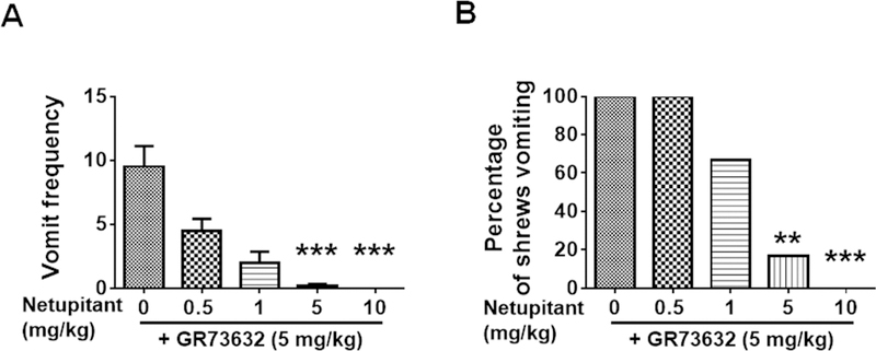
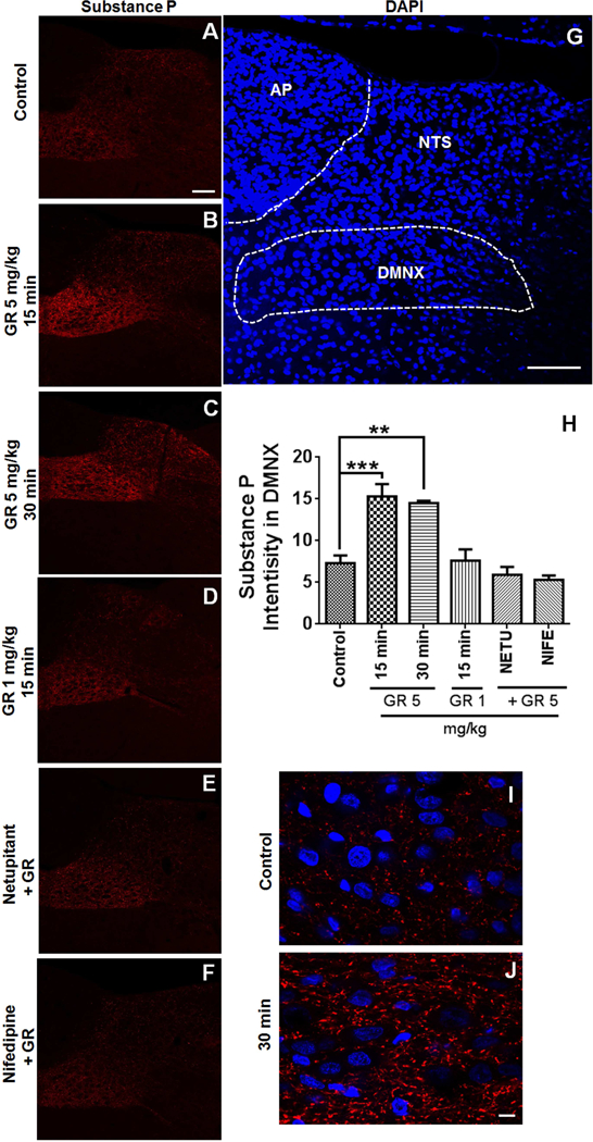
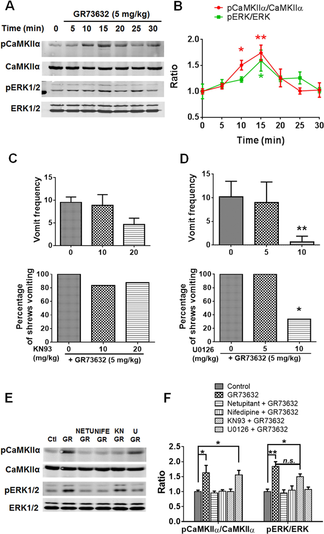
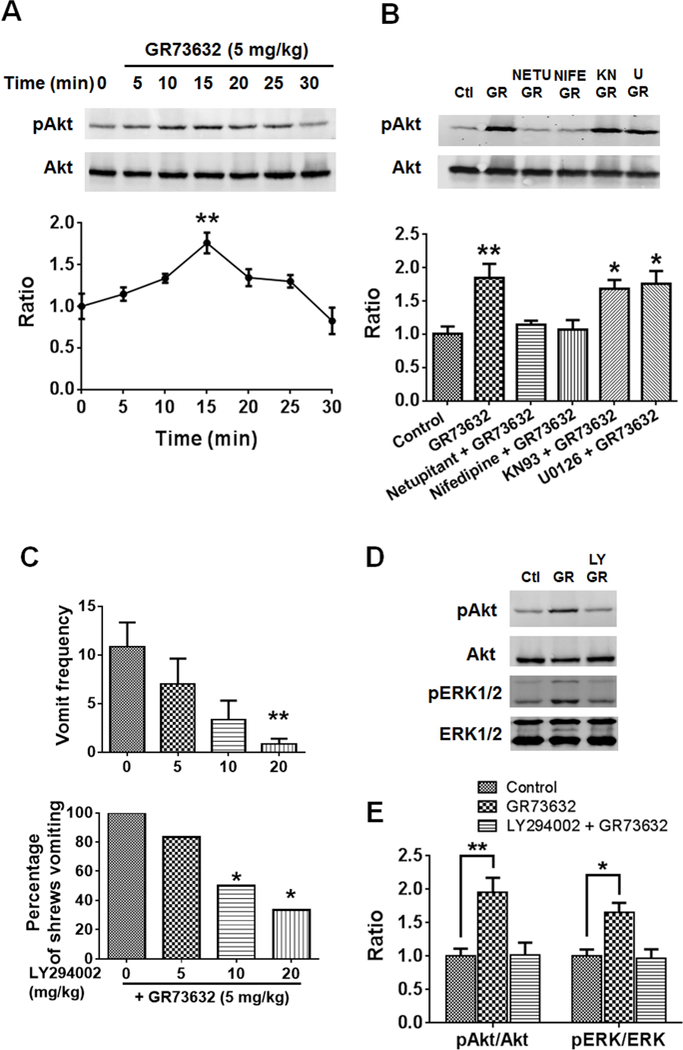
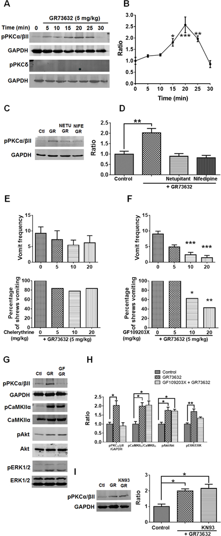
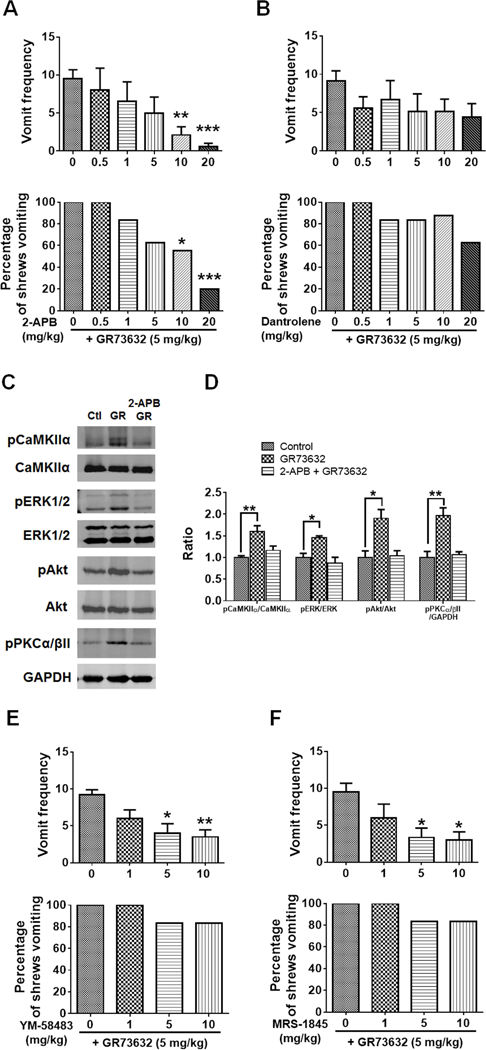
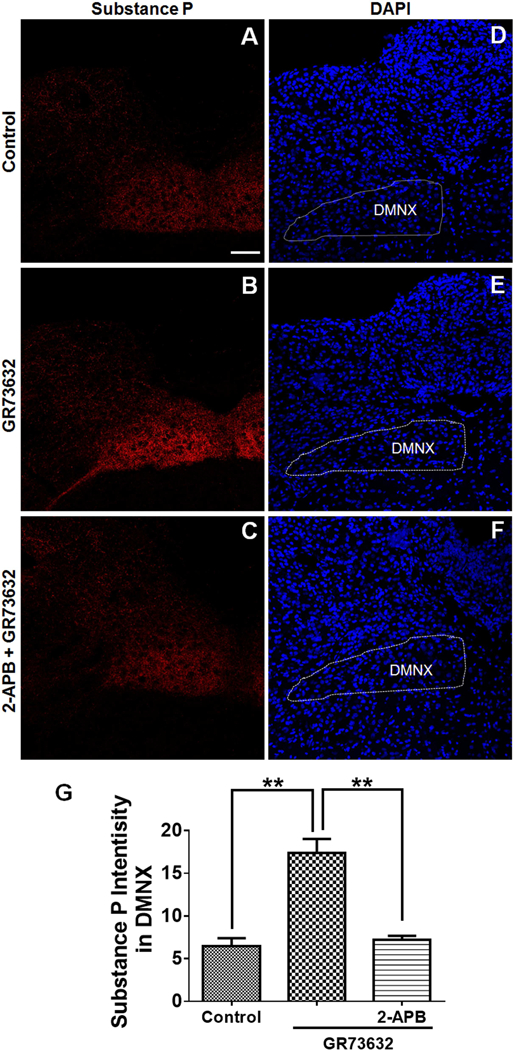
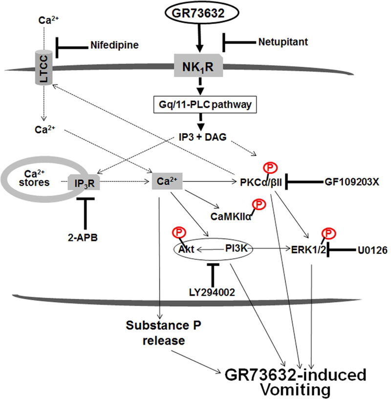
To characterize mechanisms involved in neurokinin type 1 receptor (NK1R)-mediated emesis, we investigated the brainstem emetic signaling pathways following treating least shrews with the selective NK1R agonist GR73632. In addition to episodes of vomiting over a 30-min observation period, a significant increase in substance P-immunoreactivity in the emetic brainstem dorsal motor nucleus of the vagus (DMNX) occurred at 15 min post an intraperitoneal (i.p.) injection GR73632 (5 mg/kg). In addition, time-dependent upregulation of phosphorylation of several emesis -associated protein kinases occurred in the brainstem. In fact, Western blots demonstrated significant phosphorylations of Ca2+/calmodulin kinase IIα (CaMKIIα), extracellular signal-regulated protein kinase1/2 (ERK1/2), protein kinase B (Akt) as well as α and βII isoforms of protein kinase C (PKCα/βII). Moreover, enhanced phospho-ERK1/2 immunoreactivity was also observed in both brainstem slices containing the dorsal vagal complex emetic nuclei as well as in jejunal sections from the shrew small intestine. Furthermore, our behavioral findings demonstrated that the following agents suppressed vomiting evoked by GR73632 in a dose-dependent manner: i) the NK1R antagonist netupitant (i.p.); ii) the L-type Ca2+ channel (LTCC) antagonist nifedipine (subcutaneous, s.c.); iii) the inositol trisphosphate receptor (IP3R) antagonist 2-APB (i.p.); iv) store-operated Ca2+ entry inhibitors YM-58483 and MRS-1845, (i.p.); v) the ERK1/2 pathway inhibitor U0126 (i.p.); vi) the PKC inhibitor GF109203X (i.p.); and vii) the inhibitor of phosphatidylinositol 3-kinase (PI3K)-Akt pathway LY294002 (i.p.). Moreover, NK1R, LTCC, and IP3R are required for GR73632-evoked CaMKIIα, ERK1/2, Akt and PKCα/βII phosphorylation. In addition, evoked ERK1/2 phosphorylation was sensitive to inhibitors of PKC and PI3K. These findings indicate that the LTCC/IP3R-dependent PI3K/PKCα/βII-ERK1/2 signaling pathways are involved in NK1R-mediated vomiting.

Keywords: Brainstem; ERK1/2; Emesis; GR73632; Gut; NK(1) receptor.


Conflict of interest statement

Conflicts of interest

We have no conflict of interest to declare.

Figures

Fig. 1.
Fig. 1.. Antiemetic effects of the neurokinin 1 receptor (NK1R) selective antagonist netupitant against GR73632-induced emesis in the least shrew.
Different groups of shrews received either vehicle (0 mg/kg, i.p.), or varying doses of netupitant (0.5, 1, 5 or 10 mg/kg, i.p.), 30 min prior to an emetic dose of GR73632 (5 mg/kg, i.p.). Emetic parameters were recorded for 30 min post emetic injection. A. The frequency (mean ± SEM) of emesis. n = 6. *** p < 0.001 vs netupitant 0 + GR73632, Kruskal-Wallis non-parametric one-way ANOVA and followed by Dunn’s post hoc test. B. The percentage of shrews vomiting. ** p < 0.01, chi square test.
Fig. 2.
Fig. 2.. Immunohistochemical analysis of GR73632-evoked substance P (SP) release in the least shrew brainstem.
Coronal brainstem sections (20 μm) were prepared from different groups of Shrews (n = 3 per group), including vehicle-treated control (A); 15 or 30 min post a 5 mg/kg (i.p.) injection of GR73632 (B and C); 15 min post a 1 mg/kg (i.p.) GR73632 injection (D); 15 min post GR73632 (5 mg/kg, i.p.) with 30-min pretreatment with either netupitant (10 mg/kg, i.p.) (E) or nifedipine (10 mg/kg, s.c.) (F). Sections were immunolabeled with rat SP antibody overnight followed by CY™3-conjugated donkey anti-rat secondary antibody incubation. A-F. Representative 20x images of brainstem slice showing SP immunoreactivity among the brainstem emetic nuclei, area postrema (AP), nucleus of the solitary tract (NTS) and dorsal motor nucleus of the vagus (DMNX). Scale bar, 100 μm. G. The boundary of DMNX is shown on representative section with nuclei stained with DAPI. Scale bar, 100 μm. H. Quantitative analysis of substance P immunostaining. In order to correct for section-to-section variation in staining intensity, substance P intensities determined in a 267 × 96 μm2 area in the DMNX were subtracted from the same size area bellow DMNX (known as the hypoglossal nucleus) that was negative of SP staining of the same slice. 3 images from each shrew were chosen for analysis. The average SP intensity was calculated and used as a value for an individual animal. Shown are means ± SEM of n = 3. ** p < 0.01; *** p < 0.001 vs Control, One-way ANOVA followed by Dunnett’s test. I-J. High magnification images show GR73632 enhancement of SP-immunoreactive puncta structures in the DMNX at 30 min post treatment. Bar, 10 μm.
Fig. 3.
Fig. 3.. Involvement of L-type Ca2+ channel (LTCC) in NK1R-dependent CaMKIIα and ERK1/2 activation.
A. Representative Western blots for time-course of CaMKIIα and ERK1/2 phosphorylation in the least shrew brainstems collected at indicated time points after GR73632 (5 mg/kg, i.p.) administration (n = 3 per group). Phospho-CaMKIIα T286 (pCaMKIIα), CaMKIIα, phospho-ERK1/2 (pERK1/2) and ERK1/2 were detected by Western blot. B. Quantitative analysis of Western blots as shown in A. The ratios of pCaMKIIα to CaMKIIα and pERK1/2 to ERK1/2 (pERK/ERK) were compared. All ratios were normalized to vehicle-treated control (0 min) values before analysis and expressed as fold change of control. * p < 0.05; ** p < 0.01 vs. 0 min, one-way ANOVA followed by Dunnett’s test. n = 3. C-D. Effects of either CaMKII inhibitor KN93 (C) or ERK1/2 pathway inhibitor U0126 (D) on GR73632-produced emesis. Different groups of shrews received i.p. vehicle (0 mg/kg), varying doses of KN93 (10 and 20 mg/kg, i.p. n = 6–8) or U0126 (5 and 10 mg/kg, i.p., n = 6), 30 min prior to GR73632 (5 mg/kg, i.p.). Emetic parameters were recorded for 30 min post emetic injection. Upper panel shows frequency data presented as mean (± SEM). ** p < 0.01 vs. U0126 0 + GR73632, Kruskal-Wallis non-parametric one-way ANOVA and followed by Dunnett’s post hoc test. Below panel shows the percentage of shrews vomiting. * p < 0.05 vs. U0126 0 + GR73632, chi square test. E-F. Effect of selective NK1R (netupitant) or LTCC (Nifedipine) antagonists on CaMKIIα and ERK1/2 phosphorylation evoked by GR73632 and the relationship between CaMKIIα and ERK1/2. E. Representative Western blots of CaMKIIα and ERK1/2 phosphorylation in brainstems obtained from vehicle-treated control (Ctl) or when least shrews were treated with GR73632 (5 mg/kg., i.p.) for 15 min in the absence (GR) or presence of an i.p. 10 mg/kg dose of netupitant (NETU+GR), an s.c. 10 mg/kg dose of nifedipine (NIFE+GR), i.p. 20 mg/kg KN93 (KN+GR), or an i.p. 10 mg/kg dose of U0126 (U+GR). All inhibitors were delivered 30 min prior to GR73632. Phospho-CaMKIIα T286 (pCaMKIIα), CaMKIIα, phospho-ERK1/2 (pERK1/2) and ERK1/2 were detected by Western blot. F. Quantitative analysis of Western blots of data shown in F. The ratios of pCaMKIIα to CaMKIIα and pERK1/2 to ERK1/2 (pERK/ERK) were compared. All ratios were normalized to control values before analysis and expressed as fold change of control. * p < 0.05; ** p < 0.01 vs. Control, one-way ANOVA followed by Dunnett’s test. n.s., not significantly different, unpaired t-test. n = 3.
Fig. 4.
Fig. 4.. Involvement of LTCC in NK1R-dependent PI3K-ERK1/2 activation.
A. Western blots for time-course of Akt phosphorylation in the least shrew brainstems were collected at the indicated time points after GR73632 (5 mg/kg, i.p.) administration. Phospho-Akt S473 (pAkt) and Akt were detected by Western blot. The below panel shows quantitative analysis of Western blots. The ratios of p-Akt to Akt were compared. All ratios were normalized to vehicle-treated control (0 min) values before analysis and expressed as fold change of control. ** p < 0.01 vs. 0 min, one-way ANOVA followed by Dunnett’s test. n = 3. B. Effects of NK1R, LTCC, CaMKIIα and ERK1/2 inhibitors on Akt phosphorylation evoked by GR73632. Western blots of Akt phosphorylation in brainstems obtained from vehicle-treated control (Ctl) or the least shrews treated with GR73632 (5 mg/kg., i.p.) for 15 min in the absence (GR) or presence of an i.p. 10 mg/kg dose of netupitant (NETU+GR), s.c. 10 mg/kg dose of nifedipine (NIFE+GR), i.p. 20 mg/kg dose of KN93 (KN+GR), or an i.p. 10 mg/kg dose of U0126 (U+GR). All inhibitors were delivered 30 min prior to GR73632. Phospho-Akt S473 (pAkt) and Akt were detected by Western blot. The below panel shows quantitative analysis of Western blots. The ratios of pAkt to Akt were compared. All ratios were normalized to control values before analysis and expressed as fold change of control. * p < 0.05; ** p < 0.01 vs. Control, one-way ANOVA followed by Dunnett’s test. n = 3. C. Antiemetic effects of the PI3K inhibitor LY294002 on GR73632-induced emesis. Different groups of shrews received i.p. vehicle (0 mg/kg), or varying doses of LY294002, 30 min prior to GR73632 (5 mg/kg, i.p.). Emetic parameters were recorded for 30 min post emetic injection. Upper panel shows frequency data presented as mean ± SEM. n = 6. ** p < 0.01 vs. LY294002 0 + GR73632, Kruskal-Wallis non-parametric one-way ANOVA and followed by Dunnett’s post hoc test. Below panel shows the percentage of shrews vomiting. * p < 0.05, chi square test. D. Effect of the PI3K inhibitor on Akt and ERK1/2 phosphorylation induced by GR73632. Shrews were pretreated (i.p.) for 30 min with 20 mg/kg LY294002 (LY+GR) or the vehicle (GR) and then stimulated with GR73632 (5 mg/kg, i.p.) for 15 min. Phospho-Akt S473 (pAkt), Akt, phospho-ERK1/2 (pERK1/2) and ERK1/2 in brainstems were detected by Western blot. Shown are representative western blots. E. Quantitative analysis of Western blots in D. The ratios of pAkt to Akt and pERK1/2 to ERK1/2 (pERK/ERK) were compared. All ratios were normalized to control values before analysis and expressed as fold change of control. * p < 0.05; ** p < 0.01 vs. Control, one-way ANOVA followed by Dunnett’s test. n = 3.
Fig. 5.
Fig. 5.. Involvement of LTCC in NK1R-dependent PKCα/βII-ERK1/2 activation.
A. Representative Western blots for time-course of PKC isoforms activation in the least shrew brainstems collected at indicated time points after GR73632 (5 mg/kg, i.p.) administration. Phospho-PKCα/βII Thr638/641 (pPKCα/βII), phospho-PKCδ Tyr311 (pPKCδ) and GAPDH (loading control) were detected by Western blot. B. Quantitative analysis of Western blots shown in A. The ratios of pPKCα/βII to GAPDH were compared. All ratios were normalized to vehicle-treated control (0 min) values before analysis and expressed as fold change of control. * p < 0.05; ** p < 0.01 vs. 0 min, one-way ANOVA followed by Dunnett’s test. n = 3. C. Representative Western blots of PKCα/βII phosphorylation in brainstems obtained from vehicle-treated control (Ctl) or the least shrews treated with GR73632 (5 mg/kg., i.p.) for 15 min in the absence (GR) or presence of an i.p. 10 mg/kg dose of netupitant (NETU+GR), or s.c. 10 mg/kg dose of nifedipine (NIFE+GR). All inhibitors were delivered 30 min prior to GR73632. Phospho-PKCα/βII Thr638/641 (pPKCα/βII) and GAPDH were detected by Western blot. D. Quantitative analysis of Western blots shown in C. The ratios of pPKCα/βII to GAPDH were compared. All ratios were normalized to control values before analysis and expressed as fold change of control. ** p < 0.01 vs. Control, one-way ANOVA followed by Dunnett’s test. n = 3. E-F. Antiemetic effects of PKC inhibitors on GR73632-produced emesis. Different groups of shrews received i.p. vehicle (0 mg/kg), varying doses of chelerythrine (i.p.) or GF109203X (i.p.), 30 min prior to an injection of GR73632 (5 mg/kg, i.p.). Emetic parameters were recorded for the next 30 min. Upper panels show frequency data presented as mean ± SEM. n = 6–9. *** p < 0.001 vs. 0, Kruskal-Wallis non-parametric one-way ANOVA and followed by Dunnett’s post hoc test. Below panels show the percentage of shrews vomiting. * p < 0.05; ** p < 0.01, chi square test. G-H. Effects of PKC inhibition with GF109203X on PKCα/βII, CaMKIIα, Akt and ERK1/2 phosphorylation induced by GR73632. G. Representative Western blots of PKCα/βII, CaMKIIα, Akt and ERK1/2 phosphorylation in brainstems obtained from vehicle-treated control (Ctl) or least shrews treated with GR73632 (5 mg/kg., i.p.) for 15 min in the absence (GR) or presence of an i.p. 20 mg/kg GF109203X (GF+GR). The inhibitor was delivered 30 min prior to GR73632. Phospho-PKCα/βII Thr638/641 (pPKCα/βII), GAPDH, phospho-CaMKIIα T286 (pCaMKIIα), CaMKIIα, phospho-Akt S473 (pAkt), Akt, phospho-ERK1/2 (pERK1/2) and ERK1/2 were detected by Western blot. H. Quantitative analysis of Western blots similar to those in G. The ratios of pPKCα/βII to GAPDH, pCaMKIIα to CaMKIIα, pAkt to Akt and pERK1/2 to ERK1/2 (pERK/ERK) were calculated. All ratios were normalized to control values before analysis and expressed as fold change of control. * p < 0.05; ** p < 0.01 vs. Control, one-way ANOVA followed by Dunnett’s test. n = 3. I. Effect of CaMKIIα inhibiton by KN93 on PKCα/βII phosphorylation induced by GR73632. Shrews were pretreated (i.p.) for 30 min with 20 mg/kg KN93 (KN+GR) or the vehicle (GR) and then stimulated with GR73632 (5 mg/kg, i.p.) for 15 min. Phospho-PKCα/βII Thr638/641 (pPKCα/βII) and GAPDH in brainstems were detected by Western blot. Left panel shows the representative Western blots. Right panel show quantitative analysis of Western blots. The ratio of pPKCα/βII and GAPDH Was compared. All ratios were normalized to control values before analysis and expressed as fold change of control. * p < 0.05 vs. Control, one-way ANOVA followed by Dunnett’s test. n = 3.
Fig. 6.
Fig. 6.. Role of Ca2+ channels in GR73632-induced emesis and signaling cascades.
Effects of intracellular Ca2+ modulators on GR73632-induced emesis in the least shrews. Different groups of least shrews were given an injection (i.p.) of either the corresponding vehicle, or varying doses of: 1) the inositol-1, 4, 5-triphosphate receptor IP3R antagonist 2-APB (n = 6–10) (A); 2) the ryanodine receptor RyR antagonist dantrolene (n = 6–8) (B); 3) store-operated Ca2+ entry blockers YM-58483 (n = 6, s.c.) (E) or MRS-1845 (n = 6, s.c.) (F); 30 min prior to GR73632 injection (5 mg/kg., i.p.). The vomiting responses were recorded for 30 min post GR73632 administration. Upper panel shows the frequency data presented as mean ± SEM. *** p < 0.001 compared to 2-APB 0 + GR73632 (the corresponding vehicle-pretreated control), Kruskal-Wallis non-parametric one-way ANOVA and followed by Dunnett’s post hoc test. Below panel shows the percentage of shrews vomiting. C-D. Effects of 2-APB on CaMKIIα, ERK1/2, Akt and PKCα/βII phosphorylation evoked by GR73632. C. Representative Western blots of CaMKIIα, ERK1/2, Akt and PKCα/βII phosphorylation in brainstems obtained from vehicle-treated control (Ctl) or the least shrews treated with GR73632 (5 mg/kg., i.p.) for 15 min in the absence (GR) or presence of i.p. 20 mg/kg 2-APB (2-APB+GR). 2-APB was delivered 30 min prior to GR73632. Phospho-CaMKIIα T286 (pCaMKIIα), CaMKIIα, phospho-ERK1/2 (pERK1/2), ERK1/2, phospho-Akt S473 (pAkt), Akt, phospho-PKCα/βII Thr638/641 (pPKCα/βII) and GAPDH were detected by Western blot. D. Quantitative analysis of Western blots shown in C. The ratios of pCaMKIIα to CaMKIIα, pERK1/2 to ERK1/2 (pERK/ERK), pAkt to Akt and pPKCα/βII to GAPDH were compared. All ratios were normalized to control values before analysis and expressed as fold change of control. * p < 0.05; ** p < 0.01; *** p < 0.001 vs. Control, one-way ANOVA followed by Dunnett’s test. n = 3.
Fig. 7.
Fig. 7.
Immunohistochemical analysis of the effect of 2-APB on GR73632-evoked substance P (SP) release in the least shrew brainstem. Coronal brainstem sections (20 μm) were prepared from different groups of shrews (n = 3 per group), including vehicle-treated control (A); 30 min post a 5 mg/kg (i.p.) injection of GR73632 (B); 30 min post GR73632 (5 mg/kg, i.p.) with 30-min pretreatment with 2-APB (20 mg/kg, i.p.). Sections were immunolabeled with rat SP antibody overnight followed by CYTM3-conjugated donkey anti-rat secondary antibody incubation. A-C. Representative 20x images of brainstem slice showing SP immunoreactivity among the brainstem emetic nuclei, area postrema (AP), nucleus of the solitary tract (NTS) and dorsal motor nucleus of the vagus (DMNX). Scale bar, 100 μm. D-F. Nuclei stained with DAPI in blue for A-C respectively. G. Quantitative analysis of substance P immunoreactivity among groups of A-C. Shown are means ± SEM of n = 3. ** p < 0.01 vs Control, One-way ANOVA followed by Tukey’s test.
Fig. 8.
Fig. 8.. Schematic illustration of the potential intracellular pathways in the least shrew brainstem proposed to modulate the NK1R-mediated emesis.
Stimulation of NK1R can activate the Gq/11 protein-activating phospholipase C (PLC) signaling system in which accumulation of IP3 and diacylglycerol (DAG), subsequently trigger intracellular Ca2+ mobilization and PKC activation, respectively. Activated PKC can positively modulate Ca2+ influx through LTCC channels. In this study, potential mechanisms uunderlying the NK1R selective agonist GR73632-induced vomiting, involve stimulation of NK1Rs which results in intracellular Ca2+-release through IP3Rs and as well as extracellular Ca2+-influx via LTCCs. Both Ca2+ events are responsible for increasing intracellular Ca2+ which subsequently evokes substance P release in the brainstem emetic nuclei DMNX, and the activation/phosphorylation of CaMKIIα, PI3K-Akt and PI3K/PKCα/βII-ERK1/2 signals. Blockade/inhibition of NK1R (netupitant), LTCC (nifedipine), IP3R (2-APB), ERK1/2 (U0126), PKCα/βII (GF109203X) and PI3K (LY294002) respectively suppress GR73632-evoked vomiting to different degrees in the shrew. NK1R, neurokinin type 1 receptor; LTCC, L-type Ca2+ channel; IP3, inositol 1,4,5-trisphosphate; DAG, diacylglycerol; IP3R, inositol-1, 4, 5-triphosphate receptor; CaMKIIα, Ca2+/calmodulin kinase IIα; PKCα/βII, α & βII isoforms of protein kinase C; PI3K, phosphatidylinositol 3-kinase; Akt, protein kinase B; ERK1/2, extracellular signal-regulated protein kinase1/2.

Similar articles

Cited by

References

    1. Adelson D, Lao L, Zhang G, Kim W, Marvizón JC. Substance P release and neurokinin 1 receptor activation in the rat spinal cord increase with the firing frequency of C-fibers. Neuroscience 2009; 161: 538–553. - PMC - PubMed
    1. Allen BJ, Rogers SD, Ghilardi JR, Menning PM, Kuskowski MA, Basbaum AI, Simone DA, Mantyh PW. Noxious cutaneous thermal stimuli induce a graded release of endogenous substance P in the spinal cord: imaging peptide action in vivo. J Neurosci 1997; 17: 5921–5927. - PMC - PubMed
    1. Amadoro G, Pieri M, Ciotti MT, Carunchio I, Canu N, Calissano P, Zona C, Severini C. Substance P provides neuroprotection in cerebellar granule cells through Akt and MAPK/ERK1/2 activation: evidence for the involvement of the delayed rectifier potassium current. Neuropharmacology 2007; 52: 1366–1377. - PubMed
    1. Andrews PLR, Rudd JA. The role of tachykinins and the tachykinin NK1 receptor in nausea and emesis. Handb Exp Pharmacol 2004; 164: 359–440.
    1. Asehnoune K, Strassheim D, Mitra S, Yeol Kim J, Abraham E. Involvement of PKCalpha/beta in TLR4 and TLR2 dependent activation of NF-kappaB. Cell Signal 2005; 17: 385–394. - PubMed

Publication types

MeSH terms