. 2019 Jan 15:527:47-56.
doi: 10.1016/j.virol.2018.11.003. Epub 2018 Nov 16.

Localization of the kinase Ataxia Telangiectasia Mutated to Adenovirus E4 mutant DNA replication centers is important for its inhibitory effect on viral DNA accumulation

Affiliations

Localization of the kinase Ataxia Telangiectasia Mutated to Adenovirus E4 mutant DNA replication centers is important for its inhibitory effect on viral DNA accumulation

Dipendra Gautam et al. Virology. .

Abstract

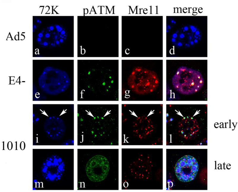
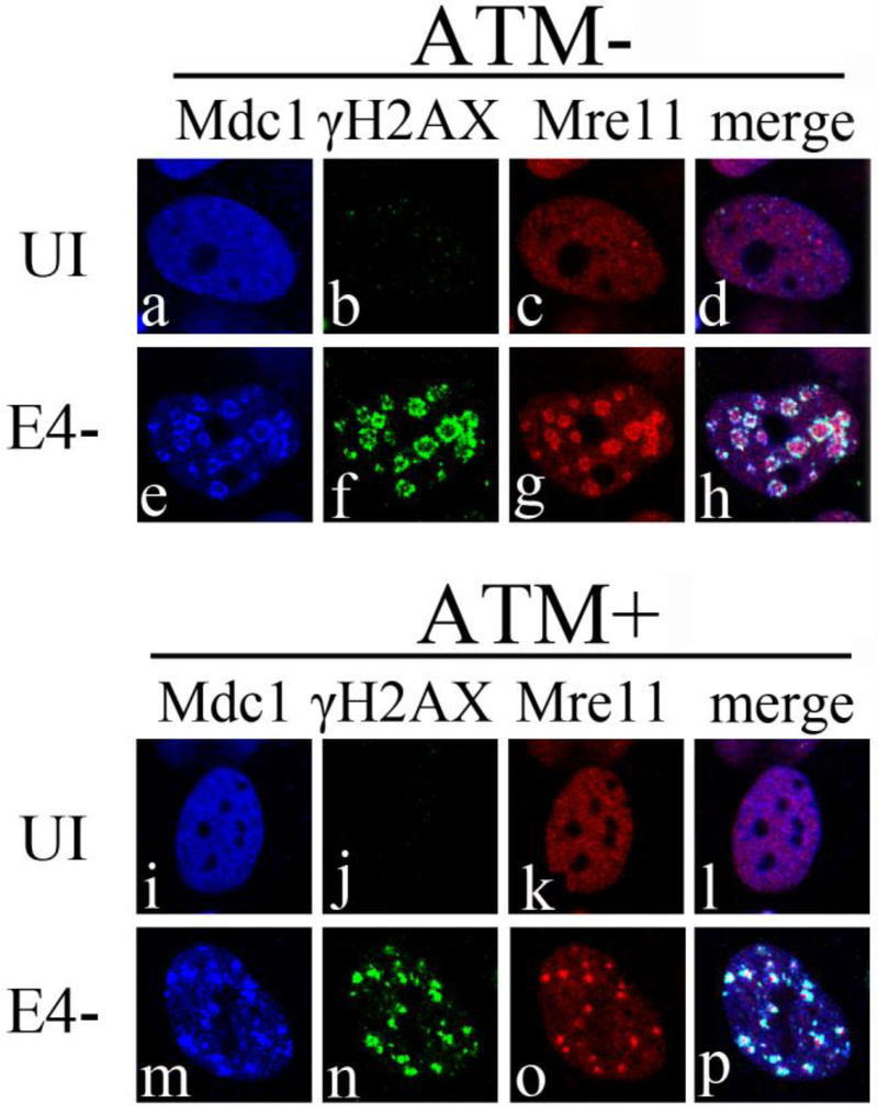
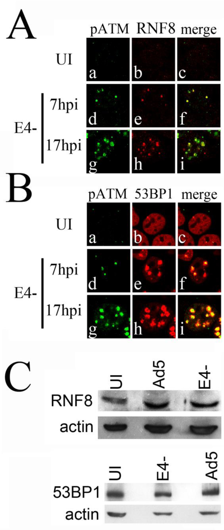
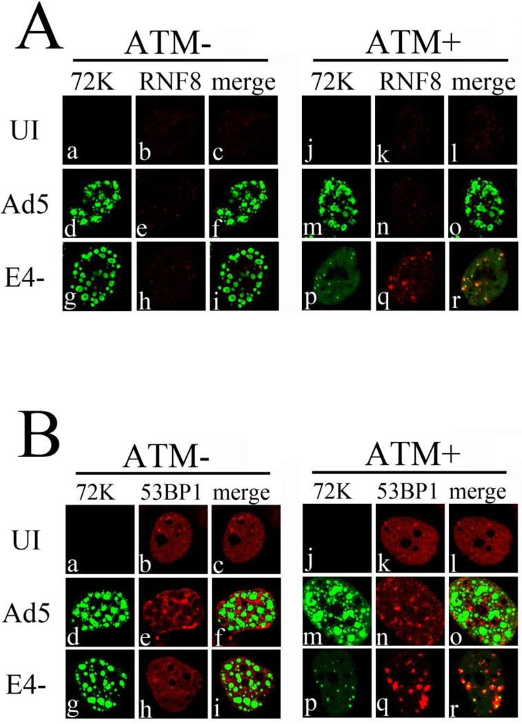
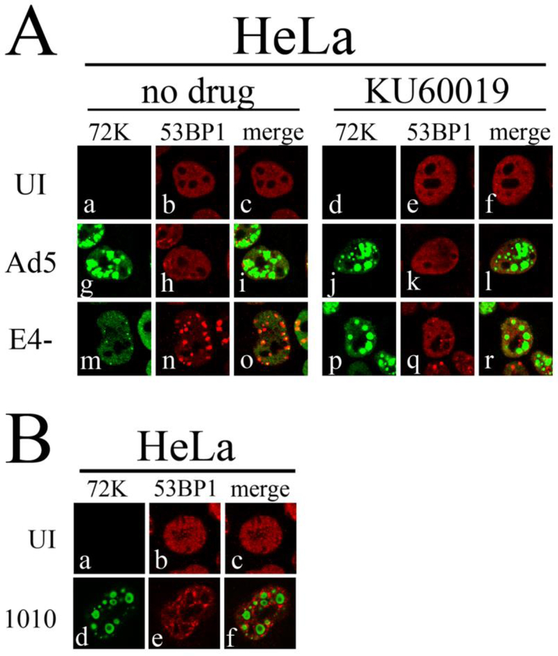
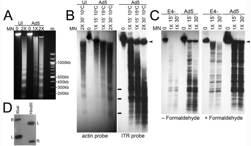
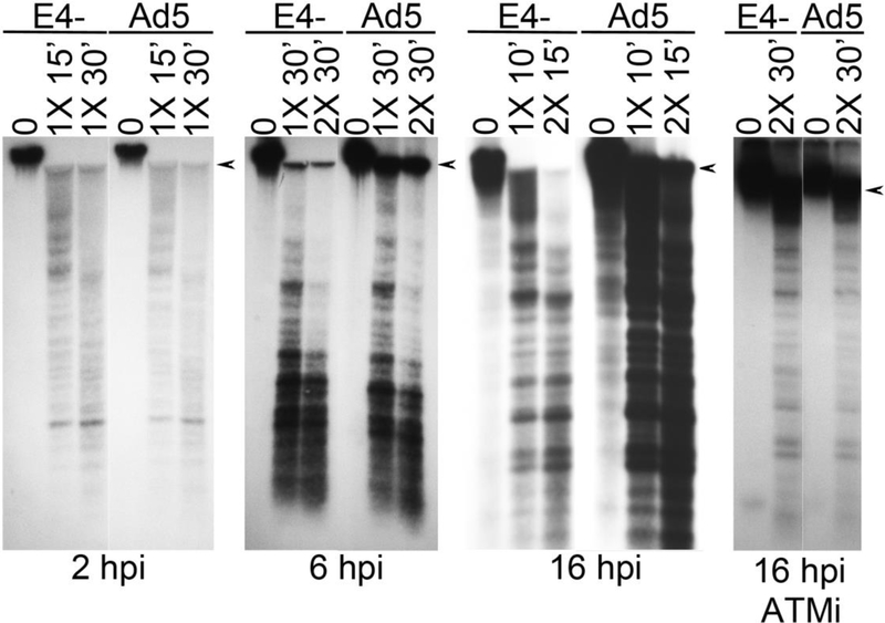
Adenovirus (Ad) type 5 (Ad5) E4 deletion mutants including H5dl1007 (E4-) induce a DNA damage response (DDR) that activates the kinase ataxia-telangiectasia mutated (ATM), which can interfere with efficient viral DNA replication. We find that localization of active phosphorylated ATM (pATM) to E4- viral replication centers (VRCs) is important for its inhibitory effect. ATM is necessary for localization of RNF8 and 53BP1 to E4 mutant VRCs, while recruitment of DDR factors Mre11, Mdc1 and γH2AX is ATM-independent, raising the possibility that ATM may affect viral chromatin at VRCs. We assessed E4- and Ad5 chromatin organization by micrococcal nuclease (MN) digestion. A significant fraction of Ad5 DNA is somewhat resistant to MN digestion, whereas E4- DNA is more susceptible. ATM inhibition increases the fraction of E4- DNA that is resistant to MN digestion. Our results address possible mechanisms through which ATM inhibits E4- DNA replication.

Keywords: 53BP1; ATM; Adenovirus; DNA damage response; E4 11 kDa; E4 34 kDa; E4 ORF 3; E4 ORF 6; Micrococcal nuclease; RNF8.


Figures

Figure 1.
Figure 1.. pATM does not colocalize with VRCs in dl1010 infections.
HeLa cells were infected with Ad5, E4-, or dl1010 at 3FFU/cell. At 17 hpi cells were fixed and stained with anti-pATM (phosphoepitope S1981), anti-Mre11 and anti-E2 72kDa antibodies. Immunofluorescence confocal micrographs showing E2 72kDa (72K) in blue, pATM in green, Mre11 in red, and merged staining patterns are presented. Cells with early and late stage replication centers were identified based on size of E2 72kDa foci. Arrows indicate the location of 72K centers that are still colocalized with Mre11 and pATM in early stages of viral infection.
Figure 2.
Figure 2.. Mdc1 and γH2AX colocalize with Mre11 at E4- VRCs independent of ATM.
ATM+ and ATM− cells were infected with E4- at an MOI of 3 FFU/cell. At 17 hpi cells were fixed and immunostained with antibodies against Mdc1, γH2AX and Mre11. Immunofluorescence confocal micrographs show Mdc1 (blue), γH2AX (green), Mre11 (red), and merged staining patterns of uninfected (UI) or E4- infected cells.
Figure 3.
Figure 3.. RNF8 and 53BP1 are recruited to pATM containing E4- VRCs.
HeLa cells were uninfected or infected with E4-, and at 7 and 17 hpi cells were fixed and immunostained with anti-pATM (phosphoepitope S1981) and anti-RNF8 (A) or 53BP1 (B) antibodies. Immunofluorescence micrographs showing pATM (green), RNF8 or 53BP1 (red), and merged staining patterns are presented. UI, uninfected cells. (C) Western blot showing levels of the RNF8 and 53BP1 proteins in uninfected (UI), Ad5 and E4- infected cells. Levels of β-actin in the samples were measured as a control.
Figure 4.
Figure 4.. Localization of RNF8 and 53BP1 to E4- VRCs requires ATM.
ATM+ and ATM− cells were infected with E4- and Ad5 viruses at an MOI of 3 FFU/cell for 17 h. Cells were then fixed and immunostained with antibodies against the viral DNA binding protein E2 72kDa (72K) and RNF8 (A) or 53BP1 (B). Immunofluorescence confocal micrographs show E2 72kDa (green), RNF8 or 53BP1 (red), and merged staining patterns of uninfected (UI), Ad5, or E4-infected cells.
Figure 5.
Figure 5.. Failure to localize pATM to VRCs results in inefficient recruitment of 53BP1.
(A) HeLa cells were uninfected (UI), or infected with Ad5 or E4-. The ATM inhibitor KU60019 (5μM) or DMSO was added to the medium at 2 hpi, and cells were fixed and stained for immunofluorescence at 17 hpi. Immunofluorescence confocal micrographs showing E2 72kDa (green), 53BP1 (red), or merged staining patterns are shown. (B) HeLa cells were uninfected or infected with dl1010 for 17 h and cells were prepared for immunostaining with anti-72K and anti-53BP1 antibodies. Immunofluorescence confocal micrographs showing E2 72kDa (green), 53BP1 (red), or merged staining patterns are shown.
Figure 6.
Figure 6.. Analysis of viral chromatin by MN digestion.
HeLa cells were infected with Ad5 or E4- at 3 FFU/cell. Nuclei were prepared and treated with MN as indicated, followed by preparation of total DNA from each sample. (A) Ethidium bromide stained gel showing nucleosome protected DNA prepared from uninfected (UI) and Ad5 infected cells. Samples were incubated with MN at the indicated amounts for 30 min at 10°C. (B) Southern blot analysis of MN digested DNAs. Samples were run on the same gel, and after transfer the membrane was cut and separately hybridized to radiolabeled β-actin (left panel) or Ad ITR probe (right panel) as indicated. The positons of bands corresponding to DNA protected by 1, 2 and 3 nucleosomes are marked with a bar. The arrowhead indicates the position of viral DNA that was not digested by MN treatment. (C) Southern blot analysis of MN treated DNA from Ad5 and E4- infected cells. Samples were treated with MN for the indicated amounts and times at 4°C. Some samples were fixed with formaldehyde prior to MN digestion as indicated. The arrowhead indicates the position of viral DNA that was not digested by MN treatment. (D) Southern blot analysis of Hind III and XbaI digested Ad5 DNA hybridized with the radiolabeled ITR probe. The probe labeled only the left (L) and right (R) terminal fragments in both digests. Sizes of the left and right terminal fragments are 1339 bp and 5469 bp for the XbaI digest, and 2804 bp and 1008 bp for the HindIII digest.
Figure 7.
Figure 7.. Analysis of MN digested viral chromatin at different time points.
HeLa cells were infected with Ad5 or E4- at 3 FFU/cell. Some cultures were treated with the ATM inhibitor KU60019 at 5 μM from 2 to 16hpi. Cells were fixed in formaldehyde and nuclei were prepared and treated with MN as indicated at 4°C, or at room temperature for 16hpi samples that were not treated with ATM inhibitor, followed by preparation of total DNA from each sample. DNA samples were then analyzed by Southern blotting using radiolabeled ITR probe. Arrowheads indicate the position of viral DNA that was not digested by MN treatment.

Similar articles

Cited by

References

    1. Araujo FD et al., 2005. Adenovirus type 5 E4orf3 protein targets the Mre11 complex to cytoplasmic aggresomes. J. Virol 79 (17) 11382–11391. - PMC - PubMed
    1. Baker A et al., 2007. Adenovirus E4 34 k and E1b 55 k oncoproteins target host DNA ligase IV for proteasomal degradation. J. Virol 81 (13), 7034–7040. - PMC - PubMed
    1. Bakkenist CJ, Kastan MB, 2003. DNA damage activates ATM through intermolecular autophosphorylation and dimer dissociation. Nature 421 (6922), 499–506. - PubMed
    1. Bekker-Jensen S, Mailand N, 2010. Assembly and function of DNA double-strand break repair foci in mammalian cells. DNA Repair (Amst). 9 (12), 1219–1228. - PubMed
    1. Blackford AN, Jackson SP, 2017. ATM, ATR, and DNA-PK: The Trinity at the Heart of the DNA Damage Response. Mol. Cell 66 (6), 801–817. - PubMed

Publication types

MeSH terms