. 2019 Feb 1;141(2):0210111-02101111.
doi: 10.1115/1.4042014.

Effects of Elastase Digestion on the Murine Vaginal Wall Biaxial Mechanical Response

Affiliations

Effects of Elastase Digestion on the Murine Vaginal Wall Biaxial Mechanical Response

Akinjide R Akintunde et al. J Biomech Eng. .

Abstract

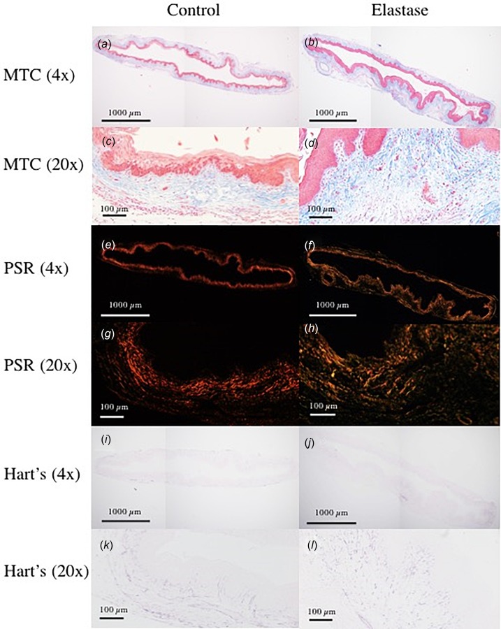
Although the underlying mechanisms of pelvic organ prolapse (POP) remain unknown, disruption of elastic fiber metabolism within the vaginal wall extracellular matrix (ECM) has been highly implicated. It has been hypothesized that elastic fiber fragmentation correlates to decreased structural integrity and increased risk of prolapse; however, the mechanisms by which elastic fiber damage may contribute to prolapse are poorly understood. Furthermore, the role of elastic fibers in normal vaginal wall mechanics has not been fully ascertained. Therefore, the objective of this study is to investigate the contribution of elastic fibers to murine vaginal wall mechanics. Vaginal tissue from C57BL/6 female mice was mechanically tested using biaxial extension-inflation protocols before and after intraluminal exposure to elastase. Elastase digestion induced marked changes in the vaginal geometry, and biaxial mechanical properties, suggesting that elastic fibers may play an important role in vaginal wall mechanical function. Additionally, a constitutive model that considered two diagonal families of collagen fibers with a slight preference toward the circumferential direction described the data reasonably well before and after digestion. The present findings may be important to determine the underlying structural and mechanical mechanisms of POP, and aid in the development of growth and remodeling models for improved assessment and prediction of changes in structure-function relationships with prolapse development.

Keywords: elastic fibers; mechanical testing; pelvic floor disorders; vaginal wall; women's health.


Figures

Fig. 1
Fig. 1
Estimation of the physiologic, in vivo axial stretch ratio for representative sample #4 in Table 1: (a) the nearly constant transducer-measured axial force occurs at the estimated physiologic (in vivo) axial stretch ratio and (b) the point of intersection of force–length (extension) tests performed at constant, multiple luminal pressures further confirms the estimated in vivo axial stretch ratio. The dashed vertical line represents the estimated in vivo axial stretch.
Fig. 2
Fig. 2
Fits of the HGO model to experimental (pre- and post-treatment with elastase) data (mean±SEM, n = 8, paired): (a) intraluminal pressure versus outer diameter: a rightward shift was observed post-treatment, indicating dilatation of the vagina, (b) wall-averaged circumferential Cauchy stress versus circumferential stretch ratio: a rise was observed post-treatment, indicating increased structural stiffness circumferentially, (c) wall-averaged axial Cauchy stress versus circumferential stretch ratio, (d) wall-averaged axial Cauchy stress versus axial stretch ratio, (e) the transducer-measured axial force versus pressure, which remained nearly constant with increasing pressure, and (f) the sum of transducer-measured force and force due to increasing intraluminal pressure versus pressure: the increased separation at higher pressures is attributable to dilated lumen (increased inner radius) of the elastase-treated vagina
Fig. 3
Fig. 3
Holzapfel–Gasser–Ogden model sensitivity in the (a) circumferential and (b) axial directions, pre-elastase (solid lines) and postelastase (dashed lines) treatment. Pre- and post-treatment, the model was most sensitive to the collagen-associated nondimensional parameter—c2 and the alignment angle-α. Relative to control, post-treatment, the influence of the two parameters increased the most in both directions. Sensitivity indices were computed using optimized model parameters for averaged data (n = 8/group, for control and elastase).
Fig. 4
Fig. 4
Vaginal tissue average compliance as a function of intraluminal pressure, data = mean±SEM. Statistically significant (*p < 0.05) decrease was observed at 5 and 10 mmHg.
Fig. 5
Fig. 5
Histological section samples stained with: (a)–(d) PSR,(e)–(h) MTC, and (i)–(l) Hart's elastin stain. Elastic fiber area fraction exhibited statistically significant decrease post-treatment. For all stains, area fraction analysis was performed using images acquired at 4× objective. In the 20× and 40× (Supplemental Fig. 3, which is available under the “Supplemental Data” tab for this paper on the ASME Digital Collection) objective images of Hart's stained section, a decrease in population and organization of elastic fibers is observable.

References

    1. DeLancey, J. O. , 2005, “ The Hidden Epidemic of Pelvic Floor Dysfunction: Achievable Goals for Improved Prevention and Treatment,” Am. J. Obstet. Gynecol., 192(5), pp. 1488–1495. 10.1016/j.ajog.2005.02.028 - DOI - PubMed
    1. Subak, L. L. , Waetjen, L. E. , van den Eeden, S. , Thom, D. H. , Vittinghoff, E. , and Brown, J. S. , 2001, “ Cost of Pelvic Organ Prolapse Surgery in the United States,” Obstet. Gynecol., 98(4), pp. 646–651. 10.1016/S0029-7844(01)01472-7 - DOI - PubMed
    1. Pizarro-Berdichevsky, J. , Clifton, M. M. , and Goldman, H. B. , 2015, “ Evaluation and Management of Pelvic Organ Prolapse in Elderly Women,” Clin. Geriatr. Med., 31(4), pp. 507–521. 10.1016/j.cger.2015.06.008 - DOI - PubMed
    1. Baah-Dwomoh, A. , McGuire, J. , Tan, T. , and De Vita, R. , 2016, “ Mechanical Properties of Female Reproductive Organs and Supporting Connective Tissues: A Review of the Current State of Knowledge,” ASME Appl. Mech. Rev., 68(6), p. 060801. 10.1115/1.4034442 - DOI
    1. Abramowitch, S. D. , Feola, A. , Jallah, Z. , and Moalli, P. A. , 2009, “ Tissue Mechanics, Animal Models, and Pelvic Organ Prolapse: A Review,” Eur. J. Obstetr. Gynecol., 144, pp. S146–S158. 10.1016/j.ejogrb.2009.02.022 - DOI - PubMed