. 2018 Nov 18;10(11):650.
doi: 10.3390/v10110650.

Host Lipid Rafts Play a Major Role in Binding and Endocytosis of Influenza A Virus

Affiliations

Host Lipid Rafts Play a Major Role in Binding and Endocytosis of Influenza A Virus

Dileep Kumar Verma et al. Viruses. .

Abstract

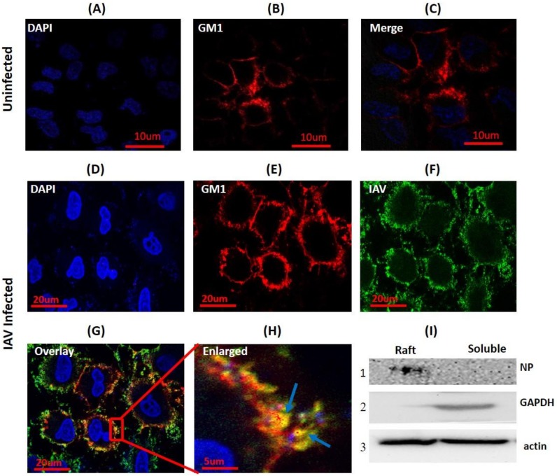
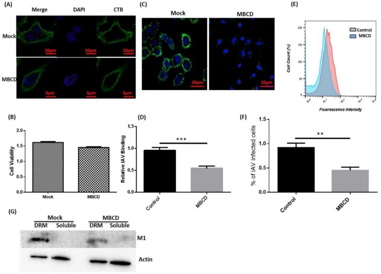
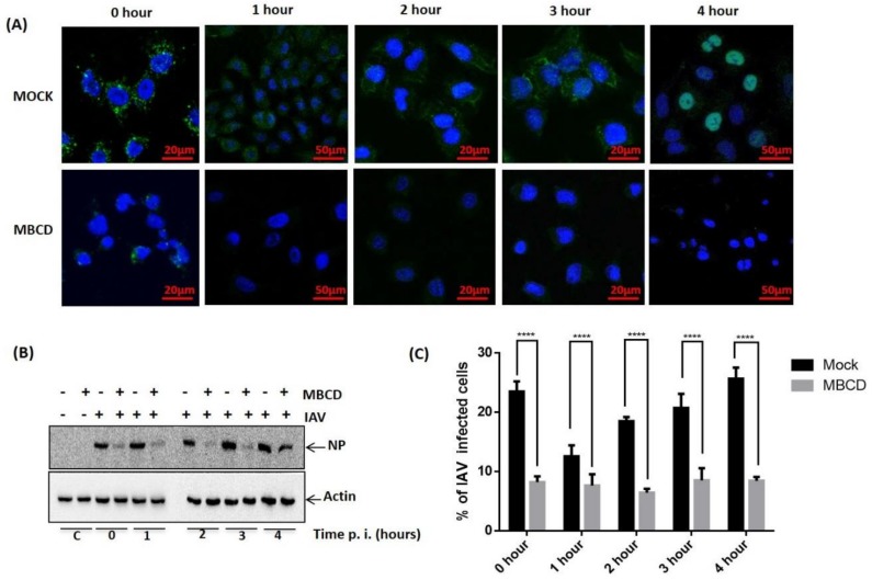
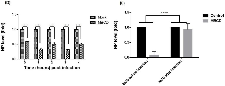
Influenza still remains one of the most challenging diseases, posing a significant threat to public health. Host lipid rafts play a critical role in influenza A virus (IAV) assembly and budding, however, their role in polyvalent IAV host binding and endocytosis had remained elusive until now. In the present study, we observed co-localization of IAV with a lipid raft marker ganglioside, GM1, on the host surface. Further, we isolated the lipid raft micro-domains from IAV infected cells and detected IAV protein in the raft fraction. Finally, raft disruption using Methyl-β-Cyclodextrin revealed significant reduction in IAV host binding, suggesting utilization of host rafts for polyvalent binding on the host cell surface. In addition to this, cyclodextrin mediated inhibition of raft-dependent endocytosis showed significantly reduced IAV internalization. Interestingly, exposure of cells to cyclodextrin two hours post-IAV binding showed no such reduction in IAV entry, indicating use of raft-dependent endocytosis for host entry. In summary, this study demonstrates that host lipid rafts are selected by IAV as a host attachment factors for multivalent binding, and IAV utilizes these micro-domains to exploit raft-dependent endocytosis for host internalization, a virus entry route previously unknown for IAV.

Keywords: Cholesterol; Ganglioside GM1; Methyl-β-Cyclodextrin; Raft-dependent endocytosis; lipid raft.


Conflict of interest statement

The authors declare no competing financial interest.

Figures

Figure 1
Figure 1
Influenza A virus (IAV) co-localizes with host membrane lipid rafts during binding. (AC) CTB-594 stained (4 °C) A549 cells (uninfected control). Patchy distribution of ganglioside 1 (GM1) (rafts) is visible (red); blue, DAPI (4′, 6-diamidino-2-phenylindole) nuclear staining. (DG) IAV incubated (4 °C) A549 cells stained with CTB-594 (4 °C). Surface bound viruses were stained with anti-NP-FITC (anti-nucleoprotein-fluorescein isothiocyanate) antibody (Green). Yellow color and arrows indicate IAV co-localization with GM1; blue, DAPI nuclear staining. (H) Enlarged view of indicated section of image G, magnification ~3X. (I) Western blot of raft proteins isolated from IAV incubated (4 °C) A549 cells. NP was detected in the raft fraction (I, panel 1). GAPDH (Glyceraldehyde 3-phosphate dehydrogenase) was used as cytosolic marker and beta-actin as internal control.
Figure 2
Figure 2
Disruption of host lipid rafts and IAV binding. (A) CTB-488 stained A549 cells (untreated and Methyl-β-Cyclodextrin (MBCD) treated). (B) Assessment of cell viability by MTT (3-4,5-Dimethylthiazol-2-yl)-2,5-Diphenyltetrazolium Bromide) assay. Error bars represent ±SD. (C) IAV incubated (4 °C) A549 cells, (D) Relative IAV host binding represented as mean fluorescence intensity (FITC). *** p < 0.0002, calculated by student’s t-test. Error bars show ±SD. (E,F) Flow-cytometric analysis showing IAV host binding. The results are a representation of three independent experiments. ** p < 0.002, calculated by student’s t-test. Error bars show ±SD. (G) Isolation of lipid rafts as detergent-resistant membranes (DRM) and detection of IAV protein (M1). Actin served as internal control.
Figure 3
Figure 3
Exploitation of raft-dependent endocytosis by IAV for host internalization. (A) Confocal images of mock or MBCD treated, IAV infected (37 °C) A549 cells with indicated time of post IAV infection. Internalized IAVs were detected by staining with anti-NP-FITC antibody. (B) Analysis of IAV internalization via Western blot in mock or MBCD treated A549 cells showing drastic reduction in IAV entry in MBCD treated cells. Actin served as an internal control. (C) Flow cytometric analysis of IAV endocytosis in mock versus MBCD treated A549 cells. **** p < 0.0001, calculated by two way ANOVA. Error bars show ±SD. (D). Relative quantitation of NP a central core of RNA genome (vRNA) by qRT-PCR. **** p < 0.0001, calculated by two way ANOVA. Error bars show ±SD. (E). Relative quantitation of NP vRNA in A549 cells that were MBCD treated before IAV infection and MBCD treated after IAV infection. **** p < 0.0001, calculated by two way ANOVA. Error bars show ±SD.
Figure 3
Figure 3
Exploitation of raft-dependent endocytosis by IAV for host internalization. (A) Confocal images of mock or MBCD treated, IAV infected (37 °C) A549 cells with indicated time of post IAV infection. Internalized IAVs were detected by staining with anti-NP-FITC antibody. (B) Analysis of IAV internalization via Western blot in mock or MBCD treated A549 cells showing drastic reduction in IAV entry in MBCD treated cells. Actin served as an internal control. (C) Flow cytometric analysis of IAV endocytosis in mock versus MBCD treated A549 cells. **** p < 0.0001, calculated by two way ANOVA. Error bars show ±SD. (D). Relative quantitation of NP a central core of RNA genome (vRNA) by qRT-PCR. **** p < 0.0001, calculated by two way ANOVA. Error bars show ±SD. (E). Relative quantitation of NP vRNA in A549 cells that were MBCD treated before IAV infection and MBCD treated after IAV infection. **** p < 0.0001, calculated by two way ANOVA. Error bars show ±SD.

References

    1. Taubenberger J.K., Morens D.M. The pathology of influenza virus infections. Annu. Rev. Pathol. 2008;3:499–522. doi: 10.1146/annurev.pathmechdis.3.121806.154316. - DOI - PMC - PubMed
    1. Bouvier N.M., Palese P. The biology of influenza viruses. Vaccine. 2008;26(Suppl. 4):D49–D53. doi: 10.1016/j.vaccine.2008.07.039. - DOI - PMC - PubMed
    1. Simons K., Sampaio J.L. Membrane organization and lipid rafts. Cold Spring Harb. Perspect. Biol. 2011;3:a004697. doi: 10.1101/cshperspect.a004697. - DOI - PMC - PubMed
    1. Simons K., Ikonen E. Functional rafts in cell membranes. Nature. 1997;387:569–572. doi: 10.1038/42408. - DOI - PubMed
    1. Chazal N., Gerlier D. Virus entry, assembly, budding, and membrane rafts. Microbiol. Mol. Biol. Rev. 2003;67:226–237. doi: 10.1128/MMBR.67.2.226-237.2003. - DOI - PMC - PubMed

Publication types

LinkOut - more resources