

A comparative study to evaluate the feasibility of preoperative percutaneous catheter drainage for the treatment of lumbar spinal tuberculosis with psoas abscess
- PMID: 30454001
- PMCID: PMC6245803
- DOI: 10.1186/s13018-018-0993-9
A comparative study to evaluate the feasibility of preoperative percutaneous catheter drainage for the treatment of lumbar spinal tuberculosis with psoas abscess
Abstract
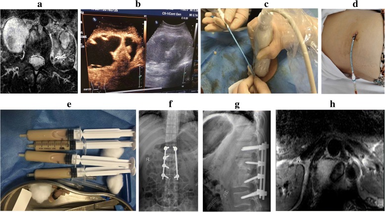
Background: Spinal tuberculosis is a frequent cause of psoas abscess (PA), and PA largely negates the efficacy of antituberculosis therapy. This study aimed to investigate the clinical outcome of preoperative percutaneous catheter drainage (PCD) in patients with lumbar spinal tuberculosis and PA.
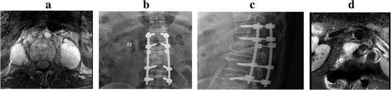
Methods: Between January 2015 and January 2017, 72 patients with lumbar spinal tuberculosis with PA were assigned to group A (preoperative PCD) and group B (n = 36 per group). All patients received posterior pedicle screw fixation and anterior focal debridement and fusion. Data on intraoperative blood loss, the duration of the surgery, and the length of the anterior incision were recorded, as well as the postoperative anal exhaust time, visual analogue scale (VAS), Cobb angle, lumbar vertebra function, erythrocyte sedimentation rate (ESR), C-reactive protein (CRP) level, and sinus tract formation.
Results: Sixty-eight patients were followed up for an average time of 13 months (range 6-21 months). Until the final follow-up, no mixed infections, recurrence of tuberculosis, pedicle screw loosening, or screw pullout had occurred. There were significant between-group differences in blood loss, surgery duration, anterior incisional length, postoperative anal exhaust time, and sinus tract formation. As compared with group B, the ESR and CRP levels of the patients in group A were markedly improved following 3 weeks of antituberculosis therapy and 1 week postsurgery.
Conclusion: Preoperative PCD helps to increase the efficacy of antituberculosis therapy prior to surgery, reduce surgical trauma, and avoid postoperative complications, making it a safe and feasible treatment option for lumbar spinal tuberculosis with PA.
Keywords: Catheter drainage; Lumbar vertebra; Psoas abscess; Spine; Tuberculosis.
Conflict of interest statement
Ethics approval and consent to participate
The study was approved by the ethics committee of Hospital of Integrated Traditional Chinese and Western medicine in Zhejiang Province.
Consent for publication
Not applicable.
Competing interests
The authors declare that they have no competing interests.
Publisher’s Note
Springer Nature remains neutral with regard to jurisdictional claims in published maps and institutional affiliations.
Figures
Similar articles
-
[Feasibility study of preoperative percutaneous catheter drainage in the treatment of lumbar tuberculosis with psoas abscess].Zhongguo Gu Shang. 2018 Nov 25;31(11):998-1004. doi: 10.3969/j.issn.1003-0034.2018.11.004. Zhongguo Gu Shang. 2018. PMID: 30514039 Chinese.
-
Surgical treatment for spinal tuberculosis with bilateral paraspinal abscess or bilateral psoas abscess: one-stage surgery.J Spinal Disord Tech. 2014 Dec;27(8):E309-14. doi: 10.1097/BSD.0000000000000120. J Spinal Disord Tech. 2014. PMID: 25093646
-
Clinical efficacy of CT-guided percutaneous huge ilio-psoas abscesses drainage combined with posterior approach surgery for the management of dorsal and lumbar spinal tuberculosis in adults.Orthop Traumatol Surg Res. 2017 Dec;103(8):1251-1255. doi: 10.1016/j.otsr.2017.07.015. Epub 2017 Sep 13. Orthop Traumatol Surg Res. 2017. PMID: 28919066
-
A case of skeletal tuberculosis and psoas abscess: disease activity evaluated using (18) F-fluorodeoxyglucose positron emission tomography-computed tomography.BMC Med Imaging. 2013 Nov 14;13:37. doi: 10.1186/1471-2342-13-37. BMC Med Imaging. 2013. PMID: 24225333 Free PMC article. Review.
-
Does computed tomography-guided percutaneous catheter drainage is effective for spinal tuberculous abscess: a midterm results.Spinal Cord Ser Cases. 2022 Feb 7;8(1):19. doi: 10.1038/s41394-022-00488-9. Spinal Cord Ser Cases. 2022. PMID: 35132064 Free PMC article. Review.
Cited by
-
A Comparative Study of Anterior and Posterior Tuberculosis Lesions for the Treatment of Thoracolumbar Tuberculosis disease: A Single Institution Experience in a Major Academic Hospital.Infect Drug Resist. 2024 Dec 4;17:5375-5386. doi: 10.2147/IDR.S495231. eCollection 2024. Infect Drug Resist. 2024. PMID: 39649429 Free PMC article.
-
Swiss Cheese Loin: A Rare Initial Presentation of Pott's Spine.Cureus. 2021 Mar 15;13(3):e13912. doi: 10.7759/cureus.13912. Cureus. 2021. PMID: 33880267 Free PMC article.
-
Comparison of Affected-Vertebra Fixation of Cortical Bone Trajectory Screw and Pedicle Screw for Lumbar Tuberculosis: A Minimum 3-Year Follow-Up.Biomed Res Int. 2022 Jul 21;2022:6312994. doi: 10.1155/2022/6312994. eCollection 2022. Biomed Res Int. 2022. PMID: 35909489 Free PMC article.
-
Vacuum Sealing Drainage for Primary Thoracolumbar Spondylodiscitis: A Technical Note.Biomed Res Int. 2022 Aug 9;2022:9248972. doi: 10.1155/2022/9248972. eCollection 2022. Biomed Res Int. 2022. Retraction in: Biomed Res Int. 2023 Jun 21;2023:9785616. doi: 10.1155/2023/9785616. PMID: 35983250 Free PMC article. Retracted.
-
Minimally invasive surgery for paravertebral or psoas abscess with spinal tuberculosis - a long-term retrospective study of 106 cases.BMC Musculoskelet Disord. 2020 Jun 6;21(1):353. doi: 10.1186/s12891-020-03344-9. BMC Musculoskelet Disord. 2020. PMID: 32505204 Free PMC article.
References
-
- Khorgade Ranjana R., Bhise Pramod R., Deshmukh Mukta M. Psoas abscess due to mycobacterium tuberculosis: a case report. International Journal of Research in Medical Sciences. 2017;5(7):3251. doi: 10.18203/2320-6012.ijrms20173025. - DOI
-
- Ding WY, Yan-Lei HE, Bao-Jun LI. Brachy segament posterior fixation combined with anterior debridement used in the treatment of lumbar tuberculosis. J Cervicodynia Lumbodynia. 2010;31(2):38–42.
Publication types
MeSH terms
Substances
LinkOut - more resources
Full Text Sources
Research Materials
Miscellaneous
