. 2018 Nov 20;10(1):115.
doi: 10.1186/s13195-018-0441-4.

Active full-length DNA Aβ42 immunization in 3xTg-AD mice reduces not only amyloid deposition but also tau pathology

Affiliations

Active full-length DNA Aβ42 immunization in 3xTg-AD mice reduces not only amyloid deposition but also tau pathology

Roger N Rosenberg et al. Alzheimers Res Ther. .

Abstract

Background: Alzheimer's disease (AD) is the most well-known and most common type of age-related dementia. Amyloid deposition and hyperphosphorylation of tau protein are both pathological hallmarks of AD. Using a triple-transgenic mouse model (3xTg-AD) that develops plaques and tangles in the brain similar to human AD, we provide evidence that active full-length DNA amyloid-β peptide 1-42 (Aβ42) trimer immunization leads to reduction of both amyloid and tau aggregation and accumulation.

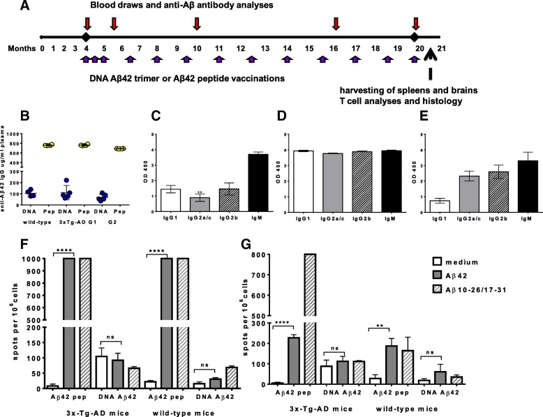
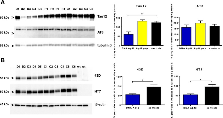
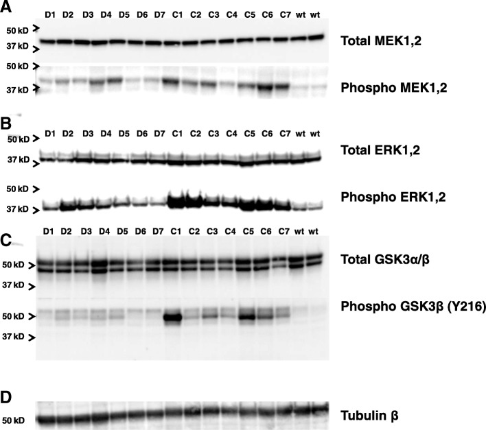
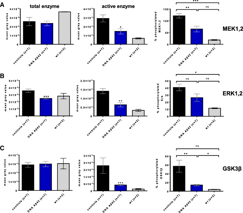
Methods: Immune responses were monitored by enzyme-linked immunosorbent assay (ELISA) (antibody production) and enzyme-linked immunospot (cellular activation, cytokine production). Brains from 20-month-old 3x Tg-AD mice that had received DNA Aβ42 immunotherapy were compared with brains from age- and gender-matched transgenic Aβ42 peptide-immunized and control mice by histology, Western blot analysis, and ELISA. Protein kinase activation and kinase levels were studied in Western blots from mouse hemibrain lysates.

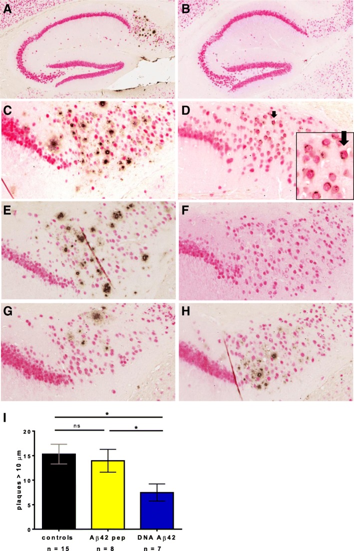
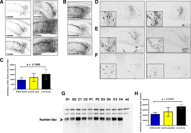
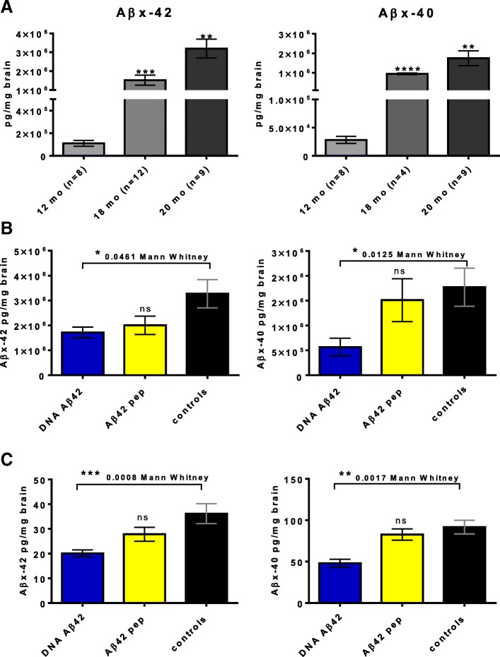
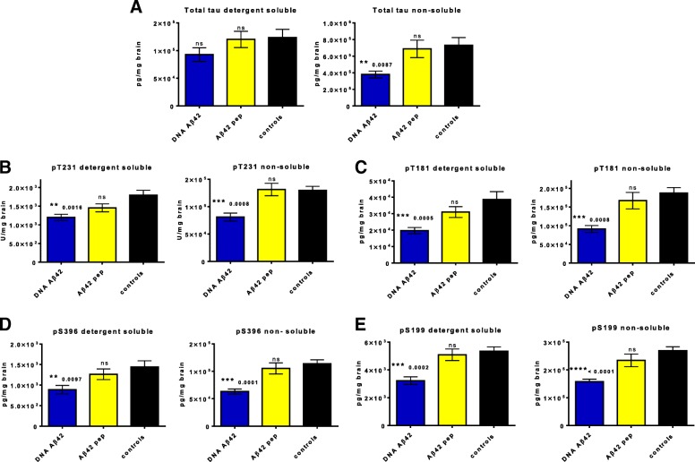
Results: Quantitative ELISA showed a 40% reduction of Aβ42 peptide and a 25-50% reduction of total tau and different phosphorylated tau molecules in the DNA Aβ42 trimer-immunized 3xTg-AD mice compared with nonimmunized 3xTg-AD control animals. Plaque and Aβ peptide reductions in the brain were due to the anti-Aβ antibodies generated following the immunizations. Reductions of tau were likely due to indirect actions such as less Aβ in the brain resulting in less tau kinase activation.

Conclusions: The significance of these findings is that DNA Aβ42 trimer immunotherapy targets two major pathologies in AD-amyloid plaques and neurofibrillary tangles-in one vaccine without inducing inflammatory T-cell responses, which carry the danger of autoimmune inflammation, as found in a clinical trial using active Aβ42 peptide immunization in patients with AD (AN1792).

Keywords: Alzheimer’s disease; Amyloid-β; Aβ oligomer; DNA vaccination; Immunotherapy; Tau; Tau kinases.


Conflict of interest statement

Ethics approval

Animal use for this study was approved by the Institutional Animal Care and Use Committee at UTSW.

Consent for publication

Not applicable.

Competing interests

The authors declare that they have no competing interests.

Publisher’s Note

Springer Nature remains neutral with regard to jurisdictional claims in published maps and institutional affiliations.

Figures

Fig. 1
Fig. 1
Timeline of the experimental procedures, anti-amyloid-β peptide 1–42 (anti-Aβ42) antibody production upon immunization with DNA Aβ42 trimer and Aβ42 peptide in triple-transgenic Alzheimer’s disease (3xTg-AD) and wild-type mice and cytokine secretion in restimulated splenocyte cultures. a Immunizations, blood draws, and final analyses are shown along the experimental timeline of 20 months. b High levels of anti-Aβ42 antibodies (micrograms per milliliter of plasma) were found in all of the immunized mouse groups following the last immunization (wild-type mice and 3xTg-AD mice). Blue symbols indicate mice that had received DNA Aβ42 trimer immunizations; yellow symbols indicate mice that had received Aβ42 peptide immunizations. Antibody levels of two groups of 20-month-old 3xTg-AD mice are shown as group 1 (G1) and group 2 (G2). Plasma samples had been used in a 1:1000 dilution. Samples were run in triplicates, and the assay was repeated twice. Antibody isotype analyses from DNA Aβ42 trimer-immunized 3xTg-AD mice (c) and Aβ42 peptide-immunized 3xTg-AD mice (d). White bars show levels of anti-Aβ42 antibodies of the immunoglobulin G1 (IgG1) isotype; gray bars show IgG2a antibody levels; hatched bars show IgG2b antibody levels; and black bars show IgM antibody levels. Differences in the amount of IgG1 (Th2) and IgG2a/c (Th1) antibody levels are statistically significant (p = 0.0068). Levels were measured as optical density at 450 nm (OD450). Plasma samples had been used in a 1:500 dilution, analyzed in triplicates, and the assay was repeated twice. e Antibody isotype profile of plasma samples from peptide-immunized mice in a 1:20,000 dilution. Interferon (IFN)-γ (f) and interleukin (IL)-17 (g) enzyme-linked immunospot analysis of splenocytes from 20-month-old 3xTg-AD mice (n = 4/group) and 129/SvJ wild-type mice (n = 4/group) that had received 13 Aβ42 peptide or 13 DNA Aβ42 trimer immunizations, respectively. No IFN-γ- or IL-17-secreting cells were found in DNA Aβ42-immunized mice, whereas high numbers of cells secreting IFN-γ and IL-17 were found in splenocytes from peptide-immunized mice upon Aβ1–42 peptide or Aβ10–26/17–31 peptide mix restimulation in vitro. **, and **** indicate p values of ≤ 0.01 and ≤ 0.001, respectively (unpaired Student's t test)
Fig. 2
Fig. 2
Amyloid-β (Aβ) immunization results in removal of amyloid plaques in brains of triple-transgenic Alzheimer’s disease (3xTg-AD) mice. Brain sections of mice aged 18 months (c and d) and 20 months (a, b, e–h) were stained with a NeuN antibody (red) to detect neurons and an anti-Aβ antibody (McSA1, brown) to detect numerous plaques in the subiculum of the hippocampus in 3xTg-AD mice. a The hippocampus of a 20-month-old female control 3xTg-AD mouse with numerous amyloid plaques is shown (5× magnification). b Hippocampus of a 20-month-old male control 3xTg-AD mouse showing no plaque pathology. c The subiculum of an 18-month-old female control 3xTg-AD mouse is shown at higher magnification (20×). d Aβ staining in the subiculum of an 18-month-old male mouse. Only intraneuronal Aβ can be detected (indicated with arrow and shown at higher magnification in inset). e Numerous plaques in the hippocampus of an untreated 20-month-old 3xTg-AD female mouse. f No plaques were found in 20-month-old wild-type mice. Both immunization regimens, Aβ42 peptide (g) and DNA Aβ42 (h), led to a reduction of plaques in 20-month-old 3xTg-AD mice compared with the control mouse (e). i Images were counted for plaques ≥ 10 μm in a 1-mm2 area of the subiculum/CA1 of the hippocampus by two blinded experimenters. Blue bars show plaque count in DNA Aβ42 trimer-immunized mice (n = 7), and yellow bars show plaque count found in brains of Aβ42 peptide-immunized mice (n = 8). Black bars show the numbers found in age- and gender-matched 3xTg-AD control mice (n = 15). * indicates p value of ≤ 0.05 (unpaired Student's t test)
Fig. 3
Fig. 3
IHC staining of T231p (AT180) and T202p/S205p (AT8) in triple-transgenic Alzheimer’s disease (3xTg-AD) mouse brains and Western blots for total tau. a The age progression of T231p (AT180) in 3xTg-AD mouse brains is shown by IHC and staining in 2-, 4-, 7-, 9-, 12-, and 18-month-old mice. b T231p staining in the hippocampus of three brains from 18-month-old 3xTg-AD mice that had received DNA amyloid-β 1–42 peptide (Aβ42) trimer immunizations is shown for comparison. c Semiquantitative analyses for pT231 staining in the hippocampus of four 18-month-old control mice, five 18-month-old DNA Aβ42 immunized mice, six 18-month-old Aβ42 peptide-immunized mice, and four wild-type mice using ImageJ software (National Institutes of Health, Bethesda, MD, USA). Blue bars show positive areas found in DNA Aβ42 trimer-immunized mice, and yellow bars show areas found in brains of Aβ42 peptide-immunized mice. Black bars show the values of age- and gender-matched 3xTg-AD control mice. df DNA Aβ42 trimer immunization decreased AT8 staining in hippocampal sections from 20-month-old 3xTg-AD mice. d Representative sections from two control mice. e Sections from Aβ42 peptide-immunized mice. f Staining of AT8+ tangles in the hippocampus of two DNA Aβ42-immunized mice. All pictures are in 10× magnification (hippocampus); insets are in 40× magnification (subiculum). g A representative Western blot from detergent-soluble brain lysates of 20-month-old 3xTg-AD control mice (labeled C1–C4), DNA Aβ42-immunized mice (labeled D1–D4), Aβ42 peptide-immunized mice (labeled P1, P2), and a wild-type control (wt) mouse is shown. h Gray value intensities of human tau bands (indicated with an arrowhead, missing in the wt control, at 50 kD) were semiquantitatively analyzed using ImageJ software. Black bars show the values in 3xTg-AD control mice; yellow bars represent the peptide-immunized mice; and blue bars show values found in DNA Aβ42-immunized mice
Fig. 4
Fig. 4
Western blot analyses for total and phosphorylated tau. Equal amounts of proteins from detergent-soluble brain lysates of 20-month-old triple-transgenic Alzheimer’s disease (3xTg-AD) mice (D1–D5 = DNA Aβ42-immunized mice, P1–P4 = amyloid-β 1–42 [Aβ42] peptide-immunized mice, C1–C5 = 3xTg-AD control mice, wt = wild-type controls) were separated by SDS-PAGE, blotted onto nitrocellulose filters, and probed using antibodies specific for total human tau (a, upper panel), and phosphorylated tau AT8 (a, middle panel), and β-tubulin as a loading control (a, bottom panel). The graph on the right-hand side of the SDS-PAGE pictures shows analyses of the band intensities performed with ImageJ software. All gray-level intensities of tau protein bands were normalized to the gray-level intensities of protein bands of the housekeeping proteins β-tubulin or β-actin, respectively. The reduction of total tau in the DNA-immunized mice compared with the 3xTg-AD control animals was highly significant (p = 0.0059). Of note, gray-level intensities for sample D2 were not included in thess calculations, because the loading control for this sample indicated a much lower protein content (a, bottom panel). b A comparison of total tau levels in DNA-immunized mice, 3xTg-AD control mice, and wt control mice in Western blots is shown using two different antibodies. In the upper panel, 43D (Tau1–100) was used for detection; in the middle panel, antibody HT7 was used; and in the lower panel, the same membrane was probed with a β-actin antibody as a protein loading control. The graph on the right-hand side of the panels shows the analyses of gray-level intensities for the protein bands with ImageJ software normalized to gray-level intensities of the housekeeping protein β-actin. Differences were statistically significant with p values of 0.0152 (HT7) and 0.0138 (43D). * and ** indicate p values of ≤ 0.05 and ≤ 0.01, respectively (Mann-Whitney U test)
Fig. 5
Fig. 5
Quantitative enzyme-linked immunosorbent assay (ELISA) analyses for amyloid-β 1–42 peptide (Aβ42) and Aβ40 in brain lysates from triple-transgenic Alzheimer’s disease (3xTg-AD) mice. a Analyses of an increase of Aβ42 and Aβ40 peptides in brains from 3xTg-AD mice with age (12-month-, 18-month-, and 20-month-old female control mice). b Reduction of Aβ42 and Aβ40 peptide concentrations in the nonsoluble fractions of the brain lysates owing to Aβ42 immunotherapy. Blue bars show Aβ42 peptide concentrations found in brains from DNA Aβ42 trimer-immunized mice; yellow bars show the concentrations found in brains from Aβ42 peptide-immunized mice. The black bars show Aβ42 peptide concentrations in age- and gender-matched 3xTg-AD control mice. The left-hand graph displays data for Aβ42 peptides, and the right-hand graph shows data for Aβ40 peptides. c Reduction of Aβ42 and Aβ40 peptide concentrations in the soluble fractions of the brain lysates owing to Aβ42 immunotherapy. The left-hand graph shows data for Aβ42 peptides, and the right-hand graph displays data for Aβ40 peptides. ELISAs for the nonsoluble brain lysates were performed three times (dilution 1:10,000), and ELISAs for the detergent-soluble brain lysates were performed twice (dilution 1:2) for this particular group of mice and confirmed the data shown. * p  ≤0.05, ** p ≤ 0.01, *** p ≤ 0.005, and **** p  ≤ 0.001 (Mann-Whitney U test)
Fig. 6
Fig. 6
DNA amyloid-β 1–42 (Aβ42) immunization reduces total and phosphorylated tau in brains of triple-transgenic Alzheimer’s disease (3xTg-AD) mice. Quantitative enzyme-linked immunosorbent assay analyses for tau in detergent-soluble and nonsoluble fractions of brain lysates from 20-month-old 3xTg-AD mice. a Analysis of total concentrations of human tau. Blue bars show concentrations found in DNA Aβ42 trimer-immunized mice, and yellow bars show concentrations found in brains of Aβ42 peptide-immunized mice. Black bars show the values of age- and gender-matched 3xTg-AD control mice. The left-hand graph shows the analyses in detergent-soluble fractions from hemibrain lysates; the right-hand graph represents the analyses from nonsoluble fractions. b Analysis of tau phosphorylated at residue T231 (pT231), c Analysis of tau phosphorylated at residue S396 (pS396). d Analysis of tau phosphorylated at residue T181 (pT181). e Analysis of tau phosphorylated at residue S199 (pS199). All data are based on the analyses and comparison of 7 DNA Aβ42 trimer-immunized mice, 9 Aβ42 peptide-immunized mice, and 14 age- and gender-matched 3xTg-AD control mice. All samples were run in duplicates, and the assay was repeated twice. * p ≤ 0.05, ** p  ≤ 0.01, and *** p  ≤ 0.005 (Mann-Whitney U test)
Fig. 7
Fig. 7
Significant changes in enzymes of the Ras/mitogen-activated protein kinase kinase (MEK)/extracellular signal-regulated kinase (ERK) signaling pathway and glycogen synthase kinase 3β (GSK3β) following DNA amyloid-β 1–42 peptide (Aβ42) immunization. Equal amounts of proteins from soluble brain lysates of 20-month-old triple-transgenic Alzheimer’s disease (3xTg-AD) mice (D1–D7 = DNA Aβ42-immunized mice, C1–C7 = 3xTg-AD control mice, wt = wild-type control mice) were separated by SDS-PAGE, blotted onto nitrocellulose filters, and probed using antibodies specific for MEK (a, upper panel) and its active form phosphorylated MEK (a lower panel), total ERK1/2 (b, upper panel) and the phosphorylated forms of ERK1/2 (b lower panel), and GSK3α/β (c, upper panel) and activated GSK3β (c, lower panel). Of note, a blot with GSK3α/β is shown in the comparison for activated GSK3β because it appears that there was weak cross-reactivity of this specific antibody with both GSK3 bands (c, lower panel), but differences were seen only for the strong reactivity with GSK3β phosphorylated at residue Y216 (46 kD band). As a loading control, the blots were reprobed with the housekeeping protein β-tubulin (d). All assays were performed three times in independent experiments. Shown are representative results from one of these assays
Fig. 8
Fig. 8
Changes in enzymes of the Ras/mitogen-activated protein kinase kinase (MEK)/extracellular signal-regulated kinase (ERK) signaling pathway and glycogen synthase kinase 3β (GSK3β) following DNA amyloid-β 1–42 peptide (Aβ42) immunization. Gray-level intensities (arbitrary units) of the protein bands from the Western blots shown in Fig. 7 were semiquantitatively analyzed using the ImageJ software package (National Institutes of Health). Black bars represent levels found in triple-transgenic Alzheimer’s disease (3xTg-AD) control mice (n = 7); blue bars represent levels found in DNA Aβ42 trimer-immunized mice (n = 7); and gray bars represent levels in wild-type mice (n = 2). a Analyses for MEK. b Analyses for ERK. c Analyses for GSK3β. The first graph in each row shows gray-level intensities for total enzyme; the second graph shows gray-level intensities for the active (phosphorylated) forms of the respective kinases; and the third graph shows the normalized data in which the levels of the phosphorylated kinases were calculated as a percentage of the total enzyme levels for each of the mouse brain lysates used. *, **, and *** indicate p values of ≤ 0.05, ≤ 0.01 and ≤ 0.005, respectively (Mann-Whitney U test)

Similar articles

Cited by

References

    1. Orgogozo JM, Gilman S, Dartigues JF, Laurent B, Puel M, Kirby LC, Jouanny P, Dubois B, Eisner L, Flitman S, Michel BF, Boada M, Frank A, Hock C. Subacute meningoencephalitis in a subset of patients with AD after Aβ42 immunization. Neurology. 2003;61(1):46–54. doi: 10.1212/01.WNL.0000073623.84147.A8. - DOI - PubMed
    1. Fox NC, Black RS, Gilman S, Rossor MN, Griffith SG, Jenkins L, Koller M, AN1792(QS-21)-201 Study Effects of Aβ immunization (AN1792) on MRI measures of cerebral volume in Alzheimer disease. Neurology. 2005;64:1563–1572. doi: 10.1212/01.WNL.0000159743.08996.99. - DOI - PubMed
    1. Gilman S, Koller M, Black RS, Jenkins L, Griffith SG, Fox NC, Eisner L, Kirby L, Rovira MB, Forette F, Orgogozo JM, AN1792(QS-21)-201 Study Team Clinical effects of Aβ immunization (AN1792) in patients with AD in an interrupted trial. Neurology. 2006;64:1553–1562. doi: 10.1212/01.WNL.0000159740.16984.3C. - DOI - PubMed
    1. Blennow K, Zetterberg H, Rinne JO, Salloway S, Wei J, Black R, Grundman M, Liu E, AAB-001 201/202 Investigators Effect of immunotherapy with bapineuzumab on cerebrospinal fluid biomarker levels in patients with mild to moderate Alzheimer disease. Arch Neurol. 2011;69(8):1002–1010. - PubMed
    1. Adolfsson O, Pihlgren M, Toni N, Varisco Y, Buccarello AL, Antoniello K, Lohmann S, Piorkowska K, Gafner V, Atwal JK, Maloney J, Chen M, Gogineni A, Weimer RM, Mortensen DL, Friesenhahn M, Ho C, Paul R, Pfeifer A, Muhs A, Watts RJ. An effector-reduced anti-β-amyloid (Aβ) antibody with unique Aβ binding properties promotes neuroprotection and glial engulfment of Aβ. J Neurosci. 2012;32(28):9677–9689. doi: 10.1523/JNEUROSCI.4742-11.2012. - DOI - PMC - PubMed

Publication types