Review
. 2018 Nov 19;6(1):124.
doi: 10.1186/s40478-018-0628-8.

The physiology of foamy phagocytes in multiple sclerosis

Affiliations
Review

The physiology of foamy phagocytes in multiple sclerosis

Elien Grajchen et al. Acta Neuropathol Commun. .

Abstract

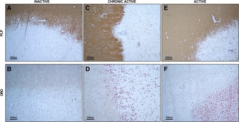
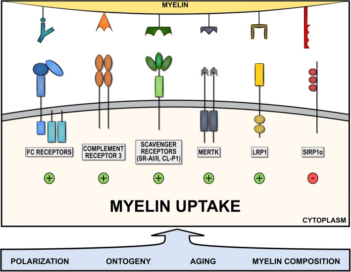
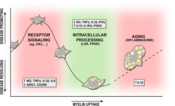
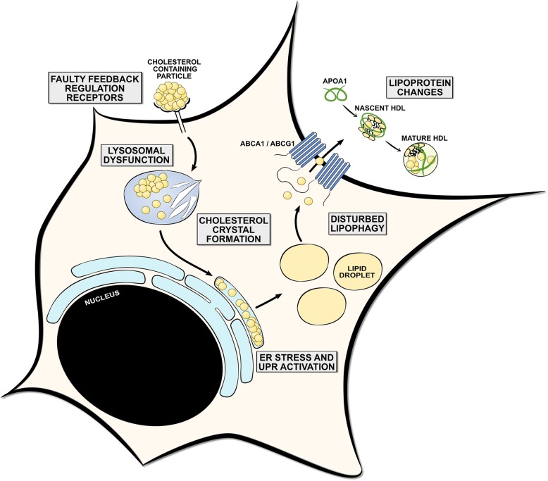
Multiple sclerosis (MS) is a chronic disease of the central nervous system characterized by massive infiltration of immune cells, demyelination, and axonal loss. Active MS lesions mainly consist of macrophages and microglia containing abundant intracellular myelin remnants. Initial studies showed that these foamy phagocytes primarily promote MS disease progression by internalizing myelin debris, presenting brain-derived autoantigens, and adopting an inflammatory phenotype. However, more recent studies indicate that phagocytes can also adopt a beneficial phenotype upon myelin internalization. In this review, we summarize and discuss the current knowledge on the spatiotemporal physiology of foamy phagocytes in MS lesions, and elaborate on extrinsic and intrinsic factors regulating their behavior. In addition, we discuss and link the physiology of myelin-containing phagocytes to that of foamy macrophages in other disorders such atherosclerosis.

Keywords: Macrophage; Microglia; Multiple sclerosis; Neuroinflammation; Polarization; Remyelination.


Conflict of interest statement

Competing interests

The authors declare that they have no competing interests.

Publisher’s Note

Springer Nature remains neutral with regard to jurisdictional claims in published maps and institutional affiliations.

Figures

Fig. 1
Fig. 1
Histopathology of inactive, chronic active, and active multiple sclerosis lesions. Inactive, chronic active, and active multiple sclerosis (MS) lesions were stained for intracellular lipid droplets (oil red o; ORO) and myelin (proteolipid protein; PLP). a and b, c and d, e and f are taken from the same lesion. Foamy phagocytes (ORO+ cells) are apparent in demyelinating chronic active and active MS lesions, but not in inactive lesions
Fig. 2
Fig. 2
Endocytosis of myelin by phagocytes. Myelin internalization by phagocytes is dependent on the receptor repertoire as well as various intrinsic and extrinsic factors. While scavenger receptors (SR-AI/II and collectin placenta 1 (CL-P1)), Fc receptors, complement receptors (CR3), mer tyrosine kinase (MerTK), and low-density lipoprotein receptor-related protein 1 (LRP1) positively regulate the uptake of myelin, ligation of signal regulatory protein α (SIRPα) inhibits myelin uptake. Cell intrinsic and extrinsic factors, such as phagocyte polarization (M1- or M2-like), phagocyte ontogeny, (hematopoietic stem cells or yolk-sac progenitors), cellular aging, and myelin composition and modifications, can impact the capacity of phagocytes to internalize myelin
Fig. 3
Fig. 3
Foamy phagocyte polarization follows a triphasic pattern. Uptake of myelin initially promotes the induction of a disease-promoting phenotype of phagocytes, characterized by an increased release of inflammatory and toxic mediators, and reduced production of anti-inflammatory factors (phase I). The induction of this phenotype likely relies on the rapid activation of the FAK/PI3K/Akt/NF-κB signaling pathway following ligation of the complement receptor 3 (CR3). In time, intracellular processing of myelin will generate lipid metabolites capable of activating the anti-inflammatory liver X receptor (LXR) and peroxisome proliferator-activated receptor (PPAR). Activation of these nuclear receptors will repress the inflammatory transcriptional profile in macrophages (phase II). With aging, an inability of phagocytes to process and efflux the enormous amounts of intracellular cholesterol-rich myelin debris results in the formation of cholesterol crystals that activate the NLRP3 inflammasome (phase III)
Fig. 4
Fig. 4
Homeostatic and dysfunctional processing of cholesterol-containing lipid particles. During homeostasis, phagocytes are well equipped to cope with relatively minor increases of cholesterol. However, sustained intracellular accumulation of cholesterol, as observed in in many peripheral pathologies and following infections with persistent pathogens, can lead to disturbances in pathways that mediate the degradation, storage, or efflux of cholesterol. First, faulty feedback regulation of phagocytic receptors may result in an uncontrolled uptake of cholesterol-containing lipid particles. Second, lysosomal cholesterol accumulation can result in lysosomal dysfunction by reducing lysosomal acidification and causing lysosomal leakiness. In addition, sustained accumulation of cholesterol can lead to the formation of cholesterol crystals that activate the caspase-1-activating NLRP3 inflammasome. Third, persistent cholesterol trafficking to ER membranes can trigger ER stress and the unfolded protein response (UPR). Fourth, dysfunctional lipophagy machinery can hamper the capacity of foamy phagocytes to process cholesterol within lipid droplets, thereby impeding the cells’ capacity to dispose of intracellular cholesterol. Finally, quantitative and qualitative changes in lipoproteins can impact the capacity of foamy phagocytes to efflux cholesterol. Altogether, disturbances in the abovementioned pathways are well-known to promote the induction of a disease-promoting phenotype of foamy phagocytes and eventually even cause apoptosis. While ample evidence suggests that faulty regulation of these pathways also occurs in myelin-containing phagocytes, more research is warranted to define to what extent they impact their inflammatory features

References

    1. Alirezaei M, Fox HS, Flynn CT, Moore CS, Hebb AL, Frausto RF, Bhan V, Kiosses WB, Whitton JL, Robertson GS, Crocker SJ. Elevated ATG5 expression in autoimmune demyelination and multiple sclerosis. Autophagy. 2009;5:152–158. doi: 10.4161/auto.5.2.7348. - DOI - PMC - PubMed
    1. Barnett MH, Parratt JD, Cho ES, Prineas JW. Immunoglobulins and complement in postmortem multiple sclerosis tissue. Ann Neurol. 2009;65:32–46. doi: 10.1002/ana.21524. - DOI - PubMed
    1. Barral P, Polzella P, Bruckbauer A, van Rooijen N, Besra GS, Cerundolo V, Batista FD. CD169(+) macrophages present lipid antigens to mediate early activation of iNKT cells in lymph nodes. Nat Immunol. 2010;11:303–312. doi: 10.1038/ni.1853. - DOI - PMC - PubMed
    1. Besler HT, Comoglu S. Lipoprotein oxidation, plasma total antioxidant capacity and homocysteine level in patients with multiple sclerosis. Nutr Neurosci. 2003;6:189–196. doi: 10.1080/1028415031000115945. - DOI - PubMed
    1. Bhattacharya A, Parillon X, Zeng S, Han S, Eissa NT. Deficiency of autophagy in dendritic cells protects against experimental autoimmune encephalomyelitis. J Biol Chem. 2014;289:26525–26532. doi: 10.1074/jbc.M114.575860. - DOI - PMC - PubMed

Publication types