. 2018 Nov 15;175(5):1418-1429.e9.
doi: 10.1016/j.cell.2018.09.050. Epub 2018 Oct 25.

Changes of Cell Biochemical States Are Revealed in Protein Homomeric Complex Dynamics

Affiliations

Changes of Cell Biochemical States Are Revealed in Protein Homomeric Complex Dynamics

Bram Stynen et al. Cell. .

Abstract

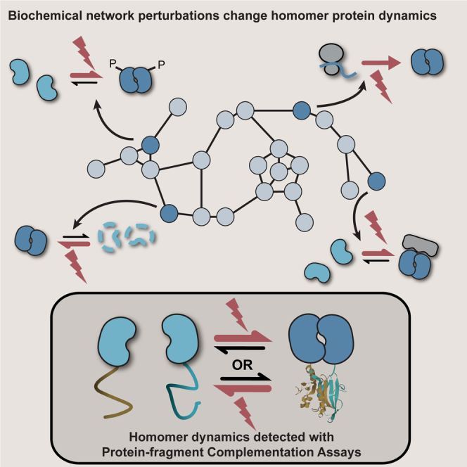
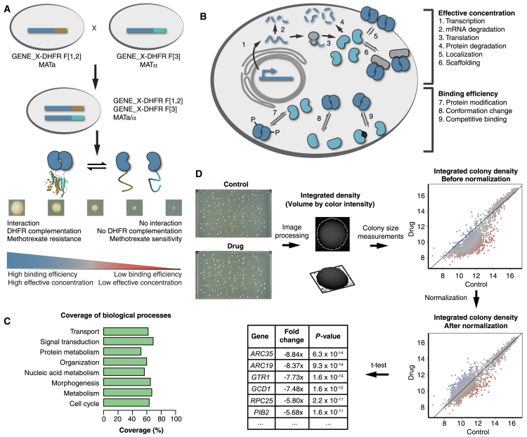
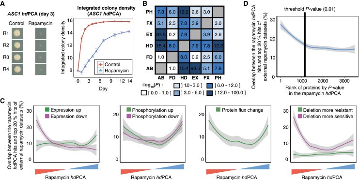
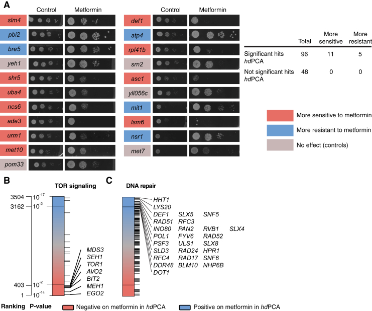
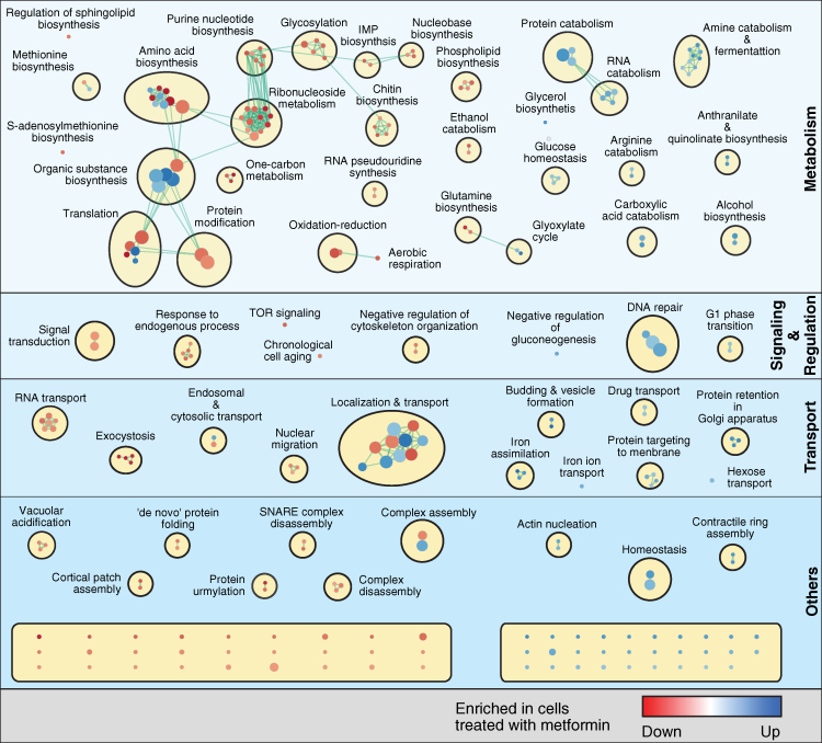
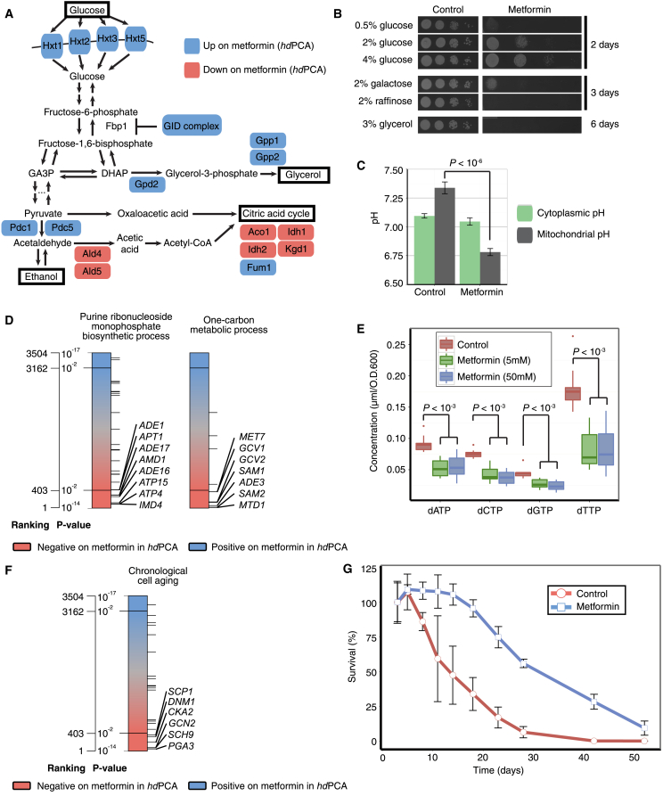
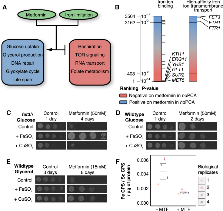
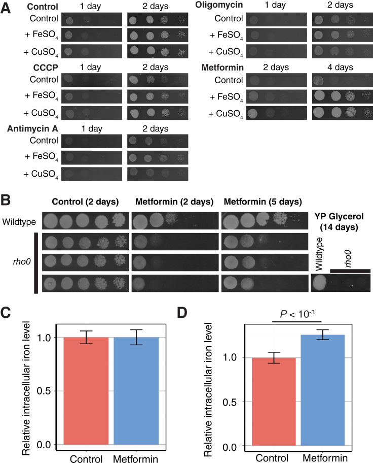
We report here a simple and global strategy to map out gene functions and target pathways of drugs, toxins, or other small molecules based on "homomer dynamics" protein-fragment complementation assays (hdPCA). hdPCA measures changes in self-association (homomerization) of over 3,500 yeast proteins in yeast grown under different conditions. hdPCA complements genetic interaction measurements while eliminating the confounding effects of gene ablation. We demonstrate that hdPCA accurately predicts the effects of two longevity and health span-affecting drugs, the immunosuppressant rapamycin and the type 2 diabetes drug metformin, on cellular pathways. We also discovered an unsuspected global cellular response to metformin that resembles iron deficiency and includes a change in protein-bound iron levels. This discovery opens a new avenue to investigate molecular mechanisms for the prevention or treatment of diabetes, cancers, and other chronic diseases of aging.

Keywords: aging; iron homeostasis; large-scale screen; metformin; protein-protein interactions; rapamycin.


Figures

None
Graphical abstract
Figure 1
Figure 1
A Homomer Dynamics DHFR PCA for the Detection of the Condition-Dependent States of Proteins (A) A library of homomer dynamics DHFR PCA (hdPCA) strains is created by mating two strains, each containing an open reading frame (ORF) of interest tagged with one of the two complementary fragments of murine dihydrofolate reductase (mDHFR, brown and light blue). Upon interaction of two molecules of the same protein, the two fragments of mDHFR fold and reconstitute into a functional enzyme. This reconstitution quantitatively correlates with growth in the presence of methotrexate (Levy et al., 2014) and is determined by effective concentration and binding efficiency. (B) The degree of homomerization (self-association) of a protein is the result of different factors, some of which influence the effective concentration (top), whereas others influence binding efficiency (bottom). (C) Coverage of gene ontology (GO) biological processes in the hdPCA, with coverage determined by the percentage of proteins associated with a GO Super-Slim biological process, that have been screened in the hdPCA. GO Super-Slim biological processes were obtained by manually condensing the standard terms in the GO Slim (available at https://www.yeastgenome.org/download-data/curation, as of July 2017) into eight GO global terms. (D) Workflow of the hdPCA.
Figure 2
Figure 2
Optimization and Validation of hdPCA with Rapamycin Data (A) Growth of a strain containing ASC1-DHFR-F[1,2] and ASC1-DHFR-F[3] during the course of an hdPCA screen with rapamycin. Four replicates (R1 to R4) were tested. Error bars indicate SD. (B) Correlations among different large-scale datasets comparing control versus rapamycin-treated yeast cells. Datasets are compared with each other for significant overlap of genes or proteins affected by rapamycin. Values correspond to –log10(P), where P is the p value of the hypergeometric test that compares significance of overlap between datasets. AB, protein abundance; FD, fitness of deletion strains; HD, hdPCA; EX, mRNA expression; FX, protein flux; PH, phosphoproteomics. (C) LOESS curves indicating the running average overlap between rapamycin hdPCA hits, ordered from most reduced to most increased hdPCA signal in the presence of rapamycin, and the top 20% hits of rapamycin data focused on expression, phosphorylation, protein flux, and deletion strain sensitivity. (D) A LOESS curve indicating the average overlap between hdPCA hits, ranked by p value, and the top 20% hits of five external rapamycin datasets (deletion screens, mRNA expression data, phosphoproteomics data, protein abundance data, and protein flux data). The enrichment found in top-ranked hdPCA hits falls to background levels close to the threshold p value of 0.01, which was chosen as a threshold value in further experiments. See also Tables S1 and S3.
Figure S1
Figure S1
Metformin hdPCA Identifies Proteins Involved in Metformin Resistance, in TOR Signaling, and in DNA Repair, Related to Figure 3 (A–C) (A) A deletion miniscreen confirms contribution of significant hits from the metformin hdPCA in metformin resistance. Significant hits from the metformin hdPCA screen were tested for their involvement in metformin sensitivity by testing their growth in the presence of the drug. Ninety-six deletion strains from significant hits were tested together with 48 negative control strains. No strains from the negative control set were affecting metformin sensitivity. Pictures for the control condition were taken after 2 days, for metformin after 5 days. A chi-square test of independence (metformin versus control) confirms that metformin hdPCA hits are more likely to modulate resistance (increased or reduced) to metformin compared to the controls (p < 0.01). Metformin hdPCA data on proteins involved in TOR signaling (B) and proteins involved in DNA repair (C).
Figure 3
Figure 3
Enrichment Map of GO Biological Processes in the Metformin hdPCA The map displays the enriched GO terms in metformin versus control (blue) and those that are enriched in control versus metformin (red). GO terms that have associated genes in common are linked with an edge. The edge width is proportional to the overlap between the linked GO terms. GO terms closer to each other in space are more functionally related than those further away from each other in space and are clustered together. Ungrouped processes not mentioned in the main text are found at the bottom (unnamed; see Table S4 for more details). The figure was generated using the Enrichment Map (GO terms cutoff, 0.05; similarity measure, Jaccard coefficient with default settings) and AutoAnnotate (default settings) plug-ins. See also Figure S1 and Tables S2, S4, S5, S6, and S7.
Figure 4
Figure 4
Metformin Influences a Range of Cellular Processes (A) Metformin increases (blue) and decreases (pink) the hdPCA signal of proteins involved in glucose uptake and catabolism. (B) Sensitivity to metformin changes with carbon source. Pictures were taken on different days to account for the basal effect of carbon source on growth. (C) The mitochondrial membrane proton gradient reverses after prolonged metformin treatment (7 hr). The pH level was determined using a pH-dependent fluorescent protein (pHluorin) with or without a mitochondrial localization sequence. Error bars indicate the SD (n = 12). (D) The hdPCA signals for proteins involved in purine ribonucleoside biosynthesis and one-carbon metabolism are reduced in the presence of metformin. Each horizontal line represents one protein member of the biological process. (E) Metformin at 5 mM and 50 mM reduces the concentration of dNTPs. Concentrations were determined by mass spectrometric analysis of the individual dNTPs. Error bars indicate SD (n = 12). (F) hdPCA results for members of the GO term “chronological cell aging.” (G) Metformin prolongs the chronological lifespan. Three cultures were incubated in the presence of metformin and three in its absence. Cell survival was tracked over time by counting the colony-forming units at regular time points. The error bars correspond to the SD of three replicates of one qualitatively representative experiment. GID, glucose-induced degradation; GA3P, glyceraldehyde-3-phosphate; DHAP, dihydroxyacetone phosphate.
Figure 5
Figure 5
Metformin Treatment Interferes with Iron Homeostasis (A) Processes affected by metformin treatment according to hdPCA data are also modulated by iron limitation. (B) Metformin hdPCA data on proteins involved in high-affinity iron ion uptake and proteins binding iron. (C) Growth of fet3Δ in the presence of metformin and CuSO4 or FeSO4. (D) Growth of wild-type yeast in the presence of metformin and CuSO4 or FeSO4. (E) Growth of wild-type yeast under respiratory conditions with metformin and CuSO4 or FeSO4. (F) Protein was extracted from yeast cultures incubated in the presence (+ MTF) or absence (− MTF) of metformin. The concentration of 56Fe bound to the protein was determined by inductively coupled plasma mass spectrometry (ICP-MS) (8 technical replicates are shown of 4 biological replicates for each condition). CPS, counts per second; Sc, scandium. See also Figure S2 and Table S8.
Figure S2
Figure S2
Metformin More Closely Relates to Iron Homeostasis than Respiration Despite the Lack of an Effect on Intracellular Iron Levels, Related to Figure 5 (A) Growth of wild-type yeast on YP raffinose (2%) in the presence of different inhibitors of oxidative phosphorylation (CCCP at 20 μM, antimycin A at 0.1 μg/ml, oligomycin at 3 μg/ml) and in the presence or absence of additional FeSO4 (500 μM) or CuSO4 (500 μM). Raffinose is a carbon source that is consumed through both fermentation and respiration. (B) Growth of wild-type yeast and three independent respiration-deficient (‘petite’) rho0 strains in the presence or absence of metformin (50 mM) on YPD. Respiration-deficient strains do not grow in respiratory conditions (YP glycerol). (C) Intracellular iron content of yeast cultures in exponential growth phase (O.D.600 = 1) after treatment with metformin, relative to the control condition. Error bars indicate standard deviation (n = 10). (D) Intracellular iron content of yeast cultures in stationary growth phase after treatment with metformin, relative to the control condition. Error bars indicate standard deviation (n = 3).

References

    1. Alam M.T., Zelezniak A., Mülleder M., Shliaha P., Schwarz R., Capuano F., Vowinckel J., Radmanesfahar E., Krüger A., Calvani E. The metabolic background is a global player in Saccharomyces gene expression epistasis. Nat. Microbiol. 2016;1:15030. - PMC - PubMed
    1. Anisimov V.N., Zabezhinski M.A., Popovich I.G., Piskunova T.S., Semenchenko A.V., Tyndyk M.L., Yurova M.N., Rosenfeld S.V., Blagosklonny M.V. Rapamycin increases lifespan and inhibits spontaneous tumorigenesis in inbred female mice. Cell Cycle. 2011;10:4230–4236. - PubMed
    1. Ansell R., Adler L. The effect of iron limitation on glycerol production and expression of the isogenes for NAD(+)-dependent glycerol 3-phosphate dehydrogenase in Saccharomyces cerevisiae. FEBS Lett. 1999;461:173–177. - PubMed
    1. Baldi P., Long A.D. A Bayesian framework for the analysis of microarray expression data: regularized t -test and statistical inferences of gene changes. Bioinformatics. 2001;17:509–519. - PubMed
    1. Barbet N.C., Schneider U., Helliwell S.B., Stansfield I., Tuite M.F., Hall M.N. TOR controls translation initiation and early G1 progression in yeast. Mol. Biol. Cell. 1996;7:25–42. - PMC - PubMed

Publication types

MeSH terms

LinkOut - more resources