

Review
doi: 10.1016/j.placenta.2018.11.003.
Epub 2018 Nov 10.
Fetal membrane architecture, aging and inflammation in pregnancy and parturition
Affiliations
- PMID: 30454905
- PMCID: PMC7041999
- DOI: 10.1016/j.placenta.2018.11.003
Item in Clipboard
Review
Fetal membrane architecture, aging and inflammation in pregnancy and parturition
Placenta.
2019 Apr.
Abstract
Preterm birth is the single major cause of infant mortality. Short and long term outcomes for infants are often worse in cases of preterm premature rupture of the fetal membranes (pPROM). Thus, increased knowledge of the structure characteristics of fetal membranes as well as the mechanisms of membrane rupture are essential if we are to develop effective treatment strategies to prevent pPROM. In this review, we focus on the role of inflammation and senescence in fetal membrane biology.
Keywords: Fetal membranes; Inflammation; Senescence; Structure.
Copyright © 2018. Published by Elsevier Ltd.
Figures
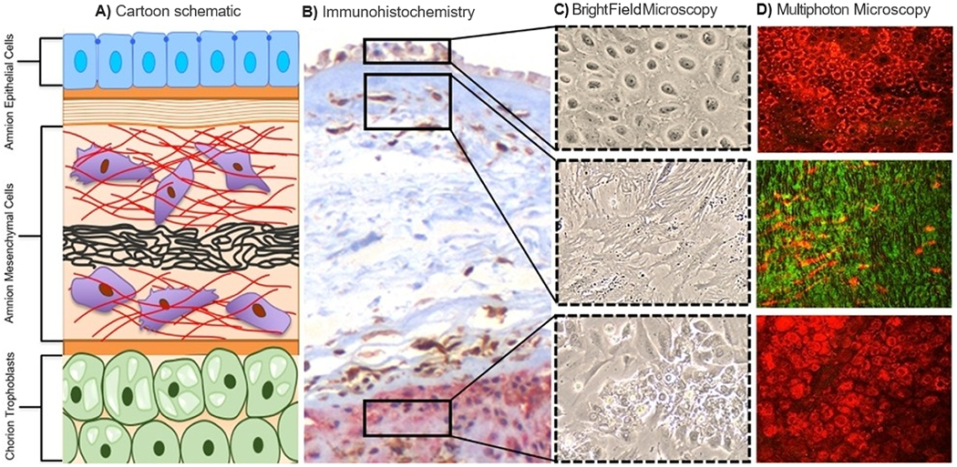
A) Proposed structures of fetal membranes. The description starts from the innermost layer (amnion) and ends at the initial layer of chorion. Amnion epithelial cells (blue) are connected to the first layer of the extracellular matrix termed the basement membrane (orange)/compact layer (orange strips). The fibroblast (top red), spongy (black), and reticular layers (bottom red) follow, containing amnion mesenchymal and stromal cells (purple). The chorion (green) is connected to the extracellular matrix through a pseudo-basement membrane (orange). The chorion is made up of two types of cells: (1) chorion leave cells that contain vacuoles (light green) and (2) the chorion trophoblast cells. The chorion interfaces with the maternal decidua, connecting the fetal to the maternal compartments of the uterus. B) Immunohistochemistry staining of fetal membranes for comparison of cellular and collagen layers. C) Bright flied microscopy of human fetal membrane primary cells reveling morphology in culture. Amnion and chorion epithelial cells express cuboidal epithelial morphology, while amnion mesenchymal cells in the extracellular matrix demonstrated elongated fibroblastoid shape with the presence of actin filaments. Images were captured at 10x and cropped. D) Multiphoton autoflorescent microscopy and second harmonic generation microscopy single-plain images confirm cellular and collagen morphology. Cellular components- red, Collagen- green. Images were captured at 25× and cropped.
References
-
- Malak TM, Ockleford CD, Bell SC, Dalgleish R, Bright N, Macvicar J, Confocal immunofluorescence localization of collagen types I, III, IV, V and VI and their ultrastructural organization in term human fetal membranes, Placenta 14(4) (1993) 385–406. - PubMed
-
- Richardson, Lauren, Vargas, Gracie, Brown, Tyra, Ochoa, Lorenzo, Trivedi J, Kacerovský, Marian, Lappas, Martha, Menon R, Redefining 3Dimensional placental membrane microarchitecture using multiphoton microscopy and optical clearing, 53(66–75) (2017). - PubMed
Publication types
MeSH terms
Grants and funding
LinkOut - more resources
Full Text Sources
Other Literature Sources
