. 2018 Dec 18;115(12):2395-2402.
doi: 10.1016/j.bpj.2018.10.013. Epub 2018 Oct 23.

Adhesion of Active Cytoskeletal Vesicles

Affiliations

Adhesion of Active Cytoskeletal Vesicles

Renu Maan et al. Biophys J. .

Abstract

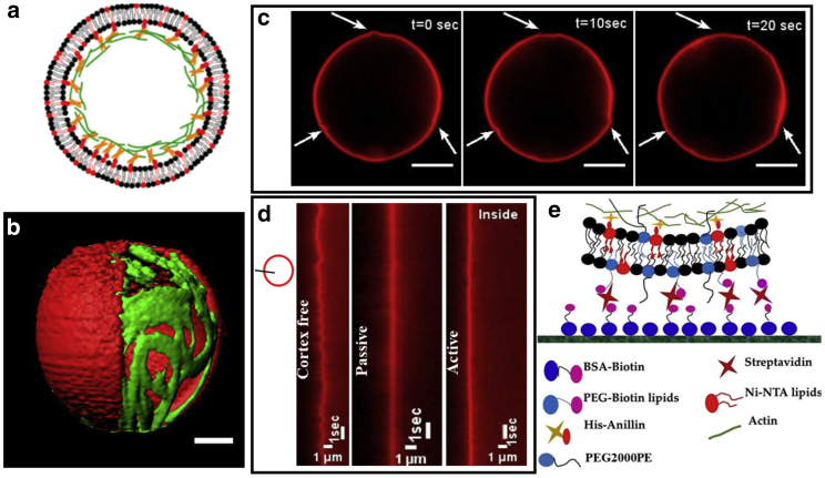
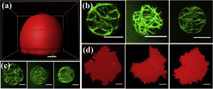
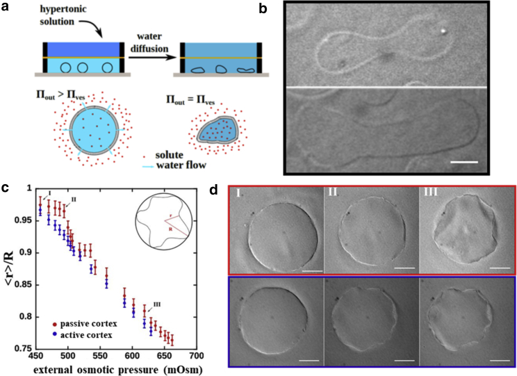
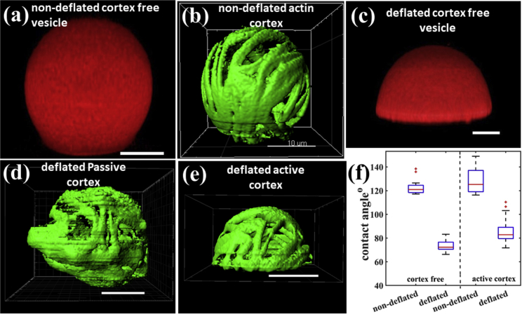
Regulation of adhesion is a ubiquitous feature of living cells, observed during processes such as motility, antigen recognition, or rigidity sensing. At the molecular scale, a myriad of mechanisms are necessary to recruit and activate the essential proteins, whereas at the cellular scale, efficient regulation of adhesion relies on the cell's ability to adapt its global shape. To understand the role of shape remodeling during adhesion, we use a synthetic biology approach to design a minimal experimental model, starting with a limited number of building blocks. We assemble cytoskeletal vesicles whose size, reduced volume, and cytoskeletal contractility can be independently tuned. We show that these cytoskeletal vesicles can sustain strong adhesion to solid substrates only if the actin cortex is actively remodeled significantly. When the cytoskeletal vesicles are deformed under hypertonic osmotic pressure, they develop a crumpled geometry with deformations. In the presence of molecular motors, these deformations are dynamic in nature, and the excess membrane area generated thereby can be used to gain adhesion energy. The cytoskeletal vesicles are able to attach to the rigid glass surfaces even under strong adhesive forces just like the cortex-free vesicles. The balance of deformability and adhesion strength is identified to be key to enable cytoskeletal vesicles to adhere to solid substrates.


Figures

Figure 1
Figure 1
(a) The lipid membrane contains a fraction of lipids functionalized with the Ni-NTA group. Elementary building blocks encapsulated in the vesicle consist of actin and polyhistidine-tagged anillin cross-linker, which is sufficient for the formation of cytoskeletal network coupled to the lipid membrane via the Ni-NTA lipid/his-anillin links. (b) A 3D reconstruction of the cytoskeletal vesicles produced using cDICE shows the close proximity of the actin cortex to the membrane. A section of the membrane has been masked to show the underlying actin cortex. The scale bar represent 5 μm. (c) When myosin motors are added to the vesicles, they induce small dynamic shape deformations, as indicated by the white arrows. A membrane labeled with Texas Red has been used in combination with epifluorescence to record these deformations. The scale bars represent 5 μm. (d) To compare the membrane fluctuations, intensity line profiles across the section of the membrane were used in the Fiji plugin to prepare the kymographs. The three kymographs show that a cortex-free vesicle has more prominent fluctuations in the membrane than a passive or active vesicle. The vesicles chosen for preparing kymographs were of size 17, 19, and 20 μm for cortex free, passive, and active, respectively. (e) The schematic shows the scheme that we have adopted to attach vesicles to the glass surface. The PEG2000PE lipids in the membrane prevent the nonspecific electrostatic interaction between the membrane and the glass surface. To see this figure in color, go online.
Figure 2
Figure 2
(a) The spherical cap shape adopted by the cortex-free vesicle under strong adhesion. The membrane was labeled with Texas Red to visualize the membrane for confocal scanning. The scale bar represents 5 μm. (b) The bottom-most slice from the z-stacks of three different active vesicles that did not burst even after 30 min of contact with the functionalized glass surface. (c) A collapsed actin structure of three different active vesicles that burst on the surface after making contact. Scale bars, 10 μm. (d) Three different active vesicles that bursted, showing the formation of supported bilayer with an irregular shape on the surface. The membrane was labeled with Texas Red. The scale bar represents 10 μm. To see this figure in color, go online.
Figure 3
Figure 3
(a) The two-leveled chamber used to deflate the vesicles. The top chamber was filled with glucose solution with a higher osmolarity than the solution encapsulated inside the vesicle. To image the vesicles, the entire chamber was closed and put on the microscope stage. (b) Deflation of the cortex-free vesicles shows the well-documented shape transformations due to the Helfrich-energy minimization. (c) For the passive cytoskeletal vesicles, the change in external osmotic pressure does not immediately cause any shape remodeling (from point I to II) but causes a continuous shape remodeling for active vesicles (from point I to III). (d) Phase-contrast images of passive (red panel) and active (blue panel) cytoskeletal vesicles at time points indicated in (c). The active vesicle is able to remodel the cytoskeleton and deforms actively while the pressure changes from I to II, whereas the passive vesicles resist the increasing osmotic pressure at first. At point III, both vesicles, the passive and the active ones, have reached a highly deformed shape with similar reduced volumes. Similar deformed shapes are observed for both the active and the passive vesicles although via different trajectories. For the passive vesicles, deformations appear abrupt, whereas for active vesicles, a continuous deformation is observed. The curves in (c) are the average values taken over five vesicles for the passive case and eight vesicles for the active case. The error bars are the SDs on the measure of the mean contracted radius <r>. We computed the Kruskal-Wallis test at the point II and second step (right before the passive vesicles crumple). The p-value comparing the data sets of active and passive vesicles are p = 0.032 and p = 0.044, respectively. Scale bars, 20 μm. To see this figure in color, go online.
Figure 4
Figure 4
(a and b) Cortex-free and passive cytoskeletal vesicle in the weakly adhered or nondeflated state, respectively. (c) The cortex-free vesicle adopts a spherical cap shape after deflation, a signature of strong adhesion. (d and e) The passive and active vesicles, respectively after deflation. The deformation leads to undefined shapes in the case of the passive vesicles and spherical cap shape in active vesicles. (f) The almost equal difference in the contact angle between nondeflated and deflated state for cortex-free and the active cortex vesicles shows that the active vesicle can use the excess membrane area developed by deflation in reducing the contact angle to spread on the surface. 20 vesicles were imaged for each vesicle type for the box-and-whisker diagram. The method we used to estimate the contact angle is shown in the Fig. S4. Scale bars, 10 μm. To see this figure in color, go online.

References

    1. Albersdörfer A., Feder T., Sackmann E. Adhesion-induced domain formation by interplay of long-range repulsion and short-range attraction force: a model membrane study. Biophys. J. 1997;73:245–257. - PMC - PubMed
    1. Cuvelier D., Nassoy P. Hidden dynamics of vesicle adhesion induced by specific stickers. Phys. Rev. Lett. 2004;93:228101. - PubMed
    1. Goennenwein S., Tanaka M., Sackmann E. Functional incorporation of integrins into solid supported membranes on ultrathin films of cellulose: impact on adhesion. Biophys. J. 2003;85:646–655. - PMC - PubMed
    1. Reister-Gottfried E., Sengupta K., Smith A.S. Dynamics of specific vesicle-substrate adhesion: from local events to global dynamics. Phys. Rev. Lett. 2008;101:208103. - PubMed
    1. Sackmann E., Smith A.S. Physics of cell adhesion: some lessons from cell-mimetic systems. Soft Matter. 2014;10:1644–1659. - PMC - PubMed

Publication types

LinkOut - more resources