. 2018 Dec 4;115(49):E11465-E11474.
doi: 10.1073/pnas.1816614115. Epub 2018 Nov 19.

Single nucleotide polymorphisms alter kinase anchoring and the subcellular targeting of A-kinase anchoring proteins

Affiliations

Single nucleotide polymorphisms alter kinase anchoring and the subcellular targeting of A-kinase anchoring proteins

F Donelson Smith et al. Proc Natl Acad Sci U S A. .

Abstract

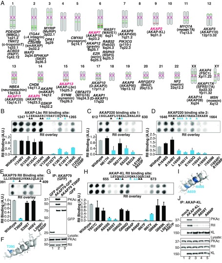
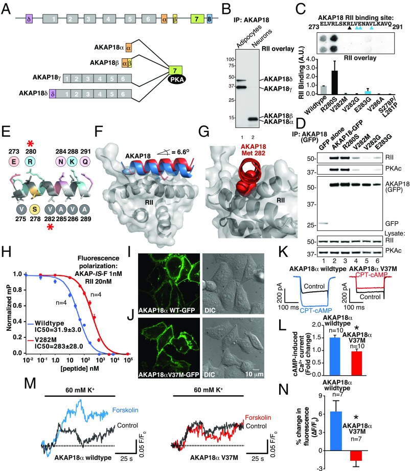
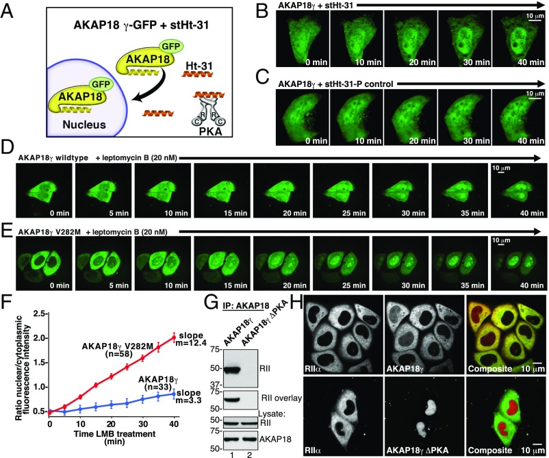
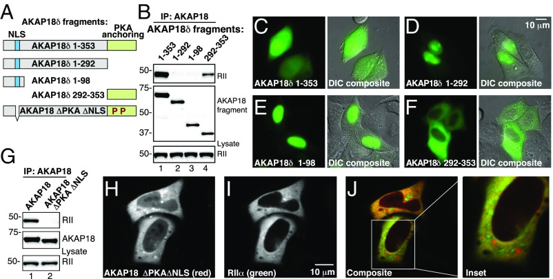
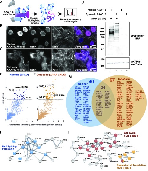
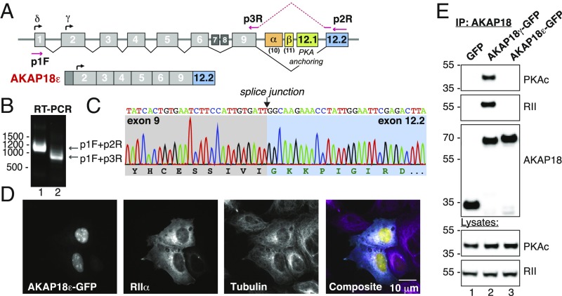
A-kinase anchoring proteins (AKAPs) shape second-messenger signaling responses by constraining protein kinase A (PKA) at precise intracellular locations. A defining feature of AKAPs is a helical region that binds to regulatory subunits (RII) of PKA. Mining patient-derived databases has identified 42 nonsynonymous SNPs in the PKA-anchoring helices of five AKAPs. Solid-phase RII binding assays confirmed that 21 of these amino acid substitutions disrupt PKA anchoring. The most deleterious side-chain modifications are situated toward C-termini of AKAP helices. More extensive analysis was conducted on a valine-to-methionine variant in the PKA-anchoring helix of AKAP18. Molecular modeling indicates that additional density provided by methionine at position 282 in the AKAP18γ isoform deflects the pitch of the helical anchoring surface outward by 6.6°. Fluorescence polarization measurements show that this subtle topological change reduces RII-binding affinity 8.8-fold and impairs cAMP responsive potentiation of L-type Ca2+ currents in situ. Live-cell imaging of AKAP18γ V282M-GFP adducts led to the unexpected discovery that loss of PKA anchoring promotes nuclear accumulation of this polymorphic variant. Targeting proceeds via a mechanism whereby association with the PKA holoenzyme masks a polybasic nuclear localization signal on the anchoring protein. This led to the discovery of AKAP18ε: an exclusively nuclear isoform that lacks a PKA-anchoring helix. Enzyme-mediated proximity-proteomics reveal that compartment-selective variants of AKAP18 associate with distinct binding partners. Thus, naturally occurring PKA-anchoring-defective AKAP variants not only perturb dissemination of local second-messenger responses, but also may influence the intracellular distribution of certain AKAP18 isoforms.

Keywords: AKAPs; PKA; kinase anchoring; nucleus; proximity labeling.


Conflict of interest statement

The authors declare no conflict of interest.

Figures

Fig. 1.
Fig. 1.
Identification and analysis of polymorphisms in PKA-anchoring domains of AKAPs. (A) Schematic of the human chromosomal locations for genes encoding 39 experimentally validated AKAPs. AKAPs harboring nonsynonymous SNPs in the PKA-anchoring helices that are investigated in this study are highlighted in red. (BE and H) Peptide SPOT arrays containing WT and variant PKA binding helices were constructed and probed by RII overlay. (Upper) Solid-phase RII binding was assessed by far-Western blotting with biotin-RII and detected by neutravidin-HRP and enhanced chemoluminescence. (Lower) Densitometric analysis of RII binding was normalized to WT signal. (B) Peptide array and amalgamated densitometric data for analysis of the AKAP-Lbc (AKAP13) RII-binding site. Densitometric analysis of RII binding was normalized to WT signal. Variants that reduce binding to RII are highlighted in blue. (C) Screening and analysis of peptide arrays representing SNP sequences for PKA binding site 1 in AKAP220 (AKAP11). (D) Screening and analysis of peptide arrays representing SNP sequences for the PKA binding site 2 in AKAP220 (AKAP11). (E) Screening and analysis of peptide arrays representing SNP sequences for the PKA binding site in AKAP79 (AKAP5). (F) Molecular modeling of the AKAP79 amphipathic helix (PDB ID code 2H9R) showing threonine 395 at the second turn of the helix (blue). (G) Immune complexes of either WT AKAP79 or AKAP79 T395P were probed for coprecipitation of PKA RII and C subunits. Introduction of the helix-breaking proline residue disrupts the AKAP79–PKA interaction. (H) Screening and analysis of peptide arrays representing SNP sequences for the PKA binding in AKAPKL (AKAP2). (I) Modeling of the AKAP-KL amphipathic helix based on coordinates for AKAP-IS (PDB ID code 2IZX), showing the residues A659 and A666 (blue). (J) Immune complexes of either WT AKAP-KL or AKAP-KL A659S, A659T, or A666P variants were probed for coprecipitation of PKA RII and C subunits. Mutation of either position drastically impairs PKA binding. IP, immunoprecipitation
Fig. 2.
Fig. 2.
Polymorphisms in AKAP18 isoforms (AKAP7) alter PKA binding and regulation of ion channels. (A) Schematic of AKAP7 gene organization depicting exon–intron structure and alternatively spliced isoforms. (B) IP-RII overlay analysis show differential expression of distinct AKAP18 isoforms in 3T3-L1 adipocytes and cultured hippocampal neurons. (C) Peptide array and amalgamated densitometric data for analysis of the AKAP18 (AKAP7) RII-binding site. Densitometric analysis of RII binding was normalized to WT signal. Variants that reduce binding to RII are highlighted in blue. (D) AKAP18 SNP variants (denoted above each lane) were immunoprecipitated and copurification of PKA regulatory (Upper) and catalytic (Upper-mid) identified by Western blotting. Expression levels of each protein are indicated in the Lower three panels. (E) Model of AKAP18 PKA binding helix highlighting selected residues (red asterisk) on either face of the amphipathic helix. (F) Structural model shows WT AKAP18 helix (blue) and AKAP18 V282M helix (red) in complex with the RII D/D domain (gray). The V282M helix (red) is oriented at a 6.6° angle from WT. (G) En face and expanded view of the AKAP18 V282M helix shows displacement of the mutant peptide by bulky methionine side chain (red). (H) Fluorescence polarization data shows AKAP18 WT and V282M peptide displacement of a fluorescent AKAP-IS peptide from RII. The IC50 values for each are indicated. (I) Fluorescent and DIC images of HeLa cells expressing WT AKAP18α-GFP. (J) Fluorescent and DIC images of HeLa cells expressing AKAP18α V37M-GFP. (Scale bar: IJ, 10 μm.) (K) Whole-cell electrophysiological recording of cAMP-potentiated Ca2+ current in HEK293 cells expressing the L-type Ca2+ channel and either AKAP18α WT or the AKAP18α V37M variant. (L) Quantification of peak current potentiation. Data are presented as mean ± SEM. *P < 0.001 by unpaired Student’s t test. (M) Fluo4-AM measurement of cAMP-potentiated Ca2+ current in mouse aortic smooth muscle cells expressing either WT AKAP18α (blue) or the AKAP18α V37M variant (red). Calcium accumulation in response to depolarization was measured before and after brief forskolin stimulation. (N) Quantification of the change in fluorescence response to K+-induced depolarization. Data are presented as mean ± SEM. *P < 0.001 by unpaired Student’s t test.
Fig. 3.
Fig. 3.
Association with the PKA holoenzyme is necessary for cytoplasmic retention of AKAP18 long forms. (A) Schematic depicting displacement of PKA from AKAP18γ by the cell-soluble PKA-anchoring disruptor peptide (stHt-31, orange) and the effect on subcellular targeting of AKAP18 long isoforms. Selected time points are shown from 0 to 40 min. (B and C) Live-cell imaging of cells expressing AKAP18γ-YFP following application of (B) stHt-31 or (C) the proline control peptide, stHt-31-P. (D and E) Live-cell imaging of cells expressing (D) WT AKAP18γ-GFP or (E) V282M AKAP18γ-GFP. Cells were treated with LMB and subcellular distribution was recorded over 40 min. (F) Amalgamated data from multiple experiments in D and E. Changes in the ratio of nuclear/cytoplasmic fluorescent signal WT AKAP18γ-GFP (n = 33 cells) and V282M AKAP18γ-GFP (n = 58 cells). (G) The PKA-binding deficient mutant AKAP18γ S278P/L281P (AKAP18γ ΔPKA) is unable coprecipitate RII or interact with it by solid-phase overlay. (H) Fluorescent confocal images of HeLa cells expressing RIIα-YFP (green) and (Lower) mCherry-AKAP18γ (red) or (Bottom) the PKA-deficient AKAP18γ mutant.
Fig. 4.
Fig. 4.
Long isoforms of AKAP18 contain a nuclear localization signal that may be masked by interaction with PKA. (A) Schematic showing fragments of YFP-tagged AKAP18δ isoforms that were used to map targeting regions within the anchoring protein. The first and last residue of each AKAP18 fragment is indicated. A polybasic NLS (blue) and PKA-anchoring regions (green) are identified. (B) IP of YFP-tagged AKAP18δ fragments. (Top) Western blot analysis of immune complexes shows PKA RII coprecipitating with full-length protein and the 292–353 fragment. (Middle) Loading control for AKAP18 fragments and (Bottom) loading controls for RIIα. (CF) Fluorescent and DIC imaging detecting the differential subcellular distribution YFP-tagged AKAP18δ fragments. (G) Characterization of WT AKAP18γ and AKAP18γ ΔPKA ΔNLS mutant immune complexes. (Top) RII coprecipitates with the WT only. (Middle) Loading control for AKAP18. (Bottom) loading control for RIIα. (H) Fluorescent confocal image of the mCherry-tagged AKAP18γ ΔPKA ΔNLS mutant shows cytoplasmic retention. (I) RIIα-YFP is also cytoplasmic. (J) Single channel and composite images show both an overlapping and distinct cytoplasmic distribution of RII (green) and AKAP18γ ΔPKA ΔNLS (red). (Inset) Magnified image showing the regional distribution of RII and AKAP18.
Fig. 5.
Fig. 5.
Proximity labeling and proteomic identification of compartment-selective AKAP18 interactors. (A) Schematic depicting the miniTurbo-based biotinylation/MS experiments. Biotin is shown as dark-blue circles. (Left) Upon biotin addition, miniTurbo biotinylates only proximal proteins. (Center) Biotinylated proteins are isolated with streptavidin pulldown and (Right) identified by MS. (B and C) Fluorescence microscopy depicting the subcellular location of (B) nuclear and (C) cytoplasmic AKAP18-miniTurbo variants and detection of compartment specific biotinylated proteins. (D) Western blot analysis of biotinylated proteins in cell lysates shows distinct and compartment-specific labeling patterns. (E and F) MS results plotted as log difference of experimental intensity minus control intensity plotted against the negative log of the P value. Selected nuclear (dark-blue) and cytoplasmic (tan) AKAP18 interacting partners are indicated. (G) Lists of statistically significant target proteins in nuclear and cytosolic preparations. Of 131 identified proteins, 40 were selectively associated with the nuclear variant, whereas 67 preferentially bound to the cytoplasmic variant. (H and I) STRING database-generated network depictions of compartment-specific proteins. Gene ontology process annotation and false-discovery rates (FDR) are listed for the highlighted nodes. Putative biological functions for each subnetwork are indicated.
Fig. 6.
Fig. 6.
Identification of an AKAP18 isoform that lacks a PKA binding helix. (A) AKAP7 intron–exon map as adapted from TCGA SpliceSeq. The start sites for the δ- and γ-isoforms are noted, as are the α- and β-isoform-specific exons. The RII anchoring site (green) is encoded in exon 12.1 and the alternate 12.2 exon (blue) are presented. The locations of the primers used for RT-PCR are marked. (B) RT-PCR from pooled human cDNA samples using primers against predicted transcripts and novel exon junctions produces the expected products where intervening exons are spliced out. (C) Sequencing chromatogram of the exon 9–12.2 splice junction shows an in-frame protein sequence corresponding to a distinct C terminus. (D) Fluorescent confocal images showing nuclear localization of EmGFP-tagged AKAP18ε and cytoplasmic labeling of RIIα-V5 and α-tubulin. (E) Immune complexes of either WT AKAP18γ or AKAP18ε were immunoprecipitated and copurification of PKA catalytic (Upper) and RII (Upper-mid) subunits were identified by Western blotting. Expression levels of both AKAP18 forms and GFP are indicated in the Lower three panels.

Similar articles

Cited by

References

    1. Lek M, et al. Exome Aggregation Consortium Analysis of protein-coding genetic variation in 60,706 humans. Nature. 2016;536:285–291. - PMC - PubMed
    1. Lemmon MA, Schlessinger J. Cell signaling by receptor tyrosine kinases. Cell. 2010;141:1117–1134. - PMC - PubMed
    1. Grabbe C, Dikic I. Cell biology. Going global on ubiquitin. Science. 2008;322:872–873. - PubMed
    1. Glusman G, et al. Mapping genetic variations to three-dimensional protein structures to enhance variant interpretation: A proposed framework. Genome Med. 2017;9:113. - PMC - PubMed
    1. Davies H, et al. Mutations of the BRAF gene in human cancer. Nature. 2002;417:949–954. - PubMed

Publication types

MeSH terms