. 2019 Jan 7;218(1):150-170.
doi: 10.1083/jcb.201805100. Epub 2018 Nov 19.

A cell separation checkpoint that enforces the proper order of late cytokinetic events

Affiliations

A cell separation checkpoint that enforces the proper order of late cytokinetic events

Jennifer L Brace et al. J Cell Biol. .

Abstract

Eukaryotic cell division requires dependency relationships in which late processes commence only after early ones are appropriately completed. We have discovered a system that blocks late events of cytokinesis until early ones are successfully accomplished. In budding yeast, cytokinetic actomyosin ring contraction and membrane ingression are coupled with deposition of an extracellular septum that is selectively degraded in its primary septum immediately after its completion by secreted enzymes. We find this secretion event is linked to septum completion and forestalled when the process is slowed. Delay of septum degradation requires Fir1, an intrinsically disordered protein localized to the cytokinesis site that is degraded upon septum completion but stabilized when septation is aberrant. Fir1 protects cytokinesis in part by inhibiting a separation-specific exocytosis function of the NDR/LATS kinase Cbk1, a key component of "hippo" signaling that induces mother-daughter separation. We term this system enforcement of cytokinesis order, a checkpoint ensuring proper temporal sequence of mechanistically incompatible processes of cytokinesis.


Figures

Figure 1.
Figure 1.
Septation mutants are sensitive to inappropriate activation of cell separation. (A) Cytokinesis in budding yeast. i: Normal, rapid cytokinesis can be separated into two broad phases: septation and cell separation. Only after completion of septation are septum-destroying enzymes secreted. To ensure this temporal order, we predict cells actively inhibit separation until septation is complete. ii: Cells with septation defects generate a remedial septum. Cells forming a slow remedial septum are particularly sensitive to premature septum degradation, and we propose cells activate a checkpoint-like mechanism to enforce the strict temporal order of septation and separation. (B) Inappropriate CTS1 expression is detrimental when septation fails. WT and inn1-AID cells transformed with an empty vector or a galactose-inducible vector expressing CTS1 were spotted in fivefold serial dilutions to plates containing glucose (CTS1 OFF) or galactose (CTS1 ON). Additionally, the plates contained 0.5 mM auxin (+Auxin) or DMSO (−Auxin). Plates were incubated at 30°C for 3 d. All strains express the E3 ligase TIR1. (C) CTS1 deletion partially restores viability to cells with disrupted septum synthesis. The indicated strains were grown on YPD plates containing 0.5 mM auxin (+Auxin) or DMSO (−Auxin) and incubated at 30°C for 3 d. All strains express the E3 ligase TIR1.
Figure 2.
Figure 2.
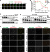
Septation failure is associated with a block in secretion of the septum degrading enzyme Cts1. (A) Cts1 transcript induction is not altered upon failed septation. CTS1 mRNA from cells in B were measured by qPCR at the time indicated after release. The fold change in CTS1 mRNA (normalized to ACT1) relative to −Auxin at time 0 is shown. A representative time course from two independent experiments is shown. (B) Cts1 secretion is blocked when septation fails. inn1-AID cells expressing HA-tagged Cts1 were synchronized in mitosis and treated with DMSO (−Auxin) or 0.5 mM auxin (+Auxin). Protein was collected at the indicated times following mitotic release at 25°C. A Western blot of secreted Cts1 (top panel) and internal pool of Cts1 (middle panel) is shown. The asterisk indicates a nonspecific band (see Fig. S1 C). Hxk2 and the cyclin Clb2 are shown as a loading and cell cycle release control, respectively (lower panels). A representative blot is shown (see also Fig. S1). (C) Western blot quantification. Fold change in the signal of secreted Cts1 (upper panel) and internal Cts1 (lower panel) relative to the maximum signal in the −Auxin control is shown. Error bars represent the SD from three independent time courses.
Figure 3.
Figure 3.
Septation mutant viability requires Fir1 to prevent inappropriate Cts1 secretion. (A) Loss of FIR1 is detrimental to cells with septation defects. Fivefold serial dilutions of the indicated strains were spotted to YPD media as in Fig. 1 C (see also Fig. S2 B). (B) CTS1 deletion rescues the lethality of inn1-AID fir1Δ. Fivefold serial dilutions of the indicated strains were spotted to YPD as in Fig. 1 C. (C) CTS1 transcript induction is not altered by lack of FIR1. CTS1 mRNA from cells in D were measured by qPCR as in Fig. 2 A. A representative time course from two independent experiments is shown. (D) Fir1 blocks Cts1 secretion when septation fails. inn1-AID fir1Δ cells expressing HA-tagged Cts1 were synchronized in mitosis and treated with DMSO (−Auxin) or 0.5 mM auxin (+Auxin). Cells were collected and processed as in Fig. 2 B. (E) Western blot quantification (as in Fig. 2 C; Fig. 3, C–E, was done in parallel with Fig. 2, A–C).
Figure 4.
Figure 4.
Fir1 prevents Cts1 secretion upon septation failure. Inhibition of Cts1 secretion upon septation failure requires Fir1. (A) Localization of Chs2-GFP-AID in the presence or absence of auxin. At 30 min after mitotic release, Chs2-GFP-AID localized to the bud neck (−Auxin) while in the presence of auxin (+Auxin) GFP signal at the bud neck could not be detected. A representative cell is shown. Bar, 2 µm. (B) chs2-AID and chs2-AID fir1Δ cells expressing HA-tagged Cts1 were synchronized in mitosis and treated with DMSO (−Auxin) or 0.5 mM auxin (+Auxin). Cells were grown at 27°C, collected, and processed as in Fig. 2 B. (C) Western blot quantification (as in Fig. 2 C). (D) FIR1 deletion enhances the lethality of chs2-AID. Fivefold serial dilutions of GalL CDC20 HA-Cts1 strains plus the indicated genotype were spotted to YP galactose plates plus or minus auxin and grown at 27°C.
Figure 5.
Figure 5.
Inappropriate Cts1 secretion causes premature septum degradation leading to failed abscission and thinned septa. (A) Inappropriate Cts1 secretion destroys chitin in the aberrant septum. The indicated genotypes, treated with or without auxin, were synchronized and released at 30°C. Cells were collected at times to enrich for cells completing cytokinesis (see Materials and methods). Calcofluor staining of fixed cells demonstrates chitin content. Enlarged region highlights bud neck chitin. Triangle highlights region of increased chitin; arrow, decreased chitin; asterisk, aberrant bud growth. Representative images are shown. Bar, 5 µm. (B) Inappropriate secretion of Cts1 leads to failed abscission and budding defects. Synchronized and auxin-treated inn1-AID and inn1-AID fir1Δ cells were released from arrest at 30°C for 3 h. Budding morphology was binned into chain-like or zygote-like, and the percentage of cells exhibiting the indicated morphology (representative image scale bar, 5 µm) is shown. Error bars represent SD of three independent experiments (n > 100 each trial); ***, P < 0.001 (two-tailed t test). (C) Strains from A were transformed to express the PH-domain of phospholipase C fused to GFP to mark the plasma membrane. Cells were collected as in A, and representative images of the indicated genotype with or without auxin are shown. The asterisk indicates a dead cell. Bar, 5 µm. (D) Cells inappropriately secreting Cts1 fail to complete septation or have thinned septal regions. Synchronized inn1-AID and inn1-AID fir1Δ cells were treated as in A and were processed for electron microscopy. Representative images of each genotype are shown. Bar, 1 µm.
Figure 6.
Figure 6.
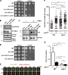
Fir1 is stabilized at the bud neck when septation is disrupted. (A) Fir1 localizes to the bud neck in late mitosis. Asynchronous cells expressing Fir1-GFP and Myo1-mCherry were subject to time-lapse microscopy. Maximum projection of serial z-stacks are shown in the xy and yz planes at the times indicated after beginning image acquisition. Bar, 1 µm. A representative cell is shown. See also Video 1. (B) Fir1 remains at the site of septation after the completion of cytokinesis. An aliquot of cells from a synchronized population was removed every 10 min after release and was imaged. The percentage of cells with bud neck localization of the indicated protein is shown (n > 20 cells per time point). (C) Fir1 is degraded before Cts1 secretion. Fir1-myc was immunoprecipitated from normalized lysates (input Hxk2 blot) following release from a synchronized culture at the times indicated at 30°C and subject to immunoblot. From the same culture, secreted Cts1 was collected and subject to immunoblot. A representative blot is shown. The asterisk indicates when peak cell separation was observed (Fig. S3 A). (D) Fir1 is not degraded when septation is disrupted. Fir1-myc was immunoprecipitated from normalized lysates (Hxk2 input) in synchronized inn1-AID cells treated with or without auxin at 30°C and subject to immunoblot. The asterisk indicates when peak cell separation was observed. Since inn1-AID cells treated with auxin do not separate, no asterisk is shown (see Fig. S3B). (E) Fir1 remains localized to the bud neck when septation is disrupted. Synchronized inn1-AID cells treated with or without auxin were released from arrest, and representative images are shown from the indicated time after release. The septin mCherry-Cdc3 was used to mark the bud neck. Bar, 5 µm (see also Fig. S3, C–E).
Figure 7.
Figure 7.
Fir1 may inhibit separation through inhibition of Cbk1. (A) CBK1 deletion rescues the growth defect of cells that fail septation. Fivefold serial dilutions of the indicated strains were spotted to YPD media as in Fig. 1 C. (B) Fir1 interaction with Cbk1 in vivo requires Fir1’s docking motif. Cbk1 was immunoprecipitated (IP Cbk1) from asynchronous cultures expressing WT Fir1 or the Cbk1 docking motif mutant (Fir1 dock*). Immunoblot of the IP sample demonstrates Fir1 co-IP with Cbk1 (Co-IP Fir1). As a control, cells lacking CBK1 were subject to the same IP and Western analysis. Total Fir1 (IP Fir1 input) was examined by IP from the same normalized lysate (Pgk1 input). (C) Cbk1 phosphorylates Fir1 in vitro. HA-tagged Fir1, Fir1 dock*, or an untagged strain were subject to Fir1 IP from asynchronous culture lysate and treated with or without bacterially purified Cbk1/Mob2 and radiolabeled 32P-ATP. An autoradiograph of Fir1 phosphorylation (upper panel) and an immunoblot showing total Fir1 (lower panel) are shown. (D) Fir1 function requires its interaction with Cbk1. Threefold serial dilutions of the indicated strains were spotted to YPD media as in Fig. 1 C (see also Fig. S4 E). (E) Cbk1 and Fir1 exhibit unique localization patterns during late mitosis. Synchronized cells expressing Fir1-GFP and Cbk1-3X mCherry were imaged every 3 min. Maximum projection of serial z-stacks is shown in the xy and yz planes at the time indicated after beginning image acquisition. Bar, 1 µm. A representative cell is shown. See also Videos 2 and 3 and Fig. S4, A and B. (F) FIR1 overexpression inhibits cell separation. WT or cbk1Δ cells expressing an inducible FIR1 overexpression vector (see Materials and methods) were grown in the absence or presence of inducer (−/+FIR1 O/E) at 30°C. The number of connected cells per cell clump (n > 100 clumps) was counted for each sample. The red line indicates the mean clump size for the indicated strain. *, P = 0.01–0.05; **, P = 0.01–0.001; ***, P < 0.001 (one-way ANOVA; see also Fig. S4 C). (G) FIR1 overexpression reduces Ace2 transcriptional output. RNA isolated from the cells in F were subject to qPCR analysis of CTS1 transcript levels (normalized to ACT1). CTS1 transcript levels are shown relative to cbk1Δ cells in the absence of inducer. Mean and SD of three independent experiments are shown. *, P = 0.01–0.05; **, P = 0.01–0.001; n.s. > 0.05 (two-tailed t test).
Figure 8.
Figure 8.
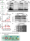
Cbk1 promotes Cts1 secretion independent of CTS1 transcription. (A) Schematic of the ACE2-GOF allele bypassing the necessity of Cbk1 to activate CTS1 transcription. The ACE2-GOF allele mimics Cbk1 phosphorylation at S122, S137, and S436 to promote CTS1 expression in the absence of CBK1. (B) Cts1 secretion is reduced in the absence of CBK1 despite normal Cts1 production. WT, cbk1Δ, ACE2-GOF, ACE2-GOF cbk1Δ cells expressing HA-tagged Cts1 were synchronized in mitosis. Protein was collected at the indicated times following mitotic release at 30°C and were processed as in Fig. 2 A (see also Fig. S5 A). (C) Western blot quantification (as in Fig. 2 B). Fold change relative to the maximum signal of WT cells is shown. (D) Fir1 primarily functions to inhibit Cbk1 to prevent Cts1 secretion. inn1-AID ACE2-GOF cells with the additional genotype indicated to the right of the panel were treated with DMSO (−Auxin) or 0.5 mM auxin (+Auxin). Secreted protein was collected at the indicated times following mitotic release at 30°C, and a representative Western blot of secreted Cts1 is shown (see also Fig. S5 B). (E) Cbk1 deletion restores viability upon septation failure despite Cts1 expression. Fivefold serial dilutions of the strains in D (all strains express PrGal-CDC20 inn1-AID ACE2-GOF TIR1) were spotted to YP galactose plates with and without the addition of 0.5 mM auxin. Plates were incubated for 2 d at 30°C. (F) ECO pathway. Upon incomplete septation, stabilized Fir1 at the bud neck inhibits Cbk1, blocking secretion of septum destroying enzymes. Activation of ECO protects cytokinesis by ensuring the strict temporal sequence of opposing processes: septation and cell separation.

Similar articles

Cited by

References

    1. Atkins B.D., Yoshida S., Saito K., Wu C.F., Lew D.J., and Pellman D.. 2013. Inhibition of Cdc42 during mitotic exit is required for cytokinesis. J. Cell Biol. 202:231–240. 10.1083/jcb.201301090 - DOI - PMC - PubMed
    1. Aulds J., Wierzbicki S., McNairn A., and Schmitt M.E.. 2012. Global identification of new substrates for the yeast endoribonuclease, RNase mitochondrial RNA processing (MRP). J. Biol. Chem. 287:37089–37097. 10.1074/jbc.M112.389023 - DOI - PMC - PubMed
    1. Balasubramanian M.K., Bi E., and Glotzer M.. 2004. Comparative analysis of cytokinesis in budding yeast, fission yeast and animal cells. Curr. Biol. 14:R806–R818. 10.1016/j.cub.2004.09.022 - DOI - PubMed
    1. Bardin A.J., and Amon A.. 2001. Men and sin: what’s the difference? Nat. Rev. Mol. Cell Biol. 2:815–826. 10.1038/35099020 - DOI - PubMed
    1. Bastos R.N., and Barr F.A.. 2010. Plk1 negatively regulates Cep55 recruitment to the midbody to ensure orderly abscission. J. Cell Biol. 191:751–760. 10.1083/jcb.201008108 - DOI - PMC - PubMed

Publication types

MeSH terms

Substances