. 2019 Jan 18;294(3):794-804.
doi: 10.1074/jbc.RA118.004038. Epub 2018 Nov 19.

Non-cryogenic structure of a chloride pump provides crucial clues to temperature-dependent channel transport efficiency

Affiliations

Non-cryogenic structure of a chloride pump provides crucial clues to temperature-dependent channel transport efficiency

Ji-Hye Yun et al. J Biol Chem. .

Abstract

Non-cryogenic protein structures determined at ambient temperature may disclose significant information about protein activity. Chloride-pumping rhodopsin (ClR) exhibits a trend to hyperactivity induced by a change in the photoreaction rate because of a gradual decrease in temperature. Here, to track the structural changes that explain the differences in CIR activity resulting from these temperature changes, we used serial femtosecond crystallography (SFX) with an X-ray free electron laser (XFEL) to determine the non-cryogenic structure of ClR at a resolution of 1.85 Å, and compared this structure with a cryogenic ClR structure obtained with synchrotron X-ray crystallography. The XFEL-derived ClR structure revealed that the all-trans retinal (ATR) region and positions of two coordinated chloride ions slightly differed from those of the synchrotron-derived structure. Moreover, the XFEL structure enabled identification of one additional water molecule forming a hydrogen bond network with a chloride ion. Analysis of the channel cavity and a difference distance matrix plot (DDMP) clearly revealed additional structural differences. B-factor information obtained from the non-cryogenic structure supported a motility change on the residual main and side chains as well as of chloride and water molecules because of temperature effects. Our results indicate that non-cryogenic structures and time-resolved XFEL experiments could contribute to a better understanding of the chloride-pumping mechanism of ClR and other ion pumps.

Keywords: X-ray crystallography; X-ray free electron laser; anion pump; chloride transport; circular dichroism (CD); light-driven chloride pump; non-cryogenic condition; rhodopsin; serial femtosecond crystallography; structure-function; temperature dependence; time-resolved XFEL; transport efficiency; ultraviolet-visible spectroscopy (UV-Vis spectroscopy).


Conflict of interest statement

The authors declare that they have no conflicts of interest with the contents of this article

Figures
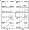
Figure 1.
Figure 1.
The anion-pumping activity of ClR depending on different temperatures. A and B, light-induced pH changes in E. coli cell suspensions expressing ClR in solution containing (A) 100 mm NaCl and (B) 100 mm NaBr observed in the absence (gray solid lines) or presence (black solid lines) of the protonophore CCCP (30 μm), or in the presence of 30 μm CCCP and 50 mm tetraphenylphosphonium ion (TPP+) (black broken lines) at different temperatures from −20 to 25 °C. ClR-expressing E. coli cells were light-induced by a green-light laser-on (540 nm) for 5 min and were dark-adapted before and after green-light laser-on for 5 min each.
Figure 2.
Figure 2.
The UV-visible absorption spectroscopy and binding affinity of ClR at 25 °C and 4 °C. A and B, the UV-visible absorption spectra of ClR following the addition of NaCl up to 1 m at (A) 4 °C and (B) 25 °C. The maximum absorption wavelength values in the absence and presence of chloride ions at 4 °C and 25 °C were 555 and 533 nm, respectively. C and D, absorption changes at 581 nm in the difference spectra plotted against chloride ion concentrations at (C) 4 °C and (D) 25 °C. E, the data were fitted using the Hill equation (solid lines) to estimate the anion affinity at 4 °C and 25 °C. All fitting parameters were normalized as Δλmax and set to 1. Error bars represent S.D. from three independent experiments. The titration experiments were performed with chloride ions and bromide ions at 4 °C and 25 °C. See also Fig. S1 and Table S2.
Figure 3.
Figure 3.
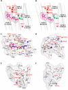
The structural comparison at two chloride-binding sites near the PSB and cytoplasmic regions. A and B, the 2Fo–DFc electron density map, contoured at 1.5 σ, near the PSB region of the (A) XFEL- and (B) synchrotron-derived ClR structures. Selected side chains close to the ATR are shown as stick models. The chloride ions are shown as green spheres. C and D, the chloride (Cl-I)-binding site near (C) ATR linked covalently to Lys235, and the Cl-II–binding site near (D) cytoplasmic region from the XFEL- and synchrotron-derived structures are shown as a stick model colored cyan and green, respectively. The two chloride ions located near the PSB and cytoplasmic regions are depicted as cyan and green spheres in the XFEL- and synchrotron-derived structures, respectively. E and F, the 2Fo–DFc electron density map, contoured at 1.5 σ, of Cl-II and two water molecules (W514 and W515) near the cytoplasmic region of the (E) XFEL- and (F) synchrotron-derived ClR structures. The B-factor values of water molecules were reflected by the electron density map.
Figure 4.
Figure 4.
Channel cavities of the XFEL- and synchrotron-derived ClR structures. A and B, the cavities embedded in the XFEL- and synchrotron-derived ClRs with water molecules are shown as cavity 1, 2, 3, and 4 marked by red, cyan, orange, and violet meshes, respectively. The volume (Å3) of individual cavities in XFEL- and synchrotron-derived ClRs are labeled. C–F, superimposed structure of ClR derived from XFEL and synchrotron in (C) cavity 1, (D) cavity 2, (E) cavity 3, and (F) cavity 4 colored by slate and orange. Participating amino acid residues for each cavity are represented by a line model. Chloride ions and water molecules from the XFEL- and synchrotron-derived structures are depicted as blue, slate, red, and orange spheres, respectively.
Figure 5.
Figure 5.
The overall differences between the XFEL- and synchrotron-derived ClR structures by the DDMP analysis. A, the DDMP according to Cα atom deviations between the XFEL- and synchrotron-derived ClR structures. Red and blue dots indicate the relative movement closer or further, respectively, with color saturation indicating a difference of 0.5 Å or more. The XFEL- and synchrotron-derived ClRs differed slightly, as shown by the regions highlighted with black dotted boxes. B, blue dotted arrows connect these blocks to a ribbon diagram, indicating the position of the highlighted regions within the structure such as helix C, helix E, helix G, the B-C loop, the C-D loop, and the C-terminal helix. C, the direction of the relative movement of the transmembrane domains based on DDMP. Opposite motional directions are indicated by red and blue arrows in the extracellular (upper) and cytoplasmic (lower) regions.

References

    1. Chae P. S., Rasmussen S. G., Rana R. R., Gotfryd K., Chandra R., Goren M. A., Kruse A. C., Nurva S., Loland C. J., Pierre Y., Drew D., Popot J. L., Picot D., Fox B. G., Guan L., et al. (2010) Maltose-neopentyl glycol (MNG) amphiphiles for solubilization, stabilization and crystallization of membrane proteins. Nat. Methods 7, 1003–1008 10.1074/jbc.RA117.001068 - DOI - PMC - PubMed
    1. Chun E., Thompson A. A., Liu W., Roth C. B., Griffith M. T., Katritch V., Kunken J., Xu F., Cherezov V., Hanson M. A., and Stevens R. C. (2012) Fusion partner toolchest for the stabilization and crystallization of G protein-coupled receptors. Structure 20, 967–976 10.1074/jbc.RA117.001068 - DOI - PMC - PubMed
    1. Privé G. G. (2007) Detergents for the stabilization and crystallization of membrane proteins. Methods 41, 388–397 10.1074/jbc.RA117.001068 - DOI - PubMed
    1. Serrano-Vega M. J., and Tate C. G. (2009) Transferability of thermostabilizing mutations between β-adrenergic receptors. Mol. Membr. Biol. 26, 385–396 10.1074/jbc.RA117.001068 - DOI - PubMed
    1. Steyaert J., and Kobilka B. (2011) Nanobody stabilization of G protein-coupled receptor conformational states. Curr. Opin. Struct. Biol. 21, 567–572 10.1074/jbc.RA117.001068 - DOI - PMC - PubMed

Publication types

MeSH terms

LinkOut - more resources