. 2018 Dec;25(12):1093-1102.
doi: 10.1038/s41594-018-0152-3. Epub 2018 Nov 19.

A PxL motif promotes timely cell cycle substrate dephosphorylation by the Cdc14 phosphatase

Affiliations

A PxL motif promotes timely cell cycle substrate dephosphorylation by the Cdc14 phosphatase

Meghna Kataria et al. Nat Struct Mol Biol. 2018 Dec.

Abstract

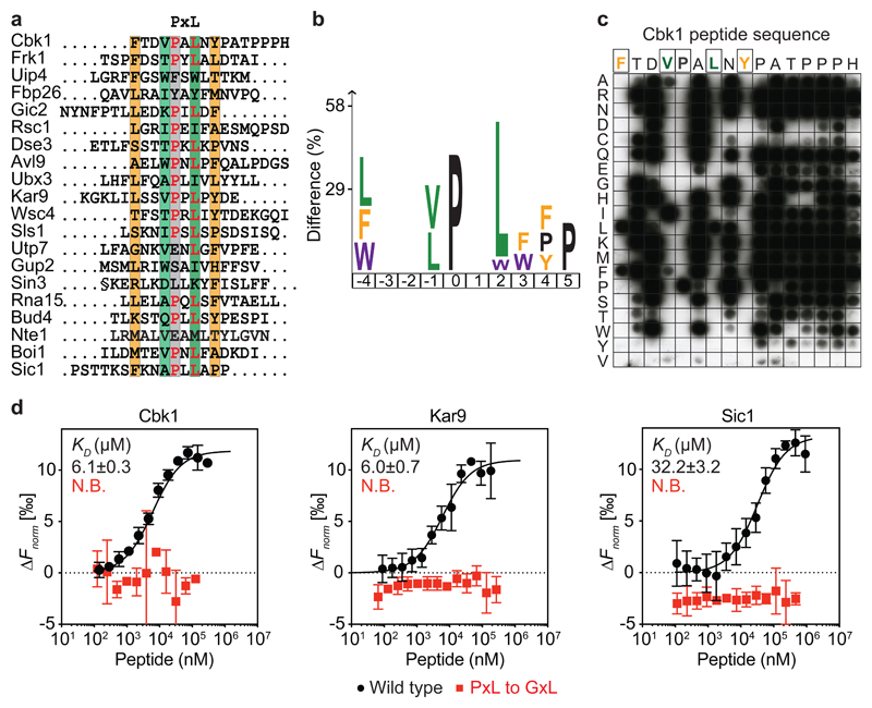
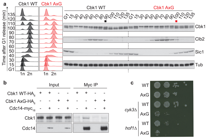
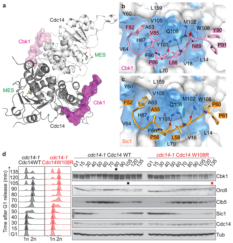
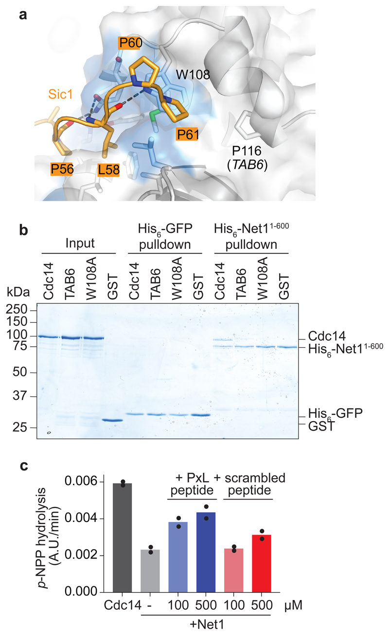
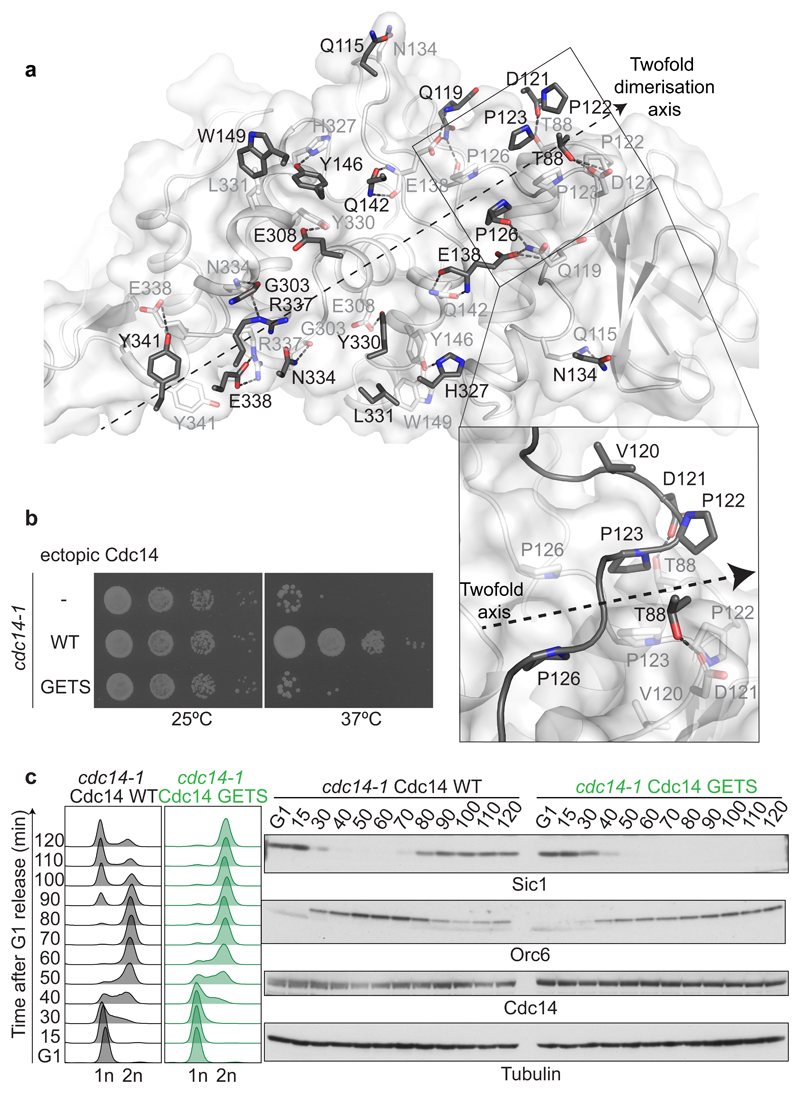
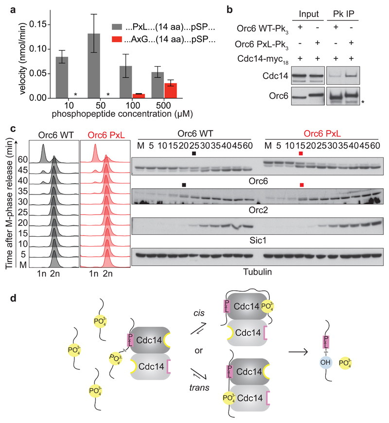
The cell division cycle consists of a series of temporally ordered events. Cell cycle kinases and phosphatases provide key regulatory input, but how the correct substrate phosphorylation and dephosphorylation timing is achieved is incompletely understood. Here we identify a PxL substrate recognition motif that instructs dephosphorylation by the budding yeast Cdc14 phosphatase during mitotic exit. The PxL motif was prevalent in Cdc14-binding peptides enriched in a phage display screen of native disordered protein regions. PxL motif removal from the Cdc14 substrate Cbk1 delays its dephosphorylation, whereas addition of the motif advances dephosphorylation of otherwise late Cdc14 substrates. Crystal structures of Cdc14 bound to three PxL motif substrate peptides provide a molecular explanation for PxL motif recognition on the phosphatase surface. Our results illustrate the sophistication of phosphatase-substrate interactions and identify them as an important determinant of ordered cell cycle progression.


Conflict of interest statement

Competing interests

The authors declare no competing interests.

Figures

Fig. 1
Fig. 1. Identification of the PxL Cdc14 docking motif.
a, Alignment of the top 20 phage display hits. Conserved positions are highlighted. b, Sequence logo representing enriched residues within the PxL motifs from all peptides with greater than five sequencing counts, generated using a detection threshold of p=0.01 in iceLogo. c, Mutational Cbk1 peptide array to probe the contribution of individual amino acid positions to Cdc14 binding. d, Microscale thermophoresis profiles of wild type (black) and mutant (red) PxL motif peptides binding to GFP-Cdc14. Shown are the means and standard deviations from three independent experiments.
Fig. 2
Fig. 2. The PxL motif promotes Cbk1 dephosphorylation.
a, Strains expressing wild type Cbk1 or Cbk1 AxG progressed synchronously through the cell cycle following α-factor arrest and release. FACS analysis of DNA content confirmed cell cycle synchrony. Western blotting was used to analyze the Cbk1 phosphorylation status, as well as Clb2 and Sic1 levels which served as additional cell cycle markers. The black and red squares mark the approximate midpoints of Cbk1 dephosphorylation. Tubulin served as a loading control. Uncropped images of all the blots are found in Supplementary Data Set 1. b, The Cdc14-Cbk1 interaction depends on the PxL motif. Cdc14-myc18 was immunoprecipitated from asynchronously growing control or Cbk1 AxG cells and Cbk1 co-purification analyzed by western blotting. c, Synthetic growth defect of Cbk1 AxG in the absence of cytokinesis factors. Ten-fold serial dilutions of the indicated strains were spotted onto YPD plates and grown at 25 °C for two days.
Fig. 3
Fig. 3. The PxL motif inserts into deep surface pockets on the dimeric Cdc14 phosphatase.
a, Structure of the Cdc14–Cbk1 peptide complex. Each Cdc14 protomer (shades of grey, cartoon representation) binds a Cbk1 peptide (shades of pink, surface representation). The enzyme active site is filled by 2-(N-morpholino) ethanesulfonic acid (MES) from the crystallization buffer. b, c, Expanded views of the interaction networks between Cbk1 (b) and Sic1-derived PxL motif peptides (c), and a hydrophobic pocket in Cdc14 lined by L14, V18, Y60, A63, V64, F66, H67, L70, Y101, M102, V105, Q106, W108, L159. d, Mitotic exit and Cbk1 dephosphorylation delays caused by a PxL binding pocket mutation. Following α-factor arrest and release, cdc14-1 cells complemented by Cdc14 or Cdc14 W108R progressed synchronously through the cell cycle at 35.5 °C. FACS analysis of DNA content was used to analyze cell cycle progression and western blotting to detect the Cbk1 and Orc6 phosphorylation status and Clb5 levels. The black and red squares mark the approximate midpoints of Orc6 and Cbk1 dephosphorylation in wild type and Cdc14 W108R cells. A Cdc14 antibody visualized ectopic Cdc14. Tubulin served as a loading control.
Fig. 4
Fig. 4. The Cdc14 inhibitor Net1 uses the PxL binding pocket.
a, Side view of the Cdc14 PxL binding pocket, bound by the Sic1 peptide. The Cdc14 W108 residue is highlighted, as well as P116, the site of the CDC14 TAB6 mutation. b, His6-pulldown assay to analyze the Net11-600-Cdc14 interaction. The input GST-Cdc14 variants and GST as a control, as well as pulldowns with His6-Net11-600, or with His6-GFP as a control, were analyzed by SDS-PAGE followed by Coomassie Blue staining. c, p-NPP hydrolysis by Cdc14 was analyzed as described in the Methods, with the addition of 200 nM Net11-600 and the indicated concentrations of the Cbk1-derived PxL motif peptide or scrambled control peptide. The results and means from two independent experiments are shown.
Fig. 5
Fig. 5. Dimerization is essential for Cdc14 function.
a, A two-fold symmetry axis along an extended dimerization interface between two Cdc14 protomers. The interacting surface of one of the protomers is shown in white with interacting residues in stick representation. The pairing residues emanating from the second protomer are shown in dark grey, its surface hidden for clarity. An enlarged view of a cluster of prolines engaged in symmetric hydrophobic interactions is shown, that is critical for dimer formation and was targeted in the dimer disrupting Cdc14 GETS mutant. b, Cdc14 GETS fails to support cell viability. Ten-fold serial dilutions of temperature sensitive cdc14-1 cells, and cdc14-1 cells complemented with wild type Cdc14 or Cdc14 GETS were spotted onto YPD plates and incubated at a permissive temperature of 25 °C (left), or at a restrictive temperature of 37 °C (right). c, Cdc14 GETS blocks mitotic exit. Following α-factor arrest, cdc14-1 cells complemented by wild type Cdc14 or Cdc14 GETS were released to progress through the cell cycle at 35.5 °C. FACS analysis of DNA content was used to analyze cell cycle progression and western blotting detected the Orc6 phosphorylation status and Sic1 levels. A Cdc14 antibody visualized ectopic Cdc14. Tubulin served as a loading control.
Fig. 6
Fig. 6. A PxL motif advances ORC dephosphorylation.
a, A PxL motif facilitates peptide dephosphorylation. Dephosphorylation velocities were determined using 100 nM Cdc14 and the indicated phosphopeptide concentrations, containing a pSPxAA site preceded by a functional or mutant PxL motif. The means and standard deviations from three independent experiments are shown. b, A PxL motif augments Orc6 interaction with Cdc14. Orc6-Pk3 or Orc6 PxL-Pk3 were immunoprecipitated from asynchronously growing cells and Cdc14-myc18 co-purification analyzed by western blotting. c, The PxL motif advances ORC dephosphorylation. Orc6 and Orc6 PxL expressing cells were arrested in metaphase by Cdc20 depletion and released to exit synchronously from mitosis by Cdc20 reinduction. FACS analysis of DNA content monitored cell cycle progression. Orc6 and Orc2 dephosphorylation was detected by western blotting. The black and red squares mark the approximate midpoints of dephosphorylation. Sic1 served as a cell cycle marker and tubulin as a loading control. d, Model for PxL motif-mediated Cdc14 substrate dephosphorylation. The PxL motif facilitates dephosphorylation in cis or in trans by increasing the local substrate concentration at both Cdc14 active sites.

References

    1. Morgan D. The cell cycle: Principles of control. New Science Press; 2007.
    1. Loog M, Morgan DO. Cyclin specificity in the phosphorylation of cyclin-dependent kinase substrates. Nature. 2005;434:104–108. - PubMed
    1. Koivomägi M, et al. Dynamics of Cdk1 substrate specificity during the cell cycle. Mol Cell. 2011;42:610–623. - PMC - PubMed
    1. Bhaduri S, Pryciak PM. Cyclin-specific docking motifs promote phosphorylation of yeast signaling proteins by G1/S Cdk complexes. Curr Biol. 2011;21:1615–1623. - PMC - PubMed
    1. Godfrey M, et al. PP2ACdc55 phosphatase imposes ordered cell-cycle phosphorylation by opposing threonine phosphorylation. Mol Cell. 2017;65:393–402. - PMC - PubMed

Publication types

Substances