. 2019 Jan;20(1):40-49.
doi: 10.1038/s41590-018-0249-1. Epub 2018 Nov 19.

DEL-1 promotes macrophage efferocytosis and clearance of inflammation

Affiliations

DEL-1 promotes macrophage efferocytosis and clearance of inflammation

Ioannis Kourtzelis et al. Nat Immunol. 2019 Jan.

Abstract

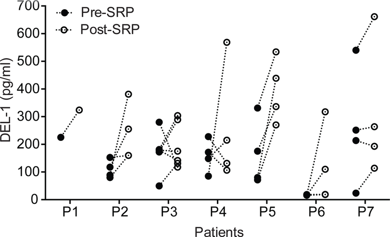
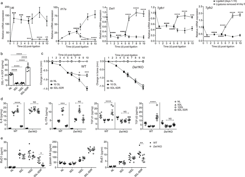
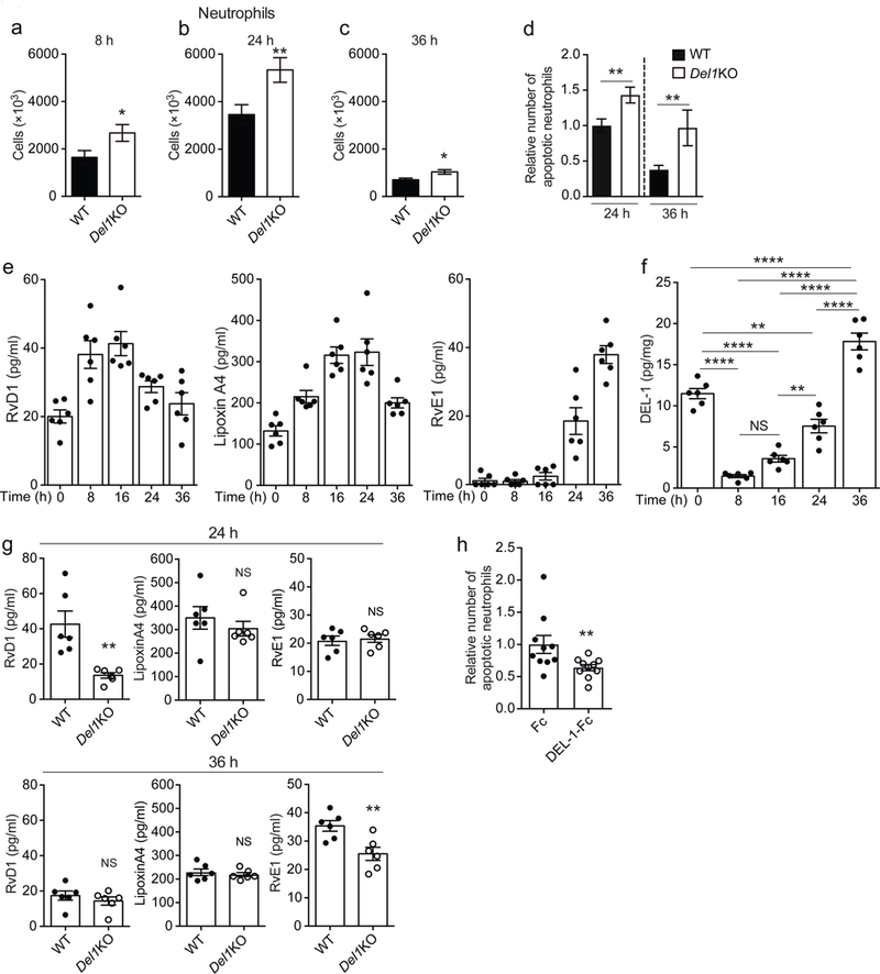
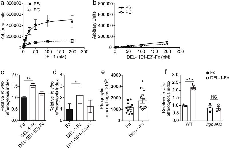
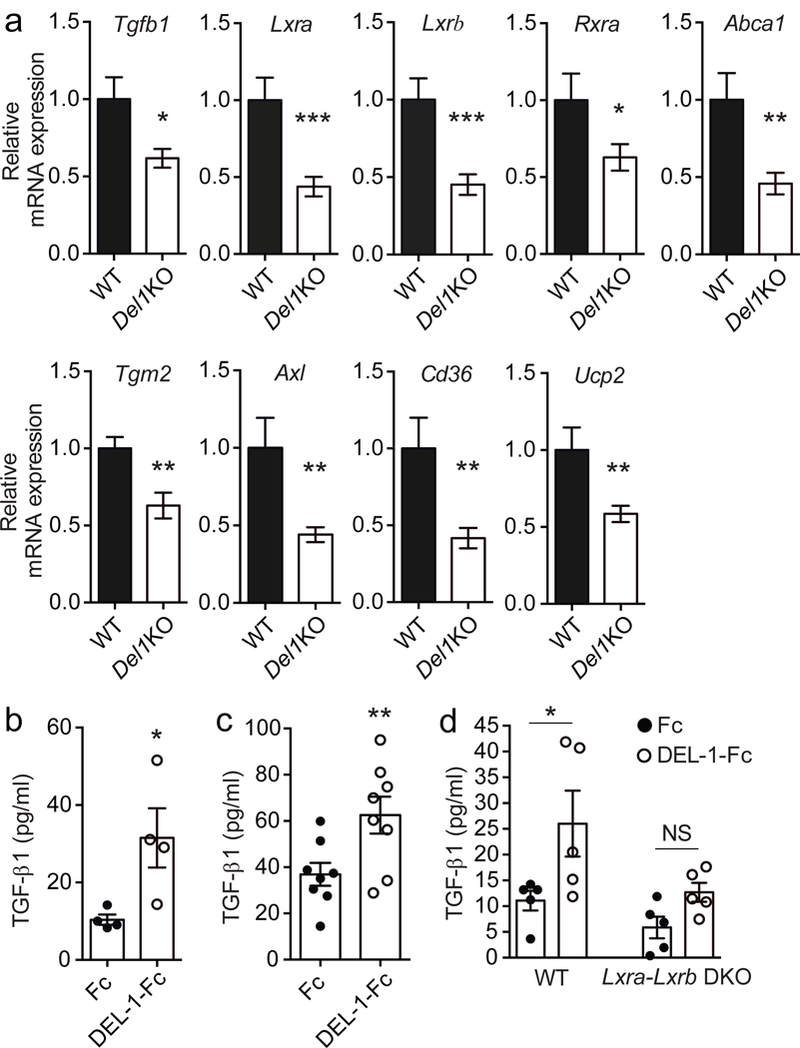
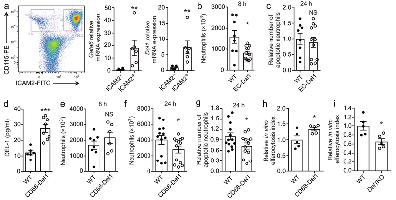
Resolution of inflammation is essential for tissue homeostasis and represents a promising approach to inflammatory disorders. Here we found that developmental endothelial locus-1 (DEL-1), a secreted protein that inhibits leukocyte-endothelial adhesion and inflammation initiation, also functions as a non-redundant downstream effector in inflammation clearance. In human and mouse periodontitis, waning of inflammation was correlated with DEL-1 upregulation, whereas resolution of experimental periodontitis failed in DEL-1 deficiency. This concept was mechanistically substantiated in acute monosodium-urate-crystal-induced inflammation, where the pro-resolution function of DEL-1 was attributed to effective apoptotic neutrophil clearance (efferocytosis). DEL-1-mediated efferocytosis induced liver X receptor-dependent macrophage reprogramming to a pro-resolving phenotype and was required for optimal production of at least certain specific pro-resolving mediators. Experiments in transgenic mice with cell-specific overexpression of DEL-1 linked its anti-leukocyte-recruitment action to endothelial cell-derived DEL-1 and its efferocytic/pro-resolving action to macrophage-derived DEL-1. Thus, the compartmentalized expression of DEL-1 facilitates distinct homeostatic functions in an appropriate context that can be harnessed therapeutically.


Conflict of interest statement

COMPETING FINANCIAL INTERESTS

All authors declare that they have no competing interests.

Figures

Fig. 1.
Fig. 1.. DEL-1 is elevated in the gingival crevicular fluid (GCF) following resolution of periodontitis due to treatment with scaling and root planing (SRP).
GCF was collected using PerioPaper strips from distinct periodontitis-involved sites from 7 patients before and after SRP. After elution in 0.1 ml buffer, the GCF was analyzed by ELISA for DEL-1 content. Data represent the concentration of DEL-1 in the eluted GCF samples. P = 0.0041 (Pre-post paired 2-way ANOVA with repeated measures by patient).
Fig. 2.
Fig. 2.. The pro-resolution effect of DEL-1 in a resolving model of murine ligature-induced periodontitis (LIP).
(a) Wild-type mice were subjected to LIP for up to 10 days and ligatures were removed or not at day 5 (n = 5 mice per group per time point). Relative mRNA expression of the indicated molecules in gingival tissue was assayed by real-time PCR. Data were normalized to Gapdh mRNA and are presented as fold change relative to baseline, set as 1. (b) Wild-type mice were subjected to LIP for up to 10 days and ligatures were removed or not at day 5 (n = 6 mice per group per time point). Protein concentration of DEL-1 was measured by ELISA in GCF that was recovered from ligatures and data were expressed as pg per mg of total protein (NL, not ligated; 5DL, 5 days ligated; 10DL, 10 days ligated; 5DL-5DR, 5 days ligated and 5 days with ligatures removed). (c) Bone loss was measured in littermate wild-type (WT) or Del1KO mice that were subjected to LIP for up to 10 days with or without ligature removal at day 5 (n = 5 mice per group). (d,e) Wild-type (WT) or Del1KO littermate mice were subjected to LIP for up to 10 days and ligatures were removed or not at day 5 (n = 6 mice per group) (NL, not ligated; 5DL, 5 days ligated; 10DL, 10 days ligated; 5DL-5DR, 5 days ligated and 5 days with ligatures removed). (d) Protein concentrations of IL-6, IL-17A, TGF-β1 and TGF- β2 were assessed in gingival tissue by ELISA (pg/mg of total protein are shown). (e) Concentrations of RvD1, Lipoxin A4 and RvE1 were measured in GCF recovered from ligatures (pg/ml are shown). ••P < 0.01, •••P < 0.001, ••••P < 0.0001 vs. day 0 (baseline) (a), *P < 0.05, **P < 0.01, ***P < 0.001, ****P < 0.0001 vs. corresponding time-point without ligature removal (a, c) or between indicated groups (b, d, e). NS, non-significant. One-way ANOVA with Dunnett’s multiple comparisons test for comparison with baseline and two-way ANOVA with Sidak’s multiple comparisons test for comparisons with corresponding time-point without ligature removal (a); one-way ANOVA with Tukey’s multiple comparisons test (b); two-tailed Student’s t-test (c); two-way ANOVA and Tukey’s multiple comparisons test (d,e). Data are means ±SEM and are derived from one experiment with independent mice at each time point (a, c) or pooled from two experiments (b, d, e).
Fig. 3.
Fig. 3.. DEL-1 modulates resolution of acute inflammation.
(a-c) Numbers of neutrophils in peritoneal exudates obtained from Del1KO and their respective wild-type (WT) mice at 8 h (a, n = 11 WT and 13 Del1KO mice), 24 h (b, n = 18 WT and 16 Del1KO mice) and 36 h (c, n = 8 mice per group) after MSU crystal-induced inflammation. (d) Relative number of apoptotic neutrophils detected in peritoneal exudates 24 h and 36 h after MSU crystal injection in wild-type (WT) or Del1KO mice. Number of apoptotic neutrophils was measured and expressed relative to the wild-type/24 h group, set as 1 (24 h, n = 18 WT and 16 Del1KO mice and 36 h, n = 8 mice per group). (e-f) Concentrations of RvD1, Lipoxin A4 and RvE1 (e) and protein concentration of DEL-1 (pg/mg of total protein) (f) were measured in the supernatant of peritoneal fluid obtained from untreated (0 h) wild-type mice or wild-type mice at 8 h, 16 h, 24 h and 36 h after MSU crystal-induced inflammation (n = 6 mice per group). (g) Concentrations of RvD1, Lipoxin A4 and RvE1 were measured in the supernatant of peritoneal exudates obtained from wild-type (WT) or Del1KO mice at 24 h and 36 h after MSU crystal-induced inflammation (n = 6 mice per group). (h) MSU crystals were injected in wild-type mice and, after 20 h, 5 μg of DEL-1-Fc or Fc control protein were administered intraperitoneally (i.p.); 4 h after the latter injection, the relative amount of apoptotic neutrophils in peritoneal exudates was assessed. Numbers of apoptotic neutrophils are expressed relative to the Fc control group, set as 1 (n = 10 mice per group). *P < 0.05, **P < 0.01, ****P < 0.0001, NS, non-significant ((a, b) two-tailed Student’s t-test; (c-d, g, h) two-tailed Mann-Whitney U-test; (f) One-way ANOVA with Tukey’s multiple comparisons test). Data are means ±SEM and are pooled from two experiments (a, c, d – 36 h time point, e-h) or three experiments (b, d – 24 h time point).
Fig. 4.
Fig. 4.. DEL-1 enhances efferocytic activity through interaction with phosphatidylserine and αvβ3 integrin.
(a, b) Saturation binding of (a) full-length DEL-1, or (b) DEL-1 protein that lacks the discoidin I-like domains expressed as an Fc fusion protein (DEL-1[E1-E3]-Fc) to liposomes composed of PC/Chol. (PC) or PC/Chol./PS (PS) (n = 3 preparations in (a) and n = 2 preparations in (b)). (c, d) Primary BMDMs from wild-type mice (n = 3 separate cell isolations) (c), or human monocyte-derived macrophages (MDMs) (n = 5 separate cell isolations) (d) were co-cultured for 30 min with apoptotic neutrophils (mouse or human, respectively) that were pre-stained with BCECF-AM ((2’,7’-Bis-(2-Carboxyethyl)-5-(and-6)-Carboxyfluorescein, Acetoxymethyl Ester) in the presence of Fc control, full-length DEL-1-Fc or DEL-1 protein lacking the discoidin I-like domains (DEL-1[E1-E3]-Fc) (each 500 ng/ml). Relative efferocytosis index is shown and was calculated as the ratio of macrophages that have phagocytosed apoptotic material to the total number of macrophages. Data are expressed relative to the Fc control group set as 1. (e) Wild-type mice were injected i.p. with thioglycollate and 72 h thereafter they received a second i.p. injection of 5 × 10⁶ pre-stained apoptotic neutrophils together with 5 μg of either Fc control or DEL-1-Fc; 3 h after the latter injection, mice were sacrificed and the number of phagocytic macrophages was analyzed (n = 11 mice for Fc control and 10 mice for Del-1-Fc). (f) In vitro phagocytosis assay was performed as described in (c). Relative efferocytosis index of BMDMs from mice deficient in β3 integrin (Itgb3KO) and of BMDMs from respective control mice (WT) in the presence of Fc control or DEL-1-Fc is shown (n = 3 separate cell isolations per genotype). *P < 0.05, **P < 0.01, ***P < 0.001, NS, non-significant (One-way ANOVA with Dunnett’s (c, d) or Tukey’s (f) multiple comparisons test; two-tailed Student’s t-test (e)). Data are means ± SEM and are derived from three experiments (a), from two experiments (b, e), from one experiment (c, f), or are pooled from five experiments (d).
Fig. 5.
Fig. 5.. DEL-1 promotes phenotypic alterations in macrophages during inflammation resolution through LXR.
(a) Relative mRNA expression of Tgfb1, Lxra, Lxrb, Rxra, Abca1, Tgm2, Axl, Cd36 and Ucp2 in peritoneal monocytes/macrophages sorted from Del1KO and their littermate wild-type (WT) mice 24 h after MSU-induced inflammation. Relative mRNA expression was normalized against 18s and was set as 1 in wild-type monocytes/macrophages (n = 7 WT and 12 Del1KO mice). (b,c) Protein concentrations of TGF-β1 were measured in efferocytosis culture supernatants of BMDMs (n = 4 separate cell isolations) (b) or isolated peritoneal macrophages (n = 8 separate cell isolations) (c) incubated for 3 h with mouse apoptotic neutrophils in the presence of Fc control or DEL-1-Fc (500 ng/ml each). (d) Protein concentrations of TGF-β1 in the supernatants of co-cultures of apoptotic neutrophils with BMDMs from mice deficient in both LXRα and LXRβ (Lxra-LxrbDKO) or BMDMs from their respective control mice (WT) for 3 h in the presence of Fc control or DEL-1-Fc (each 500 ng/ml) (n = 5 separate cell isolations per genotype). *P < 0.05, **P < 0.01, ***P < 0.001, NS, non-significant ((a,b,c) two-tailed Student’s t-test, (d) One way ANOVA with Tukey’s multiple comparisons test). Data are means ± SEM and are pooled from two experiments (a, c), are from one experiment representative of two experiments (b) or are from one experiment (d).
Fig. 6.
Fig. 6.. Compartmentalized expression of DEL-1 regulates resolution of acute inflammation.
(a) Representative FACS plot and relative mRNA expression of Gata6 and Del1 in CD115+ICAM2- macrophages and CD115+ICAM2+ resident macrophages sorted from peritoneum of wild-type mice at steady-state (n = 6 mice) are shown. Relative mRNA expression was normalized against 18s and was set as 1 in CD115+ICAM2- macrophages. (b) Neutrophil numbers in peritoneal exudates collected 8 h after MSU crystal injection in mice overexpressing DEL-1 in the endothelium (EC-Del1 mice) or their littermate wild-type (WT) mice (n = 8 WT and 12 EC-Del1 mice). (c) Relative number of apoptotic neutrophils detected in peritoneal exudates 24 h after MSU crystal injection in littermate wild-type (WT) or EC-Del1 mice. Number of apoptotic neutrophils was measured and expressed relative to the WT group, set as 1 (n = 8 wild-type and 11 EC-Del1 mice). (d) DEL-1 protein concentration in the supernatant of isolated peritoneal macrophages derived from untreated (not subjected to MSU crystal-induced inflammation) mice that overexpress DEL-1 under control of the CD68 promoter (CD68-Del1) or their littermate wild-type (WT) mice (n = 6 separate cell isolations for WT mice and 8 for CD68-Del1 mice). (e, f) Numbers of neutrophils in peritoneal exudates obtained from CD68-Del1 mice or littermate wild-type (WT) mice at 8 h (e) and 24 h (f) after MSU crystal-induced inflammation ((e) n = 8 WT and 6 CD68-Del1 mice, (f) n = 13 mice per group). (g) Relative numbers of apoptotic neutrophils in peritoneal exudates obtained from wild-type (WT) and CD68-Del1 mice 24 h after MSU crystal-induced inflammation. Numbers of apoptotic neutrophils are expressed relative to the WT group, set as 1 (n = 13 mice per group). (h, i) Isolated peritoneal macrophages from untreated CD68-Del1 mice (h), or untreated Del1KO mice (i), and from their untreated respective wild-type (WT) mice were incubated for 30 min with apoptotic neutrophils that were pre-stained with BCECF-AM. Relative efferocytosis index is shown and was calculated as the ratio of macrophages that have phagocytosed apoptotic material to the total number of macrophages. Data are expressed relative to the respective WT group, set as 1 (n = 5 separate cell isolations per genotype). *P < 0.05, **P < 0.01, ***P < 0.001. NS, non-significant ((b, c, g) two-tailed Student’s t-test, (a, d, e, f, h, i) two-tailed Mann-Whitney U-test). Data are means ± SEM and are from one experiment representative of two (a) or are pooled from two experiments (b-g) or from three experiments (h, i).

Comment in

References

    1. Fullerton JN & Gilroy DW Resolution of inflammation: a new therapeutic frontier. Nat Rev Drug Discov 15, 551–567 (2016). - PubMed
    1. Serhan CN Pro-resolving lipid mediators are leads for resolution physiology. Nature 510, 92–101 (2014). - PMC - PubMed
    1. Serhan CN, Chiang N & Dalli J The resolution code of acute inflammation: Novel pro-resolving lipid mediators in resolution. Semin. Immunol 27, 200–215 (2015). - PMC - PubMed
    1. Greenlee-Wacker MC Clearance of apoptotic neutrophils and resolution of inflammation. Immunol. Rev 273, 357–370 (2016). - PMC - PubMed
    1. Birge RB et al. Phosphatidylserine is a global immunosuppressive signal in efferocytosis, infectious disease, and cancer. Cell Death Differ 23, 962–978 (2016). - PMC - PubMed

Publication types

MeSH terms