. 2018 Dec;13(12):2991-3017.
doi: 10.1038/s41596-018-0075-9.

Lentiviral transduction of mammalian cells for fast, scalable and high-level production of soluble and membrane proteins

Affiliations

Lentiviral transduction of mammalian cells for fast, scalable and high-level production of soluble and membrane proteins

Jonathan Elegheert et al. Nat Protoc. 2018 Dec.

Abstract

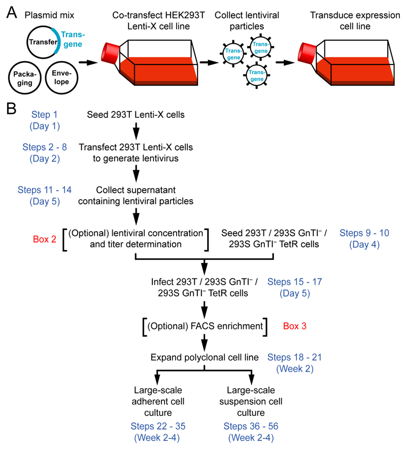
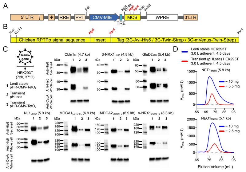
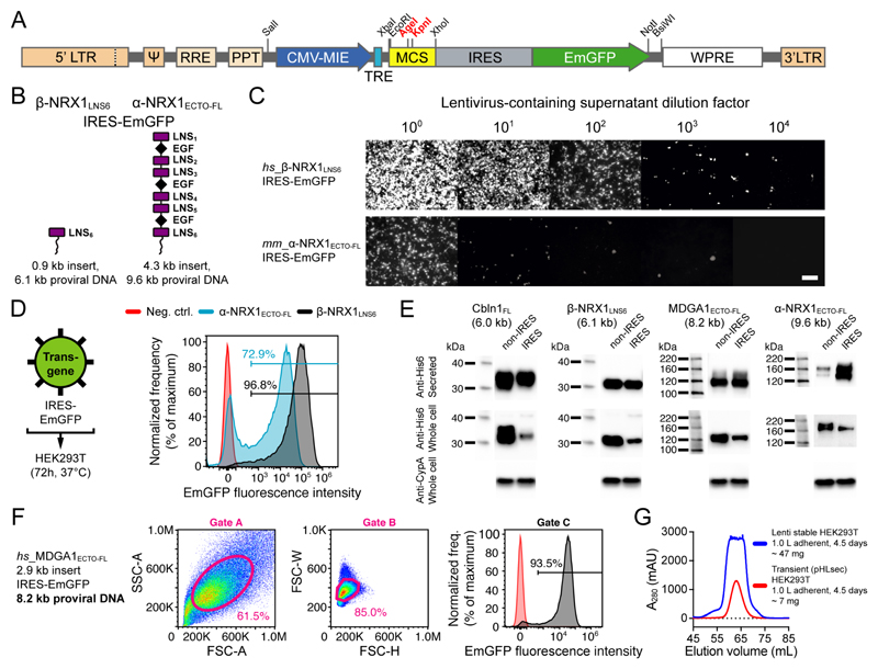
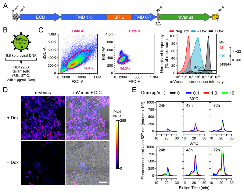
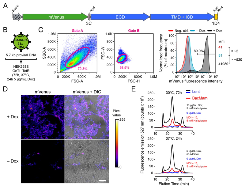
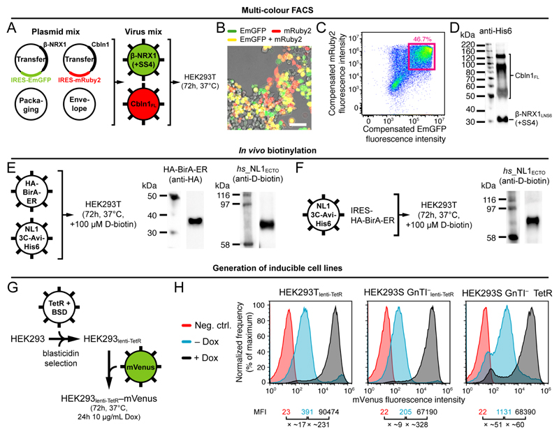
Structural, biochemical and biophysical studies of eukaryotic soluble and membrane proteins require their production in milligram quantities. Although large-scale protein expression strategies based on transient or stable transfection of mammalian cells are well established, they are associated with high consumable costs, limited transfection efficiency or long and tedious selection of clonal cell lines. Lentiviral transduction is an efficient method for the delivery of transgenes to mammalian cells and unifies the ease of use and speed of transient transfection with the robust expression of stable cell lines. In this protocol, we describe the design and step-by-step application of a lentiviral plasmid suite, termed pHR-CMV-TetO2, for the constitutive or inducible large-scale production of soluble and membrane proteins in HEK293 cell lines. Optional features include bicistronic co-expression of fluorescent marker proteins for enrichment of co-transduced cells using cell sorting and of biotin ligase for in vivo biotinylation. We demonstrate the efficacy of the method for a set of soluble proteins and for the G-protein-coupled receptor (GPCR) Smoothened (SMO). We further compare this method with baculovirus transduction of mammalian cells (BacMam), using the type-A γ-aminobutyric acid receptor (GABAAR) β3 homopentamer as a test case. The protocols described here are optimized for simplicity, speed and affordability; lead to a stable polyclonal cell line and milligram-scale amounts of protein in 3-4 weeks; and routinely achieve an approximately three- to tenfold improvement in protein production yield per cell as compared to transient transduction or transfection.


Conflict of interest statement

Competing financial interests

The authors declare no competing financial interests.

Figures

Figure 1
Figure 1. Large-scale expression of soluble or membrane proteins using lentiviral transduction.
(A) Graphical summary of the procedure. (B) Flowchart for the protocol.
Figure 2
Figure 2. The pHR-CMV-TetO2 transfer plasmid for lentiviral stable expression in HEK293 cells.
(A) Genetic elements between the 5’ and 3’ long terminal repeats (LTRs) are packaged in lentiviral particles and integrated in the expression cell genome as proviral DNA. Total proviral DNA size in the empty pHR-CMV-TetO2 plasmid is 4.2 kb. ψ: psi packaging signal; RRE: Rev response element; PPT: polypurine tract; CMV-MIE: major immediate early cytomegalovirus enhancer and promoter; TRE: tetracycline response element; MCS: multiple cloning site; WPRE: Woodchuck Hepatitis Virus posttranscriptional regulatory element. (B) The pHR-CMV-TetO2 multiple cloning site (MCS). (C) Non-concentrated lentiviral particles were used to infect HEK293T cells and protein was expressed for 72 h. His6-tagged proteins were detected using Western blotting (mouse anti-His6 primary antibody: 1:5,000 dilution, HRP-conjugated goat anti-mouse secondary antibody: 1:5,000 dilution, rabbit anti-Cyclophilin A (CypA) primary antibody: 0.5 μg/mL, HRP-conjugated goat anti-rabbit secondary antibody: 1:5,000 dilution). Proviral DNA sizes are noted in brackets. (D) Comparative size-exclusion chromatograms of secreted NET1ΔNTR and NEO1FN456, showing improved secreted protein yields from lentiviral transduction (blue curves) as compared to transient transfection (red curves) of adherent HEK293T cells.
Figure 3
Figure 3. The pHR-CMV-TetO2_IRES-EmGFP transfer plasmid for determination of transduction efficiency and enrichment of transduced cells using FACS.
(A) Schematic representation of the pHR-CMV-TetO2_IRES-EmGFP proviral DNA (5.5 kb) for bicistronic expression of the transgene of interest and free cytosolic EmGFP. (B) Schematic representation of the β-NRX1LNS6 and α-NRX1ECTO-FL constructs. (C) Endpoint dilution assay to estimate functional titer of non-concentrated lentiviral particles encoding β-NRX1LNS6_IRES-EmGFP and α-NRX1ECTO-FL_IRES-EmGFP. Every image was individually enhanced using ImageJ to reveal all cells with fluorescence. Scale bar: 100 μm. (D) Non-concentrated lentiviral particles were used to infect HEK293T cells and protein was expressed for 72 h. Comparison of transduction efficiency of β-NRX1LNS6_IRES-EmGFP and α-NRX1ECTO-FL_IRES-EmGFP. (E) Western blot detection of His6-tagged proteins produced using the pHR-CMV-TetO2_3C-Avi-His6 or pHR-CMV-TetO2_3C-Avi-His6_IRES-EmGFP transfer plasmids (mouse anti-His6 primary antibody: 1:5,000 dilution, HRP-conjugated goat anti-mouse secondary antibody: 1:5,000 dilution, rabbit anti-Cyclophilin A (CypA) primary antibody: 0.5 μg/mL, HRP-conjugated goat anti-rabbit secondary antibody: 1:5,000 dilution). Proviral DNA sizes are noted in brackets. (F) MDGA1ECTO-FL_IRES-EmGFP: forward and side scatter area (A), height (H) and width (W), and fluorescence gates. (G) Comparative size-exclusion chromatograms show secreted MDGA1ECTO-FL yields from 1.0 L (4 roller bottles) of adherent HEK293T cell culture, either transiently transfected (red curve; 6.7 mg total protein) or stably transduced using lentivirus (blue curve; 46.3 mg total protein). Transduced cells were enriched using FACS by applying sorting gates A, B and C from panel F.
Figure 4
Figure 4. Lentiviral transduction, inducible expression and FSEC screening of the GPCR Smoothened (SMO) in HEK293S GnTI TetR cells.
(A) The SMOXTAL-3C-mVenus-Twin-Strep construct (6.8 kb proviral DNA). SS: signal sequence; ECD: extracellular domain; TMD: transmembrane domain; BRIL: thermostabilized apocytochrome b562; 3C: 3C protease cleavage site; TS: Twin-Strep tag. (B) Non-concentrated lentiviral particles encoding SMOXTAL-3C-mVenus-Twin-Strep were used to infect adherent HEK293S GnTI TetR cells. (C) Flow cytometry analysis of non-transduced (negative control) and transduced (– Dox and + Dox) cells after trypsinization and suspension growth. Dox (1 μg/mL Dox for 24 h) was added when the suspension cell density reached ~1.0 ×10⁶ cells/mL. MFI: mean fluorescence intensity. (D) Fluorescence microscopic imaging of transduced (+ Dox and – Dox) adherent cells. DIC; differential interference contrast. Images were taken on a Leica SP8 SMD X confocal microscope. Fluorescence emission was detected using hybrid detectors (HyD) operated in photon-counting mode, meaning that every incident photon gave rise to one gray value. Scale bar: 100 μm. (E) FSEC screening of detergent-solubilized SMOXTAL-3C-mVenus-Twin-Strep with varied expression temperature, collection time and Dox concentration.
Figure 5
Figure 5. Lentiviral transduction and inducible expression of the GABAA receptor β3 homopentamer (GABAAR-β3FL) in HEK293S GnTI TetR cells, and comparison with BacMam.
(A) The mVenus-GABAAR-β3FL-1D4 construct (5.7 kb proviral DNA). SS: signal sequence; 3C: 3C protease cleavage site; ECD: extracellular domain; TMD: transmembrane domain; ICD: intracellular domain; 1D4: 1D4 tag. (B) Non-concentrated lentiviral particles encoding mVenus-GABAAR-β3FL-1D4 were used to infect adherent HEK293S GnTI TetR cells. (C) Flow cytometry analysis of non-transduced (negative control) and transduced (– Dox and + Dox) cells after trypsinization and suspension growth. Dox (5 μg/mL Dox for 24h) was added when the suspension cell density reached ~1.0 ×10⁶ cells/mL. MFI: mean fluorescence intensity. (D) Fluorescence microscopic imaging of transduced (+ Dox and – Dox) adherent cells. DIC; differential interference contrast. Images were taken on a Leica SP8 SMD X confocal microscope. Fluorescence emission was detected using hybrid detectors (HyD) operated in photon-counting mode, meaning that every incident photon gave rise to one gray value. Scale bar: 100 μm. (E) Comparative lentivirus vs. BacMam FSEC screening. mVenus-GABAAR-β3FL-1D4 yield using lentiviral transduction is ~5-fold greater than using BacMam when comparing the best respective conditions (Supplementary Fig. 7A,B). MOI: multiplicity of infection.
Figure 6
Figure 6. Tools for multi-color FACS, in vivo biotinylation, and the generation of inducible cell lines.
(A-D) Multi-color FACS. (A) Non-concentrated lentiviral particles encoding β-NRX1LNS6_IRES-EmGFP (6.1 kb proviral DNA) and Cbln1FL_IRES-mRuby2 (6.0 kb proviral DNA) were used to co-infect HEK293T cells. (B) Fluorescent imaging indicated a mixture of non-, single- and double-transduced cells. The image was taken on a Leica SP8 SMD X confocal microscope. Scale bar: 50 μm. (C) Two-dimensional compensated fluorescence plot. (D) Western blot detection of His6-tagged β-NRX1LNS6(+SS4) and Cbln1FL secreted from co-infected cells after cell sorting (mouse anti-His6 primary antibody: 1:5,000 dilution, HRP-conjugated goat anti-mouse secondary antibody: 1:5,000 dilution). (E-F) In vivo biotinylation. (E) Non-concentrated lentiviral particles encoding either HA-BirA-ER (5.1 kb proviral DNA) or 3C-Avi-His6-tagged NL1ECTO (5.9 kb proviral DNA) were used to co-infect HEK293T cells. (F) Non-concentrated lentiviral particles encoding 3C-Avi-His6-tagged NL1ECTO and IRES_HA-BirA-ER (7.6 kb proviral DNA) were used to infect HEK293T cells. A D-biotin concentration of 100 μM was maintained in the DMEM/F-12/2%FBS medium. Anti-D-biotin western blot detection of in vivo biotinylated NL1ECTO (high sensitivity streptavidin-HRP conjugate; 1:4000 dilution). Anti-HA western blot detection of HA-tagged TetR (mouse anti-HA primary antibody: 1:5,000 dilution, HRP-conjugated goat anti-mouse secondary antibody: 1:5,000 dilution). (G-H) Generation of inducible cell lines. (G) Non-concentrated lentiviral particles encoding TetR-HA-NLS-P2A-BSD-Myc (5.1 kb proviral DNA) and mVenus (4.7 kb proviral DNA) were used to infect HEK293T and HEK293S GnTI cells. (H) Flow cytometry analysis of the resulting HEK293Tlenti-TetR and HEK293S GnTIlenti-TetR inducible cell lines, and comparison with the HEK293S GnTI TetR cell line . MFI: mean fluorescence intensity.

References

    1. Aricescu AR, Owens RJ. Expression of recombinant glycoproteins in mammalian cells: towards an integrative approach to structural biology. Curr Opin Struc Biol. 2013;23:345–356. - PMC - PubMed
    1. Chang VT, et al. Glycoprotein structural genomics: Solving the glycosylation problem. Structure. 2007;15:267–273. - PMC - PubMed
    1. Aricescu AR, Lu W, Jones EY. A time- and cost-efficient system for high-level protein production in mammalian cells. Acta crystallographica. Section D, Biological crystallography. 2006;62:1243–1250. - PubMed
    1. Chaudhary S, et al. Overexpressing human membrane proteins in stably transfected and clonal human embryonic kidney 293S cells. Nat Protoc. 2012;7:453–466. - PMC - PubMed
    1. Naldini L, Trono D, Verma IM. Lentiviral vectors, two decades later. Science. 2016;353:1101–1102. - PubMed

Publication types

Substances