

Convolutional Neural Networks-Based MRI Image Analysis for the Alzheimer's Disease Prediction From Mild Cognitive Impairment
- PMID: 30455622
- PMCID: PMC6231297
- DOI: 10.3389/fnins.2018.00777
Convolutional Neural Networks-Based MRI Image Analysis for the Alzheimer's Disease Prediction From Mild Cognitive Impairment
Abstract
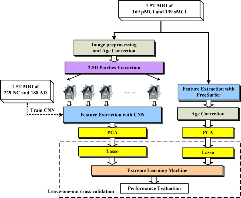
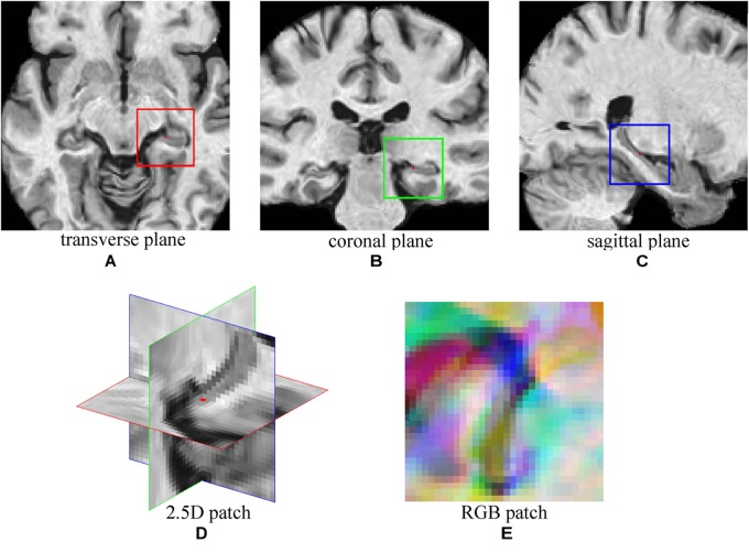
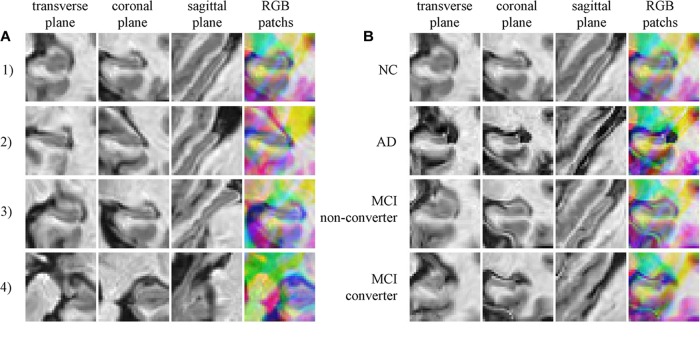
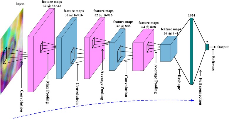
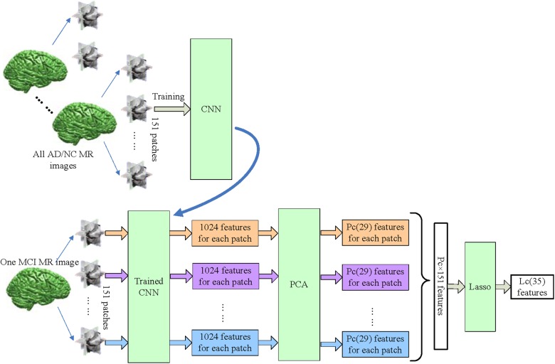
Mild cognitive impairment (MCI) is the prodromal stage of Alzheimer's disease (AD). Identifying MCI subjects who are at high risk of converting to AD is crucial for effective treatments. In this study, a deep learning approach based on convolutional neural networks (CNN), is designed to accurately predict MCI-to-AD conversion with magnetic resonance imaging (MRI) data. First, MRI images are prepared with age-correction and other processing. Second, local patches, which are assembled into 2.5 dimensions, are extracted from these images. Then, the patches from AD and normal controls (NC) are used to train a CNN to identify deep learning features of MCI subjects. After that, structural brain image features are mined with FreeSurfer to assist CNN. Finally, both types of features are fed into an extreme learning machine classifier to predict the AD conversion. The proposed approach is validated on the standardized MRI datasets from the Alzheimer's Disease Neuroimaging Initiative (ADNI) project. This approach achieves an accuracy of 79.9% and an area under the receiver operating characteristic curve (AUC) of 86.1% in leave-one-out cross validations. Compared with other state-of-the-art methods, the proposed one outperforms others with higher accuracy and AUC, while keeping a good balance between the sensitivity and specificity. Results demonstrate great potentials of the proposed CNN-based approach for the prediction of MCI-to-AD conversion with solely MRI data. Age correction and assisted structural brain image features can boost the prediction performance of CNN.
Keywords: Alzheimer’s disease; convolutional neural networks; deep learning; magnetic resonance imaging; mild cognitive impairment.
Figures

References
-
- Avci E., Turkoglu I. (2009). An intelligent diagnosis system based on principle component analysis and ANFIS for the heart valve diseases. Expert Syst. Appl. 36 2873–2878. 10.1016/j.eswa.2008.01.030 - DOI
-
- Babaoğlu I., Fındık O., Bayrak M. (2010). Effects of principle component analysis on assessment of coronary artery diseases using support vector machine. Expert Syst. Appl. 37 2182–2185. 10.1016/j.eswa.2009.07.055 - DOI
-
- Beheshti I., Demirel H., Matsuda H. and Alzheimer’s Disease Neuroimaging Initiative (2017). Classification of Alzheimer’s disease and prediction of mild cognitive impairment-to-Alzheimer’s conversion from structural magnetic resource imaging using feature ranking and a genetic algorithm. Comput. Biol. Med. 83 109–119. 10.1016/j.compbiomed.2017.02.011 - DOI - PubMed
-
- Cao P., Shan X., Zhao D., Huang M., Zaiane O. (2017). Sparse shared structure based multi-task learning for MRI based cognitive performance prediction of Alzheimer’s disease. Pattern Recognit. 72 219–235. 10.1016/j.patcog.2017.07.018 - DOI
LinkOut - more resources
Full Text Sources
Other Literature Sources