. 2018 Oct 31:16:443-449.
doi: 10.1016/j.csbj.2018.10.018. eCollection 2018.

Copper Chaperone Atox1 Interacts with Cell Cycle Proteins

Affiliations

Copper Chaperone Atox1 Interacts with Cell Cycle Proteins

Maria Matson Dzebo et al. Comput Struct Biotechnol J. .

Abstract

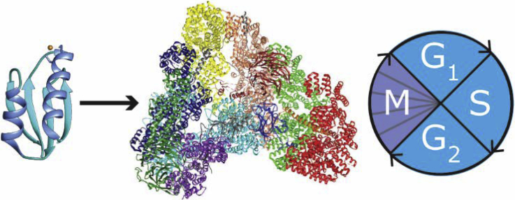
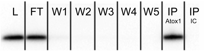
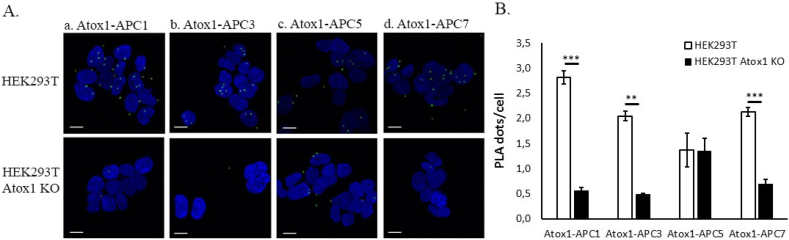
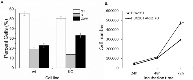
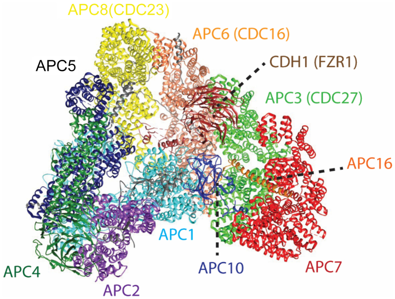
The anaphase-promoting complex (APC) is involved in several processes in the cell cycle, most prominently it facilitates the separation of the sister chromatids during mitosis, before cell division. Because of the key role in the cell cycle, APC is suggested as a putative target for anticancer agents. We here show that the copper chaperone Atox1, known for shuttling copper in the cytoplasm from Ctr1 to ATP7A/B in the secretory pathway, interacts with several APC subunits. Atox1 interactions with APC subunits were discovered by mass spectrometry of co-immunoprecipitated samples and further confirmed using proximity ligation assays in HEK293T cells. Upon comparing wild-type cells with those in which the Atox1 gene had been knocked out, we found that in the absence of Atox1 protein, cells have prolonged G2/M phases and a slower proliferation rate. Thus, in addition to copper transport for loading of copper-dependent enzymes, Atox1 may modulate the cell cycle by interacting with APC subunits.


Figures

Unlabelled Image
Graphical abstract
Fig. 1
Fig. 1
Western blot detection of Atox1 in co-IP samples: blot. cell lysate (L), flow through (FT), washes (W1–5) and immunoprecipitates (IP) with an Atox1-antibody as bait as well as using an isotype control antibody (IC). See also Fig. S2.
Fig. 2
Fig. 2
A. Representative confocal maximum intensity projection images of the in situ proximity ligation assay (PLA) between (a) Atox1 and APC1, (b) Atox1 and APC3, (c) Atox1 and APC5, and (d) Atox1 and APC7 in the wt and Atox1 KO HEK293T cells (upper and lower panel, respectively). A positive PLA signal appears as a green dot. Cell nuclei are stained with DAPI (blue color). Scale bars indicate 10 μm. B. Quantitative analysis of the PLA signals using the confocal images in ImageJ software. Results are shown as mean ± SEM. Statistical significance is indicated as follows: *p < 0.05, **p < 0.01, ***p < 0.001 (using the two-sided, unpaired t-test).
Fig. 3
Fig. 3
A. Cell cycle distribution of wt and Atox1 KO HEK293T cells by PI staining and flow cytometry analysis. The percent cells in S phase was decreased (p < 0.01) whereas the amount cells with G2/M DNA content was increased for the KO cells (p < 0.01). The error bars are standard deviation of mean (n = 3). B. Cell proliferation of wt and Atox1 KO HEK293T cells by measurement of total cell number at 24 h, 48 h and 72 h after seeding (40.000 cells/well at time zero). Decreased proliferation rate for Atox1 KO in comparison to wt HEK293T cells. Standard error indicates standard error of the mean, and * indicates p < 0.001 with two-sided t-test (3 independent experiments with three replicas in each).
Fig. 4
Fig. 4
Structure of anaphase-promoting complex (APC) with the subunits, identified in the co-IP-MS with at least 4 unique peptides, in color. Subunits APC11, APC13, APC15, and F-box only protein 5 were not found in the analysis of the Atox1 co-IP using nano-LC MS/MS and are shown in grey; also in grey is APC12 which was identified in the Atox1 immunoprecipitation experiment with only 2 unique peptides. The subunits identified with largest number of unique peptides are all located in the same area, APC7 (red), APC1 (cyan) and APC3 (light green, also called CDC27). Image made using PDB ID 4UI9 [23] and UCSF Chimera 1.11.2 software [37].

Similar articles

Cited by

References

    1. Huffman D.L., O'Halloran T.V. Function, structure, and mechanism of intracellular copper trafficking proteins. Annu Rev Biochem. 2001;70:677–701. - PubMed
    1. Puig S., Thiele D.J. Molecular mechanisms of copper uptake and distribution. Curr Opin Chem Biol. 2002;6:171–180. - PubMed
    1. Harris E.D. Basic and clinical aspects of copper. Crit Rev Clin Lab Sci. 2003;40:547–586. - PubMed
    1. Valko M., Morris H., Cronin M.T. Metals, toxicity and oxidative stress. Curr Med Chem. 2005;12:1161–1208. - PubMed
    1. O'Halloran T.V., Culotta V.C. Metallochaperones, an intracellular shuttle service for metal ions. J Biol Chem. 2000;275:25057–25060. - PubMed