Review
. 2018 Oct;7(5):814-823.
doi: 10.21037/tau.2018.08.04.

Diffusion-weighted imaging (DWI) in lymph node staging for prostate cancer

Affiliations
Review

Diffusion-weighted imaging (DWI) in lymph node staging for prostate cancer

Iztok Caglic et al. Transl Androl Urol. 2018 Oct.

Abstract

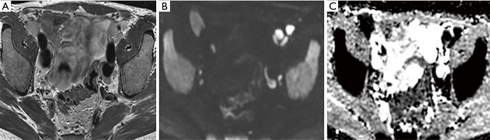
In patients with prostate cancer, the presence of lymph node (LN) metastases is a critical prognostic factor and is essential for treatment planning. Conventional cross-sectional imaging performs poorly for nodal staging as both computed tomography (CT) and magnetic resonance imaging (MRI) are mainly dependent on size and basic morphological criteria. Therefore, extended pelvic LN dissection (ePLND) remains the gold standard for LN staging, however, it is an invasive procedure with its own drawbacks, thus creating a need for accurate preoperative imaging test. Incorporating functional MRI by using diffusion-weighted MRI has proven superior to conventional MRI protocol by means of both qualitative and quantitative assessment. Currently, the increased diagnostic performance remains insufficient to replace ePLND and the future role of DWI may be through combination with MR lymphangiography or with novel positron emission tomography (PET) tracers. In this article, the current state of data supporting DWI in LN staging of patients with prostate cancer is discussed.

Keywords: Diffusion-weighted imaging (DWI); lymph nodes (LNs); magnetic resonance imaging (MRI); prostate cancer (PCa); staging.


Conflict of interest statement

Conflicts of Interest: The authors have no conflicts of interest to declare.

Figures

Figure 1
Figure 1
A 68-year-old, PSA 6.3 ng/mL referred for MRI pre-biopsy. No primary tumour detected within the gland. Multiple small volume normal nodes are identified (arrows) on T2-weighted imaging (A), with the nodes being more easily appreciated on the b-1400 diffusion-weighted imaging (B). PSA, prostate-specific antigen; MRI, magnetic resonance imaging.
Figure 2
Figure 2
A 74-year-old, PSA 11 ng/mL referred for MRI pre-biopsy. No primary tumour detected, gland volume =125.7 mL. Bilateral external iliac nodes demonstrate benign features with an ovoid shape and fatty hila on T2 (A), b-1400 DWI shows high signal (B), confirmed as “T2-shine through effect” with no restricted diffusion on ADC maps (arrows in C). PSA, prostate-specific antigen; MRI, magnetic resonance imaging; ADC, apparent diffusion coefficient; DWI, diffusion-weighted imaging.
Figure 3
Figure 3
A 71-year-old, PSA 51 ng/mL referred for MRI pre-biopsy. Large volume tumour with extra-capsular extension (not shown), Gleason 4+5 on targeted biopsy. Right and left internal iliac nodes involved, depicted on T2 and b-1400 imaging (A,B), with marked restricted diffusion on ADC maps, value 0.457×10⁻³ mm2/s (arrow) (C). PSA, prostate-specific antigen; MRI, magnetic resonance imaging; ADC, apparent diffusion coefficient.
Figure 4
Figure 4
Staging MRI in a 64-year-old patient with biopsy-proven Gleason 4+4 disease. T2-weighted imaging shows a cluster of enlarged left external and internal iliac nodes (A) with restricted diffusion (B,C). Note the high-b-value DWI also shows increased conspicuity of bone metastases to the sacrum (arrows). MRI, magnetic resonance imaging; DWI, diffusion-weighted imaging.
Figure 5
Figure 5
A 64-year-old patient on active surveillance with stable PSA. Transperineal biopsy 2 years prior showed 1/24 cores positive for Gleason 3+3 disease (<5%). No convincing lesion demonstrated in the prostate on MRI. T2-weighted imaging shows new adenopathy (A) with associated restricted diffusion (B,C) and heterogeneous bone marrow change. Nodal distribution involving inguinal nodes and iliac chains but not obturator regions. Nodal biopsy showed B-cell lymphoma with immunophenotype in keeping with CLL/SLL. MRI, magnetic resonance imaging; PSA, prostate-specific antigen; CLL, chronic lymphocytic leukemia; SLL, small lymphocytic lymphoma.

References

    1. Bader P, Burkhard FC, Markwalder R, et al. Disease progression and survival of patients with positive lymph nodes after radical prostatectomy. Is there a chance of cure? J Urol 2003;169:849-54. 10.1097/01.ju.0000049032.38743.c7 - DOI - PubMed
    1. Verhagen PC, Schröder FH, Collette L, et al. Does local treatment of the prostate in advanced and/or lymph node metastatic disease improve efficacy of androgen-deprivation therapy? A systematic review. Eur Urol 2010;58:261-9. 10.1016/j.eururo.2010.05.027 - DOI - PubMed
    1. Eggener SE, Scardino PT, Walsh PC, et al. Predicting 15-year prostate cancer specific mortality after radical prostatectomy. J Urol 2011;185:869-75. 10.1016/j.juro.2010.10.057 - DOI - PMC - PubMed
    1. Touijer KA, Karnes RJ, Passoni N, et al. Survival Outcomes of Men with Lymph Node-positive Prostate Cancer After Radical Prostatectomy: A Comparative Analysis of Different Postoperative Management Strategies. Eur Urol 2018;73:890-6. 10.1016/j.eururo.2017.09.027 - DOI - PMC - PubMed
    1. Ledezma RA, Negron E, Razmaria AA, et al. Robotic-assisted pelvic lymph node dis- section for prostate cancer: frequency of nodal metastases and oncological outcomes. World J Urol 2015;33:1689-94. 10.1007/s00345-015-1515-6 - DOI - PubMed