

Introduction to reflectance confocal microscopy and its use in clinical practice
- PMID: 30456275
- PMCID: PMC6232695
- DOI: 10.1016/j.jdcr.2018.09.019
Introduction to reflectance confocal microscopy and its use in clinical practice
Abstract
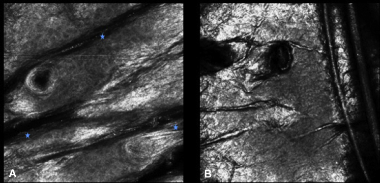
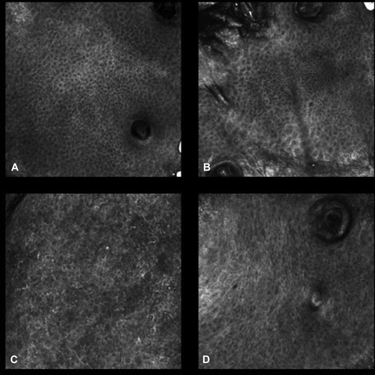
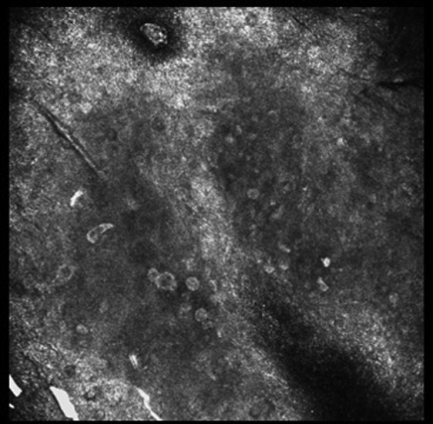
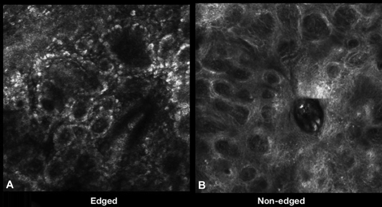
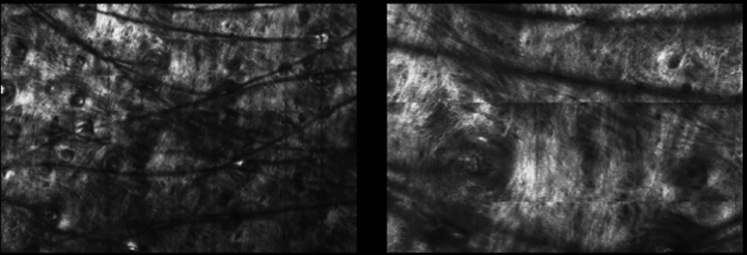
Reflectance confocal microscopy (RCM) is a novel technology that provides noninvasive, in vivo imaging of the skin at nearly histologic resolution. In 2016, the US Centers for Medicare and Medicaid Services (CMS) established reimbursement codes for RCM image acquisition and for the reading and interpretation of images. The combination of RCM imaging with dermoscopy has improved the accuracy of skin cancer diagnosis while reducing the number of biopsies of benign skin lesions. With that, we are starting to see more dermatologists and dermatopathologists using RCM in clinical practice. This editorial is to serve as an introduction on RCM imaging with a focus on its usefulness in both the diagnosis and management of skin cancers. We end by briefly describing the characteristic RCM features of normal skin to serve as a building block for later cases that will explore both the benefits and drawbacks of incorporating RCM imaging for benign and malignant lesions.
Keywords: CMS, US Centers for Medicare and Medicaid Services; CPT, current procedural terminology; LM, lentigo maligna; NNT, number needed to treat; RCM, reflectance confocal microscopy; innovative technology; lentigo maligna; melanoma; noninvasive imaging; nonmelanoma skin cancer; reflectance confocal microscopy; skin cancer.
Figures


Similar articles
-
In vivo reflectance confocal microscopy image interpretation for the dermatopathologist.J Cutan Pathol. 2018 Mar;45(3):187-197. doi: 10.1111/cup.13084. Epub 2018 Jan 3. J Cutan Pathol. 2018. PMID: 29178501 Review.
-
Role of In Vivo Reflectance Confocal Microscopy in the Analysis of Melanocytic Lesions.Acta Dermatovenerol Croat. 2018 Apr;26(1):64-67. Acta Dermatovenerol Croat. 2018. PMID: 29782304 Review.
-
Concordance of handheld reflectance confocal microscopy (RCM) with histopathology in the diagnosis of lentigo maligna (LM): A prospective study.J Am Acad Dermatol. 2016 Jun;74(6):1114-20. doi: 10.1016/j.jaad.2015.12.045. Epub 2016 Jan 27. J Am Acad Dermatol. 2016. PMID: 26826051
-
Dermoscopy vs. reflectance confocal microscopy for the diagnosis of lentigo maligna.J Eur Acad Dermatol Venereol. 2018 Aug;32(8):1284-1291. doi: 10.1111/jdv.14791. Epub 2018 Feb 6. J Eur Acad Dermatol Venereol. 2018. PMID: 29341263
-
Histopathologic and Immunohistochemical Correlates of Confocal Descriptors in Pigmented Facial Macules on Photodamaged Skin.JAMA Dermatol. 2017 Aug 1;153(8):771-780. doi: 10.1001/jamadermatol.2017.1323. JAMA Dermatol. 2017. PMID: 28564685 Free PMC article.
Cited by
-
1.7-Micron Optical Coherence Tomography Angiography for Characterization of Skin Lesions-A Feasibility Study.IEEE Trans Med Imaging. 2021 Sep;40(9):2507-2512. doi: 10.1109/TMI.2021.3081066. Epub 2021 Aug 31. IEEE Trans Med Imaging. 2021. PMID: 33999817 Free PMC article.
-
Polarimetric imaging combining optical parameters for classification of mice non-melanoma skin cancer tissue using machine learning.Heliyon. 2023 Nov 7;9(11):e22081. doi: 10.1016/j.heliyon.2023.e22081. eCollection 2023 Nov. Heliyon. 2023. PMID: 38034801 Free PMC article.
-
Research Techniques Made Simple: Emerging Imaging Technologies for Noninvasive Optical Biopsy of Human Skin.J Invest Dermatol. 2022 May;142(5):1243-1252.e1. doi: 10.1016/j.jid.2022.01.016. J Invest Dermatol. 2022. PMID: 35461534 Free PMC article.
-
Guideline for in vivo assessment of adherent and rolling leukocytes in human skin microvasculature via reflectance confocal videomicroscopy.Microcirculation. 2021 Nov;28(8):e12725. doi: 10.1111/micc.12725. Epub 2021 Sep 17. Microcirculation. 2021. PMID: 34409720 Free PMC article.
-
Multimodal Imaging Detection of Difficult Mammary Paget Disease: Dermoscopy, Reflectance Confocal Microscopy, and Line-Field Confocal-Optical Coherence Tomography.Diagnostics (Basel). 2025 Jul 29;15(15):1898. doi: 10.3390/diagnostics15151898. Diagnostics (Basel). 2025. PMID: 40804863 Free PMC article.
References
-
- Centers for Medicare & Medicaid Services. Physician Fee Schedule Search. CMS website. Available at: https://www.cms.gov/apps/physician-fee-schedule/search/search-results.as.... Published 2016.
-
- González S., Swindells K., Rajadhyaksha M., Torres A. Changing paradigms in dermatology: confocal microscopy in clinical and surgical dermatology. Clin Dermatol. 2003;21(5):359–369. - PubMed
-
- Grant-Kels J.M., Rabinovitz H.S., Oliviero M., Scope A. In vivo diagnosis of skin cancer through reflectance. Clin Oncol. 2016;1:1021.
-
- Levine A., Markowitz O. In vivo reflectance confocal microscopy. Cutis. 2017;99(6):399–402. http://www.ncbi.nlm.nih.gov/pubmed/28686758 Available at: - PubMed
LinkOut - more resources
Full Text Sources