Comparative Study
. 2019 Mar;33(3):160-168.
doi: 10.1007/s12149-018-1319-z. Epub 2018 Nov 19.

Comparison between single photon emission computed tomography with computed tomography and planar scintigraphy in sentinel node biopsy in breast cancer patients

Affiliations
Comparative Study

Comparison between single photon emission computed tomography with computed tomography and planar scintigraphy in sentinel node biopsy in breast cancer patients

Mitsuru Koizumi et al. Ann Nucl Med. 2019 Mar.

Abstract

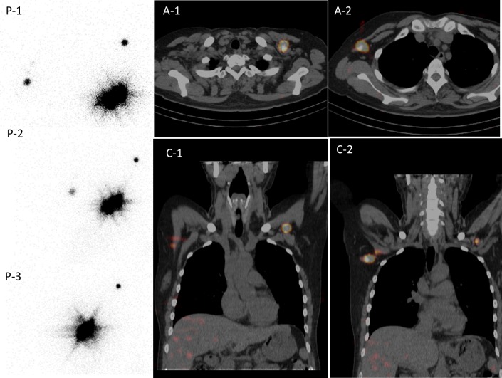
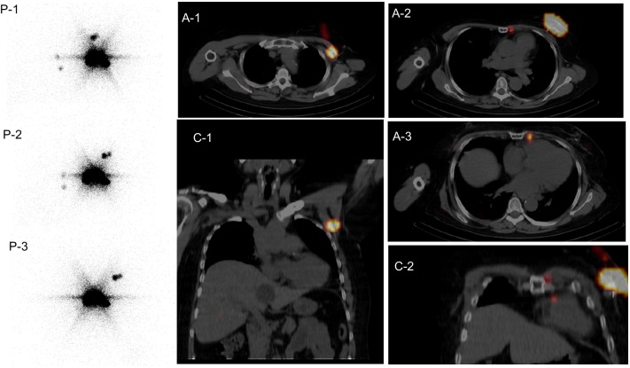
Objective: Radio-guided sentinel node (SN) biopsy is a standard method used in the treatment of early breast cancer. Single photon emission computed tomography with computed tomography (SPECT/CT) has been commonly used for SN detection. SPECT/CT adds precise anatomical information of SN sites, and it is reported that more SNs may be detectable on SPECT/CT than on planar imaging. We here investigate which breast cancer patients have benefited from SPECT/CT over planar imaging.

Methods: A total of 273 breast cancer patients including 80 with ipsilateral breast tumor relapse (IBTR) underwent both multiple-view planar imaging and SPECT/CT for SN detection. The number of SNs, the patients who had benefitted from SPECT/CT, and the SN procedure failure rate were compared between SPECT/CT and planar imaging. Factors influencing the visualization of para-sternal and ipsilateral level II, III nodes, and contralateral axillary nodes were also analyzed using logistic regression analysis.

Results: The number of hot spots did not differ between SPECT/CT and multiple-view planar imaging. Eight contaminated patients and 52 patients with visualized extra-level I axillary nodes benefited from identifying precise anatomical sites. Even though radioactive nodes could be harvested in most (192/193) of the non-IBTR patients (7/8 in non-SN visible patients), no radioactive nodes could be found during surgery in 11 of 80 IBTR patients. Axillary surgery (dissection) increased the visualization of para-sternal and level II, III axillary nodes, and previous irradiation increased the visualization of contralateral axillary nodes.

Conclusion: Multiple-view planar imaging was equivalent to SPECT/CT for depicting hot nodes for radio-guided SN detection in breast cancer. SPECT/CT was useful when precise anatomical information was necessary, especially regarding sentinel lymph nodes other than ipsilateral axilla. Logistic regression analysis revealed that axillary surgery (dissection) increased the visualization of para-sternal and level II, III axillary nodes, and the only relevant factor influencing visualization of contralateral axillary SNs was previous radiation to the breast.

Keywords: Breast cancer; Contra-axilla visualization; Planar scintigraphy; Radio-guided; SPECT/CT; Sentinel node detection.


Conflict of interest statement

No potential conflicts of interest were disclosed.

Figures

Fig. 1
Fig. 1
A 42-year-old woman received radio-guided sentinel node (SN) detection on both planar images and SPECT/CT images the day before left breast surgery for breast cancer. She had received ipsilateral breast surgery for breast cancer 3 years before; total mastectomy, SN biopsy followed by adjuvant tamoxifen therapy. Left breast recurrent tumor was diagnosed, and left breast re-surgery was planned. Planar images (P-1: anterior view, P-2: left anterior oblique, 30°, and P-3: left anterior oblique, 60°) and SPECT/CT (A-1,2: axial fusion images, C-1,2: coronal fusion images) had been shown. Planar image (P-1) illustrated contralateral axilla and ipsilateral axilla. SPECT/CT images clearly specified level I contralateral axilla and ipsilateral level II axilla (Rotter). On surgery, both lymph nodes were harvested and revealed to have cancer metastasis. Despite adjuvant hormone therapy after re-surgery, she developed local, lymph node, and distant metastasis 2 years later
Fig. 2
Fig. 2
A 57-year-old woman received radio-guided sentinel node (SN) detection on both planar images and SPECT/CT images the day before left breast surgery for breast cancer. She had a long history, with right breast surgery (lumpectomy and axillary dissection) at 38 years of age, and left breast surgery (lumpectomy and axillary dissection) at 41 years of age. She developed new left breast cancer at 57 years of age. Planar images (P-1: anterior view, P-2: left anterior oblique, 30°, and P-3: left anterior oblique, 60°) and SPECT/CT (A-1,2,3: axial fusion images, C-1,2: coronal fusion images) had been shown. Planar image (P-1) illustrated two ipsilateral axillary nodes and two para-sternal nodes. SPECT/CT images specified one level II (Rotter) axillary node (A-1, C-1) and two para-sternal nodes (A-2,3, C-2). Left total mastectomy was performed and two Rotter nodes were harvested at surgery without cancer metastasis. Para-sternal nodes were not manipulated. She received adjuvant chemotherapy after surgery, and has remained without recurrence for 1 year

References

    1. Krag D, Weaver D, Ashikaga T, Moffat F, Kimberg VS, Shiriver C, et al. The sentinel node in breast cancer—a multicenter validation study. N Engl J Med. 1998;339:941–946. doi: 10.1056/NEJM199810013391401. - DOI - PubMed
    1. Veronesi U, Paganelli G, Viale G, Luini A, Zurrida S, Galimberti V, et al. A randomized comparison of sentinel-node biopsy with routine axillary dissection in breast cancer. N Engl J Med. 2003;349:546–553. doi: 10.1056/NEJMoa012782. - DOI - PubMed
    1. Koizumi M, Nomura E, Yamada Y, Takiguchi T, Makita M, Iwase T, et al. Radioguided sentinel node detection in breast cancer patients: comparison of 99mTc phytate and 99mTc rhenium colloid efficacy. Nucl Med Commun. 2004;25:1031–1037. doi: 10.1097/00006231-200410000-00007. - DOI - PubMed
    1. Keidar Z, Israel O, Krausz Y. SPECT/CT in tumor imaging: technical aspects and clinical applications. Sem Nucl Med. 2003;33:205–218. doi: 10.1053/snuc.2003.127310. - DOI - PubMed
    1. Lerman H, Metser U, Lievshitz G, Sperber F, Shneebaum S, Even-Sapir E. Lymphoscintigraphic sentinel node identification in patients with breast cancer: the role of SPECT/CT. Eur J Nucl Med Mol Imaging. 2006;33:329–337. doi: 10.1007/s00259-005-1927-4. - DOI - PubMed

Publication types