. 2019 Feb;12(1):5-17.
doi: 10.1007/s12265-018-9842-9. Epub 2018 Nov 19.

Cardiac Progenitor Cell-Derived Extracellular Vesicles Reduce Infarct Size and Associate with Increased Cardiovascular Cell Proliferation

Affiliations

Cardiac Progenitor Cell-Derived Extracellular Vesicles Reduce Infarct Size and Associate with Increased Cardiovascular Cell Proliferation

Janita A Maring et al. J Cardiovasc Transl Res. 2019 Feb.

Abstract

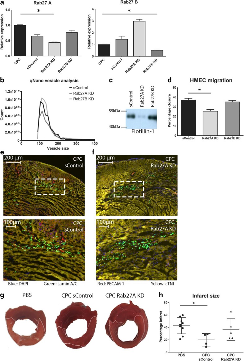
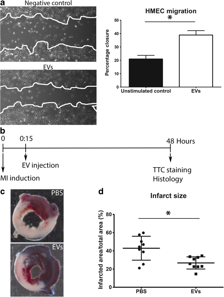
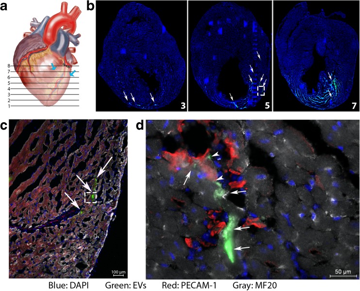
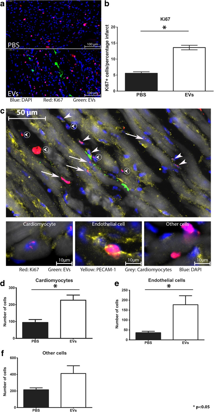
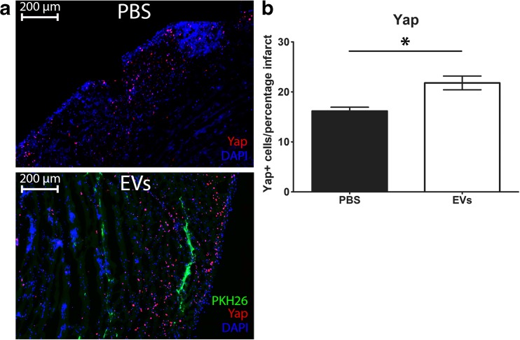
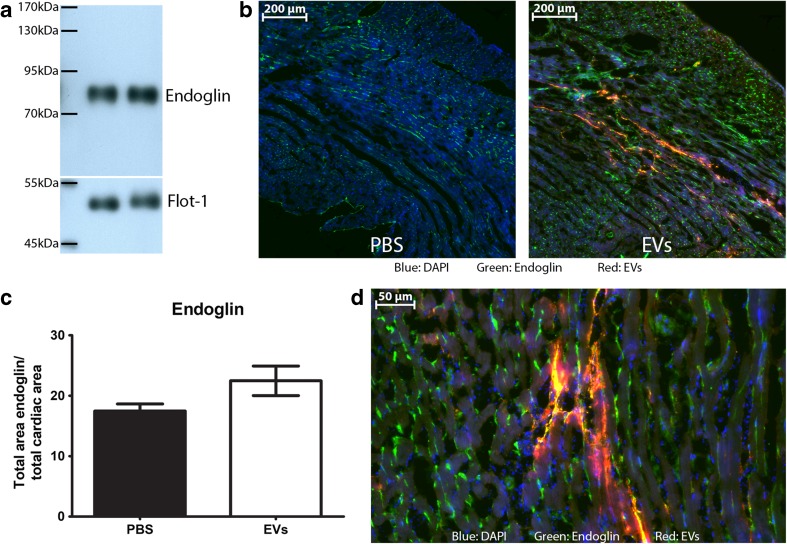
Cell transplantation studies have shown that injection of progenitor cells can improve cardiac function after myocardial infarction (MI). Transplantation of human cardiac progenitor cells (hCPCs) results in an increased ejection fraction, but survival and integration are low. Therefore, paracrine factors including extracellular vesicles (EVs) are likely to contribute to the beneficial effects. We investigated the contribution of EVs by transplanting hCPCs with reduced EV secretion. Interestingly, these hCPCs were unable to reduce infarct size post-MI. Moreover, injection of hCPC-EVs did significantly reduce infarct size. Analysis of EV uptake showed cardiomyocytes and endothelial cells primarily positive and a higher Ki67 expression in these cell types. Yes-associated protein (YAP), a proliferation marker associated with Ki67, was also increased in the entire infarcted area. In summary, our data suggest that EV secretion is the driving force behind the short-term beneficial effect of hCPC transplantation on cardiac recovery after MI.

Keywords: Angiogenesis; Cardiac progenitor cells; Cardiomyocytes; Endoglin; Extracellular vesicles; Myocardial infarction; Proliferation.


Conflict of interest statement

The authors declare that they have no conflict of interest.

Figures

Fig. 1
Fig. 1
Rab knockdown reduces the secretion of EVs as well as their positive effect in vitro and in vivo. a Rab knockdown analysis by qPCR. Both Rab27A and Rab27B are effectively knocked down after lentiviral shRNA transduction. b qNano analysis of vesicles in the EV isolate by sControl-hCPCs, and hCPCs with Rab27A and Rab27B knockdown. Only Rab27A KD reduces the number of EVs compared to sControl-hCPCs. c Western blot for flotillin-1 confirms reduction of EVs by Rab27A knockdown, while no effect is seen with Rab27B knockdown. d Effect of EV isolate on migration of endothelial cells after scratch assay. EV isolate from Rab27A KD has reduced capacity to induce migration compared to sControl-hCPCs EV isolate (EV isolates of equal cell numbers). e, f Both sControl-hCPCs (e) and Rab27 KD-hCPCs (f) are observed in the heart after 48 h (blue: DAPI, green: human Lamin A/C, red: PECAM-1, yellow: cardiac troponin I (cTNI)). g Representative pictures of TTC analysis after injection of PBS, sControl-hCPCs, and Rab27A KD-hCPCs. h Quantification of the infarcted area in hearts injected with PBS, sControl-hCPCs, and Rab27A-KD hCPCs (PBS, n = 9; control-hCPC, n = 4; Rab27A KD-hCPC, n = 5)
Fig. 2
Fig. 2
hCPC-derived EVs reduce infarct size. a EVs stimulate wound closure in a HMEC-1 scratch assay. Percentage of closure of the wounded area was determined after 24 h. b Timeline of in vivo procedures. c Representative images of TTC stained hearts injected with PBS or exosomes, 48 h after MI. White area represents the infarcted region. d Quantification of the infarcted area as a percentage of the total area of the left ventricle (n = 9)
Fig. 3
Fig. 3
EV distribution in the heart. a Schematic representation of the analysis of infarcted heart. Blue arrows indicate the EV injection site. b Analysis of the entire heart showed the uptake of PKH67 from the loaded EVs in the heart from level 3 to 7/8. White arrows point at positive cells. c Higher magnification of insert in B, showing uptake of EVs in the infarct zone. d Higher magnification of insert in C, which shows uptake of EVs by cardiomyocytes (arrows) and endothelial cells (arrowheads)
Fig. 4
Fig. 4
hCPC-derived EVs induce Ki67 expression in the left ventricle after MI. a Ki67 (red) in the heart in the presence of EVs (green). Nuclei are stained with DAPI (blue). b Quantification of Ki67-positive cells in the infarct and border zone, relative to the area of the infarct, shows that EVs increase the number of Ki67-positive cells (n = 3). c Ki67 staining in the heart after EV injection shows proliferation in various cell types (arrows: positive cardiomyocytes; arrowheads: positive endothelial cells; circled arrowheads: other cells). Smaller pictures show examples of individual cell types. Cardiomyocytes were identified through autofluorescence. df Graphs depicting the number of proliferating cardiomyocytes (d), endothelial cells (e), and other cells (f) in the heart, in layers 5 and 6, after EV and PBS injection. A significant increase was found in cardiomyocyte proliferation (n = 3)
Fig. 5
Fig. 5
YAP expression after MI. a Representative pictures of YAP expression in the border zone of PBS and EV injected hearts. b Quantitative analysis shows that the number of YAP-positive cells is higher in the infarct zone of hearts injected with hCPC-EVs compared to PBS-injected hearts. Numbers of positive cells are normalized for infarct size and shown as number of cells per percentage infarct
Fig. 6
Fig. 6
Endoglin levels in the heart. a Endoglin is present in EVs from hCPCs, two individual isolations are shown. b After injection of PKH-labeled EVs, areas positive for EV uptake are also highly positive for endoglin, compared to PBS injection. c Quantification of endoglin levels in the entire heart after PBS or EV injection. d Endoglin signal is mainly found in endothelial cells and cells positive for EV uptake

Comment in

References

    1. (2018). WHO | Global Health Observatory (GHO) data. WHO.
    1. Goumans M-J, de Boer TP, Smits AM, et al. TGF-beta1 induces efficient differentiation of human cardiomyocyte progenitor cells into functional cardiomyocytes in vitro. Stem Cell Research. 2007;1:138–149. doi: 10.1016/j.scr.2008.02.003. - DOI - PubMed
    1. Smith RR, Barile L, Cho HC, et al. Regenerative potential of cardiosphere-derived cells expanded. Circulation. 2007;115:896–908. doi: 10.1161/CIRCULATIONAHA.106.655209. - DOI - PubMed
    1. Smits AM, van Laake LW, den Ouden K, et al. Human cardiomyocyte progenitor cell transplantation preserves long-term function of the infarcted mouse myocardium. Cardiovascular Research. 2009;83:527–535. doi: 10.1093/cvr/cvp146. - DOI - PubMed
    1. den Haan MC, Grauss RW, Smits AM, et al. Cardiomyogenic differentiation-independent improvement of cardiac function by human cardiomyocyte progenitor cell injection in ischaemic mouse hearts. Journal of Cellular and Molecular Medicine. 2012;16:1508–1521. doi: 10.1111/j.1582-4934.2011.01468.x. - DOI - PMC - PubMed

Publication types

MeSH terms