. 2019 Feb;8(2):124-137.
doi: 10.1002/sctm.18-0084. Epub 2018 Nov 19.

Validation of Current Good Manufacturing Practice Compliant Human Pluripotent Stem Cell-Derived Hepatocytes for Cell-Based Therapy

Affiliations

Validation of Current Good Manufacturing Practice Compliant Human Pluripotent Stem Cell-Derived Hepatocytes for Cell-Based Therapy

Samuel J I Blackford et al. Stem Cells Transl Med. 2019 Feb.

Abstract

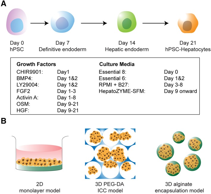
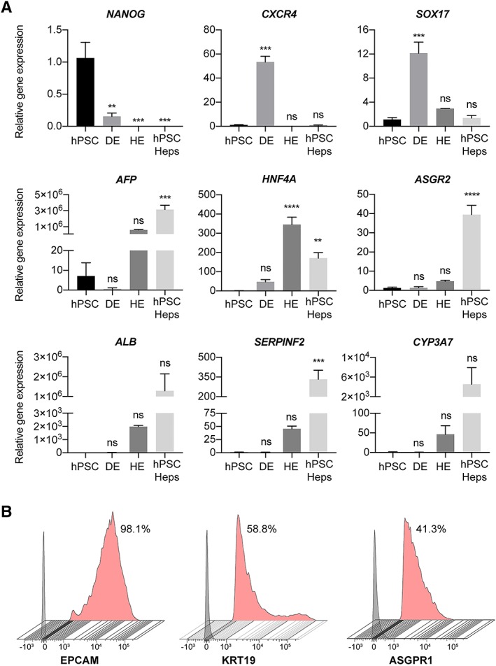
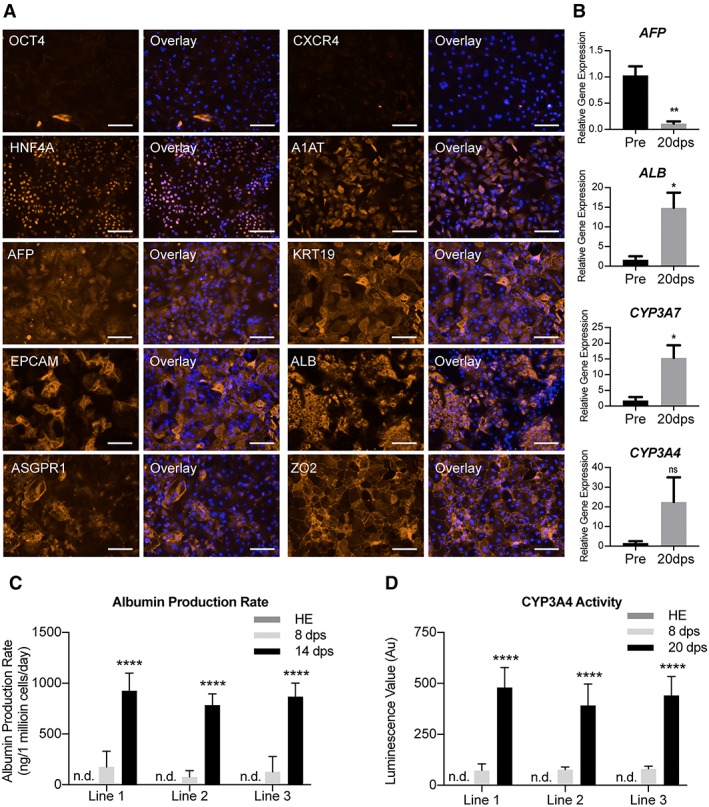
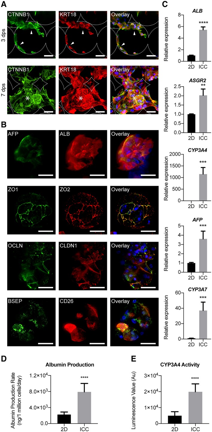
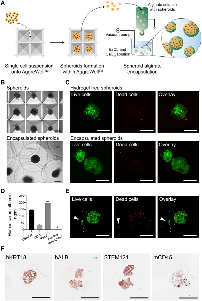
Recent advancements in the production of hepatocytes from human pluripotent stem cells (hPSC-Heps) afford tremendous possibilities for treatment of patients with liver disease. Validated current good manufacturing practice (cGMP) lines are an essential prerequisite for such applications but have only recently been established. Whether such cGMP lines are capable of hepatic differentiation is not known. To address this knowledge gap, we examined the proficiency of three recently derived cGMP lines (two hiPSC and one hESC) to differentiate into hepatocytes and their suitability for therapy. hPSC-Heps generated using a chemically defined four-step hepatic differentiation protocol uniformly demonstrated highly reproducible phenotypes and functionality. Seeding into a 3D poly(ethylene glycol)-diacrylate fabricated inverted colloid crystal scaffold converted these immature progenitors into more advanced hepatic tissue structures. Hepatic constructs could also be successfully encapsulated into the immune-privileged material alginate and remained viable as well as functional upon transplantation into immune competent mice. This is the first report we are aware of demonstrating cGMP-compliant hPSCs can generate cells with advanced hepatic function potentially suitable for future therapeutic applications. Stem Cells Translational Medicine 2019;8:124&14.

Keywords: Bioengineering; Cell transplantation; Cellular therapy; Hepatocyte differentiation; Hepatocytes; Liver therapy; Pluripotent stem cells; cGMP; hESC; iPSC.


Figures

Figure 1
Figure 1
Generation of human pluripotent stem cell‐derived hepatocytes and their therapeutic potential in various model systems. (A): Four‐stage hepatic differentiation from human pluripotent stem cells (hPSC) to definitive endoderm, hepatic endoderm, and subsequently hepatocytes (hPSC‐Heps) over a 21‐day protocol by a defined cocktail of growth factors and small molecules (in the box). (B): Schematic of further maturation of hPSC‐Heps in two‐dimensional culture dish coated with type I collagen, three‐dimensional (3D) inverse colloidal crystal scaffold coated with type I collagen (inverse colloidal crystal) and encapsulation within 3D alginate microspheres.
Figure 2
Figure 2
Morphological characterization of hepatic differentiated cells. (A): Brightfield microscopy images revealing the morphological transformation from day 0 pluripotent stem cell colony to day 17 polyhedral hepatocytes. (B): Representative images of day 21 human pluripotent stem cells‐Heps generated from three different current good manufacturing practice‐compliant lines. Scale bars: 100 μm.
Figure 3
Figure 3
Phenotypical characterization of hepatic differentiated cells. (A): Differential expression of selected genes reveals progressive maturation of human pluripotent stem cells (hPSCs) to definitive endoderm, hepatic endoderm and then hepatocytes (hPSC‐Heps). Expression relative to the housekeeping gene, and normalized against the average expression of hPSCs; n = 3 experiments, 1 cell line. Data are mean ± SEM, ordinary one‐way ANOVA followed by Dunnett post hoc test to compare the mean of each group to hPSC expression. **, p < .005; ***, p < .0005; ****, p < .0001; ns: nonsignificant. Data shown for cell line 1. (B): Flow cytometry analysis of surface marker expression on hPSC‐Heps at day 21 of differentiation. Data shows the percentage of positive cells from the live cell population. Gray histogram represents fluorescence minus one (FMO) control used to establish the gate, red histogram represents stained hPSC‐Heps. All flow cytometry analysis is representative of at least three independent experiments. Data shown for cell line 3.
Figure 4
Figure 4
Characterization of human pluripotent stem cells (hPSC)‐derived hepatocyte maturation in two‐dimensional (2D) model. (A): Immunofluorescent images revealing the transition of pluripotency (OCT4), endodermal (CXCR4), and hepatic specification (HNF4A, A1AT, AFP, KRT19, and EPCAM) and mature hepatic (ALB, ASGPR1, and ZO‐2) expression in hPSC‐Heps after 20 days postseeding on 2D model. Representative images selected from each of the three lines. Scale bar: 100 μm. (B): Differential gene expression showing the relative expression of four key hepatic genes (AFP, ALB, CYP3A7, and CYP3A4) in preseeding (pre) and 20 days postseeding (20 dps) into 2D model. Statistical significance determined by Student's t test (two‐tailed); n = 3 experiments. Data shown for cell line 1. Data are mean ± SEM, *, p < .05; **, p < .005; ns: nonsignificant. (C): Albumin production rate of hPSC‐derived hepatic endoderm (HE), and hPSC‐Heps 8 and 14 days postseeding (8 and 14 dps) into 2D model; n.d.: not detected; n = 6 experiments per cell line. Data are mean ± SD, ****, p < .0001. (D): Cytochrome P450 3A4 enzyme activity of hPSC‐derived HE, and hPSC‐Heps 8 and 20 days postseeding into 2D maturation culture, n.d.: not detected; n = 3 (mean luminescence value [n = 6] of 3 independent experiments). Data are mean ± SD, ****, p < .0001.
Figure 5
Figure 5
Characterization of human pluripotent stem cells (hPSC)‐derived hepatocyte maturation within three‐dimensional (3D) inverse colloidal crystal (ICC) model. (A): Immunofluorescent confocal images of hPSC‐Heps demonstrating two distinguished morphological phases inside the ICC scaffold. Three days postseeding an adhered lining across the hydrogel pores is observed, before the hPSC‐Heps morph into interconnected 3D clusters from 7 days postseeding onward. Arrowheads indicate cells lining the ICC scaffold surface; asterisks represent cells forming 3D clusters. Scale bar, 100 μm. (B): Immunofluorescent confocal images highlighting hepatic (AFP and ALB) and polarity (ZO‐1, ZO‐2, OCLN, CLDN1, BSEP, and CD26) proteins known to be present in adult human hepatocytes. Scale bar, 100 μm. Staining was performed on cell clusters after hPSC‐Heps had been cultured for 2 weeks in 3D. (C): Real‐time polymerase chain reaction showing the relative expression of five major hepatic genes (ALB, ASGR2, CYP3A4, AFP, and CYP3A7); n = 4 experiments, one cell line. (D): Albumin production rate of hPSC‐Heps cultured in 2D versus ICC models; n = 4 experiments, one cell line. (E): CYP3A4 basal activity of hPSC‐Heps cultured in 2D versus ICC scaffolds; n = 4 experiments, one cell line. Data are mean ± SD. Student's t test (two‐tailed) analysis. **, p < .005; ***, p < .0005; ****, p < .0001. Data shown for cell line 1.
Figure 6
Figure 6
Generation of alginate encapsulated hepatocyte spheroids suitable for acute liver failure bridging therapy. (A): Schematic illustrating the high throughout generation of uniform hepatocyte spheroids made up from around 250 human pluripotent stem cells (hPSC)‐Heps using multi bioinert V‐bottom microwells and electrostatic alginate microsphere encapsulation within a BaCl2 and CaCl2 solution bath. (B): Brightfield images showing hepatocyte spheroids inside AggreWell microwells and alginate microsphere encapsulated spheroids. (C): Confocal images revealing the live/dead staining of hPSC‐Heps as hydrogel‐free spheroids, and within alginate microspheres, 6‐hours post encapsulation. (D): Human albumin detected within the blood serum of mice intraperitoneally xenotransplanted with alginate microspheres containing hepatocyte spheroids. (E): Confocal images revealing the live/dead staining of spheroids within microspheres recovered 3 days post‐transplantation. (F): Immunohistochemical staining of recovered microspheres showing cells positive for human hepatic markers (hKRT18 and hALB) human specific STEM121, and negative for murine/host immune marker (mCD45) at day 3 post‐transplantation. Data shown for cell line 2. Scale bars, 100 μm.

References

    1. Kim PTW, Testa G. Living donor liver transplantation in the USA. Hepatobiliary Surg Nutr 2016;5:133–140. - PMC - PubMed
    1. Dhawan A, Mitry RR, Hughes RD et al. Hepatocyte transplantation for inherited factor VII deficiency. Transplantation 2004;78:1812–1814. - PubMed
    1. Meyburg J, Das AM, Hoerster F et al. One liver for four children: First clinical series of liver cell transplantation for severe neonatal urea cycle defects. Transplantation 2009;87:636–641. - PubMed
    1. Beck BB, Habbig S, Dittrich K et al. Liver cell transplantation in severe infantile oxalosisa potential bridging procedure to orthotopic liver transplantation? Nephrol Dial Transplant 2012;27:2984–2989. - PubMed
    1. Iansante V, Mitry RR, Filippi C et al. Human hepatocyte transplantation for liver disease: Current status and future perspectives. Pediatr Res 2017;83:232–240. - PubMed

Publication types