Review
. 2020;27(21):3412-3447.
doi: 10.2174/0929867325666181120101147.

Sulfated Non-Saccharide Glycosaminoglycan Mimetics as Novel Drug Discovery Platform for Various Pathologies

Affiliations
Review

Sulfated Non-Saccharide Glycosaminoglycan Mimetics as Novel Drug Discovery Platform for Various Pathologies

Daniel K Afosah et al. Curr Med Chem. 2020.

Abstract

Glycosaminoglycans (GAGs) are very complex, natural anionic polysaccharides. They are polymers of repeating disaccharide units of uronic acid and hexosamine residues. Owing to their template-free, spatiotemporally-controlled, and enzyme-mediated biosyntheses, GAGs possess enormous polydispersity, heterogeneity, and structural diversity which often translate into multiple biological roles. It is well documented that GAGs contribute to physiological and pathological processes by binding to proteins including serine proteases, serpins, chemokines, growth factors, and microbial proteins. Despite advances in the GAG field, the GAG-protein interface remains largely unexploited by drug discovery programs. Thus, Non-Saccharide Glycosaminoglycan Mimetics (NSGMs) have been rationally developed as a novel class of sulfated molecules that modulate GAG-protein interface to promote various biological outcomes of substantial benefit to human health. In this review, we describe the chemical, biochemical, and pharmacological aspects of recently reported NSGMs and highlight their therapeutic potentials as structurally and mechanistically novel anti-coagulants, anti-cancer agents, anti-emphysema agents, and anti-viral agents. We also describe the challenges that complicate their advancement and describe ongoing efforts to overcome these challenges with the aim of advancing the novel platform of NSGMs to clinical use.

Keywords: Glycosaminoglycans; anticancer; anticoagulants; antivirals; non-saccharide glycosaminoglycan mimetics; sulfated molecules..


Conflict of interest statement

CONFLICT OF INTEREST

Authors declare no competing financial conflict of interest.

Figures

Figure 1
Figure 1
The chemical structures of the predominant repeating disaccharide units and the glycosidic bonds based on which GAGs have been classified into heparin/heparan sulfate, chondroitin sulfates, hyaluronic acid/hyaluronan, and keratan sulfates. Heparin is the most acidic biomacromolecule in human physiology whereas hyaluronic acid is the only GAG that lacks sulfate groups. The chemical structures of keratan sulfates are not shown.
Figure 2
Figure 2
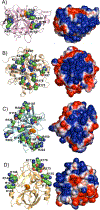
Cartoon representations and electrostatic potential surface maps of GAG-binding sites of four serine proteases [plasmin (3UIR) (A), thrombin (1XMN) (B), factor Xa (2GD4) (C), and factor XIa (1ZHM) (D)]. Basic residues in each site are shown as spheres (carbon atom is depicted in green color) and the active site serine is shown as spheres (orange color). The electrostatic potential surface was calculated using APBS tool. Electropositive surface is coded blue, while electronegative surface is in red. These sites are targeted by different sulfated NSGMs. The maps exhibit strong positive charge density for all four proteases but with significant structural differences, which justify the potential of developing selective NSGM-based inhibitors.
Figure 3
Figure 3
The crystal structure of heparin pentasaccharide DEFGH binding to the heparin-binding site on ATIII (PDB ID: 1E03) showing the interacting partners i.e. the negatively charged groups of sulfate/carboxylate of DEFGH with the positively charged amino acids of ATIII i.e. Arg and Lys residues.
Figure 4
Figure 4
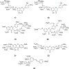
The chemical structures of saccharide and nonsaccharide ATIII activators. A) Structure-activity relationship studies revealed that the trisaccharide unit (DEF; 2) from the nonreducing end of the pentasaccharide (DEFGH; 1) is critical for both the initial recognition and the conformational activation processes. The trisaccharide DEF binds to ATIII with a KD value of 2 µM (pH 6.0) and accelerates ATIII-mediated inhibition of FXa nearly 300-fold which is equivalent to the acceleration obtained by the pentasaccharide DEFGH. B) The first generation of flavonoid-based sulfated NSGMs (38) was computationally designed to promote ATIII-mediated FXa inhibition. First generation molecules bind to ATIII with KD values of 3.5 – 26 µM (pH 6.0) and accelerate FXa inhibition by 8–22-fold.
Figure 5
Figure 5
The chemical structures of the second generation of THIQ-based ATIII activators (912). Pharmacophore approach was exploited to design this generation. Structural variations were as follows: positions of the two sulfate groups on THIQ scaffold (5,6-, 6,7-, and 5,8-), the presence or absence of carboxylate group at position-3, the linker length between the bicyclic and monocyclic units (1-, 2-, 3-, and 4-atom linkers), and the number and position of sulfate groups on the monocyclic unit (2 or 3 sulfates) and (2,5-disulfate, 3,4-disulfate, and 3,4,5-trisulfate groups). Considering the acceleration potential, THIQ-N-acyl derivative 10 accelerated ATIII inhibition of FXa by ~80-fold, only 3.75-fold less than that achieved by DEF unit under similar condition.
Figure 6
Figure 6
The chemical structures of polymeric NSGMs. A) Polyacrylic acid polymers 13 were found to bind ATIII with KD values of 1.0–2.3 µM at pH 6.0 and 34–180 µM at pH 7.4. The polymers accelerated ATIII-mediated FXa inhibition much better at pH 6.0 (15– 284-fold) than at pH 7.4 (6.2–17-fold). The polymers also accelerated ATIII-mediated thrombin inhibition in comparable fashion at the two pHs; pH 6.0 (24–1392-fold) and pH 7.4 (114–1109-fold). Polyacrylic acid polymers have opened up an opportunity to developing orally bioavailable, carboxylate-based ATIII activators. B) Chemo-enzymatically prepared oligomers of 4-hydroxycinnamic acids, known as dehydrogenation polymers (DHPs) displayed interesting anti-coagulant (thrombin inhibitors and ATIII activators), anti-fibrinolytic (plasmin inhibitors), and anti-emphysema (inhibitors of pulmonary elastolysis, oxidation, and inflammation) properties. The oligomers were prepared by peroxidase-catalyzed oxidative coupling of caffeic 14 (CDSO3), ferulic 15 (FDSO3), and sinapic 16 (SDSO3) acids, followed by sulfation using SO3–NEt3 complex. Various analytical studies suggested that the DHPs are heterogeneous and polydisperse preparations that are composed of inter-monomer linkages similar to those found in natural lignins including β−5, β-O-4, β-β and β−5. DHPs were later named as low molecular weight lignins (LMWLs).
Figure 7
Figure 7
The chemical structures of flavonoid-based (17-20), xanthone-based (21 and 22), and resveratrol-based (23) GAG mimetics. The seven mimetics demonstrated significant antithrombotic activity by inhibiting the coagulation proteins and/or inhibiting the arachidonic acid-and adenosine diphosphate-induced platelet aggregation. Molecules 19-21 are direct FXa inhibitors whereas molecules 17, 18, and 22 exhibit indirect inhibition of FXa by activating ATIII.
Figure 8
Figure 8
The chemical structures of various allosteric inhibitors of thrombin. A) Monosulfated benzofuran dimers (24-26) and trimers (27) were designed considering the structural and mechanistic aspects of LMWLs. Extensive biochemical work revealed that sulfated NSGMs bind to exosite II of thrombin in similar fashion to heparin, yet in contrast to heparin, they allosterically disrupt the active site’s catalytic triad. Trimer 27 is the most potent inhibitor in this class of sulfated NSGMs with IC50 value of 0.67 µM and efficacy of 79%. A 2-fold increase in APTT and PT required 139 and 568 μM of inhibitor 27, respectively. B) A sulfated β-O4 lignin (SbO4L) 28 was intuitively designed as a mimic of the sulfated tyrosine sequence of GPIbα. Extensive biochemical studies established that this polymeric sulfated NSGMs is an allosteric, exosite II-binding inhibitor of human thrombin and a competitive inhibitor of GPIbα-mediated platelet aggregation. SbO4L has presented unique anticoagulant and antiplatelet properties which were confirmed by studies in human plasma, human whole blood, FeCl3-induced carotid arterial thrombosis model in mice, and Rose Bengal–laser injury model of arterial thrombosis in mice. C) The chemical structures of a new generation of sulfated coumarin dimers (2931) that partially and allosterically modulate the catalytic activity of human thrombin. This was established by extensive biochemical studies. Dimer 31 is the most potent inhibitor with an IC50 value of 0.2 µM (KD=143 nM) and efficacy of 47%.
Figure 9
Figure 9
Chemical structures of allosteric inhibitors of FXIa. A) SPGG2 32 is a sulfated galloid-based NSGM. SPGG2 variant is the first small molecule allosteric inhibitor of FXIa. SPGG2 inhibited human FXIa with an IC50 value of 0.5 µM and a selectivity index of at least 200-fold against a range of coagulation, fibrinolysis, and digestive proteins. Extensive biochemical studies established the structural and mechanistic properties of this molecule. SPGG2 binds to the anion-binding site in the catalytic domain of FXIa. Its anticoagulant activity was confirmed in human normal and deficient plasmas as well as in human whole blood using thrombo-elastography technology. B) Sulfated quinazolinone dimers 33 and 34 were reported as allosteric inhibitors of FXIa. They were structurally designed using the dual-element recognition approach. Sulfated quinazolinone homodimers were identified to have micromolar potencies toward FXIa and significant selectivity against thrombin, FXa, chymotrypsin, and trypsin. In different studies, NSGM 32 inhibited the HSV-1 entry into multiple cell lines demonstrating antiviral activity that was comparable to acyclovir.
Figure 10
Figure 10
The chemical structures of flavonoid-based sulfated NSGMs that allosterically inhibit human plasmin and human FXIIIa. Pentasulfated flavonoid-quinazolinone heterodimer 35 was initially discovered as allosteric and marginally selective inhibitor of human plasmin. Subsequent efforts led to the identification of octasulfated flavonoid homodimer 36 inhibited human full-length plasmin with an IC50 value of 7.2 µM (KD=0.7 µM) and displayed significant selectivity against factors Xa, XIa, IXa, XIIa, trypsin, and chymotrypsin. Sulfated diflavonoid 36 also inhibited the physiologic function of plasmin i.e. clot lysis under physiological conditions in a dose-dependent manner with an IC50 value of 8.8 µM and efficacy of 69%. Interestingly, homologation of the sulfated diflavonoid 36 led to the undecasulfated flavonoid trimer 37 which was the first allosteric inhibitor of FXIIIa, a coagulation transglutaminase. NSGM 37 inhibited FXIIIa with an IC50 value of 36 µM (KD = 25.3 µM) and efficacy of 98%. The molecule exhibited good selectivity against thrombin, FXa, and papain. It also inhibited the physiological function of FXIIIa as measured by the cross-linking of fibrin monomers.
Figure 11
Figure 11
The chemical structures of sulfated NSGMs (3842) that inhibited angiogenesis in an in vitro tube formation assay. Minimal ‘pharmacophore’ appears to be two sulfate groups at an optimal distance of 5–10 Å.
Figure 12
Figure 12
The chemical structures of sulfated NSGMs 43 and 44, two molecules identified in a dual-filter screening protocol as selective inhibitors/antagonists of colon cancer stem cells in HT29 and HCT116 cell lines. Molecule 43 is a dodecasulfated hexasubstituted inositol derivative whereas molecule 44 is an octasulfated flavonoid homodimer derivative. These molecules, in addition to 32, downregulated several cancer stem cell markers and self-renewal factors. A three-step molecular dynamics-based algorithm indicated that the dimeric flavonoid-based NSGM 44 mimics hexameric GAG sequences including HS06 45 in the protein-bound state which selectively inhibited cancer stem cells self-renewal and induced apoptosis in colorectal, pancreatic, and breast cancer stem cells through a specific early and sustained activation of p38α/β MAPK.
Figure 13
Figure 13
The chemical structures of the monosulfated zosteric acid 46 and trisulfated gallic acid 47, both of which are NSGMs which demonstrate significant antifouling activities. Zosteric acid also inhibits the viral entry of dengue virus into host cells. The figure also depicts the chemical structure of 3-carboxy-4-hydroxybenzenesulfonate 48 which potentially inhibits CCL5-mediated leukocyte recruitment to the peritoneal cavity in the thiogallate-induce peritonitis mice model. Chemical structures of acamprosate 49, tramiprosate 50, eprodisate 51, and suramin 52 are also depicted.

Similar articles

Cited by

References

    1. Gandhi NS; Mancera RL The structure of glycosaminoglycans and their interactions with proteins. Chem. Biol. Drug Des, 2008, 72, 455–482. - PubMed
    1. Esko JD; Kimata K; Lindahl U Proteoglycans and Sulfated Glycosaminoglycans, In: Essentials of Glycobiology; Varki Ajit, Executive Editor, Cummings Richard D, Esko Jeffrey D, Stanley Pamela, Hart Gerald W, Aebi Markus, Darvill Alan G, Kinoshita Taroh, Packer Nicolle H, Prestegard James H, Schnaar Ronald L, and Seeberger Peter H., Ed.; Cold Spring Harbor (NY): Cold Spring Harbor Laboratory Press, 2009; pp. 784. - PubMed
    1. Imberty A; Lortat-Jacob H; Pérez S Structural view of glycosaminoglycan-protein interactions. Carbohydr. Res, 2007, 342, 430–439. - PubMed
    1. Pomin VH; Mulloy B Glycosaminoglycans and proteoglycans. Pharmaceuticals (Basel), 2018, 11(1), 27. - PMC - PubMed
    1. Sasisekharan R; Venkataraman G Heparin and heparan sulfate: Biosynthesis, structure and function. Curr. Opin. Chem. Biol, 2000, 4, 626–631. - PubMed