. 2019 Mar;32(2):303-314.
doi: 10.1111/pcmr.12751. Epub 2018 Dec 16.

The lncRNA RMEL3 protects immortalized cells from serum withdrawal-induced growth arrest and promotes melanoma cell proliferation and tumor growth

Affiliations

The lncRNA RMEL3 protects immortalized cells from serum withdrawal-induced growth arrest and promotes melanoma cell proliferation and tumor growth

Cibele Cardoso et al. Pigment Cell Melanoma Res. 2019 Mar.

Erratum in

  • Corrigendum.
    [No authors listed] [No authors listed] Pigment Cell Melanoma Res. 2020 May;33(3):520. doi: 10.1111/pcmr.12875. Pigment Cell Melanoma Res. 2020. PMID: 32319742 No abstract available.

Abstract

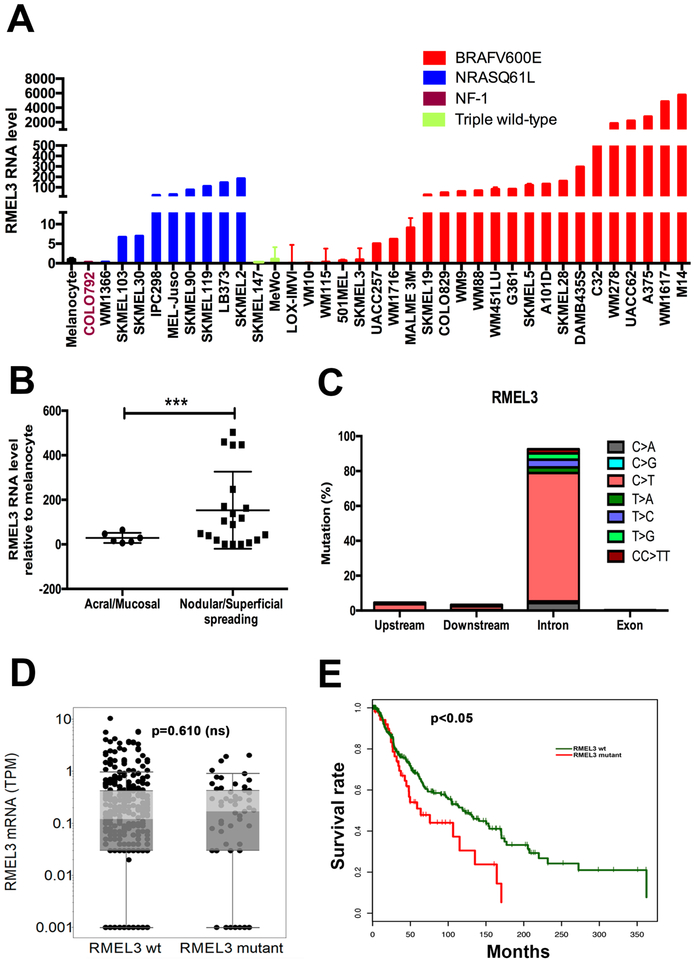
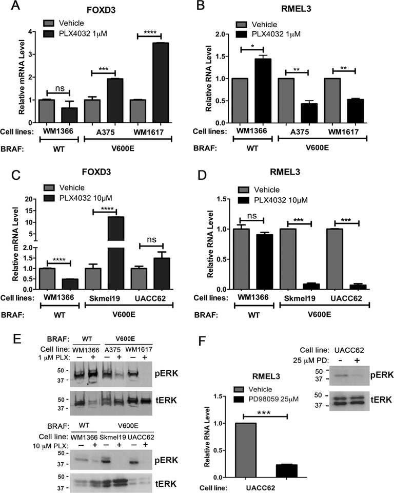
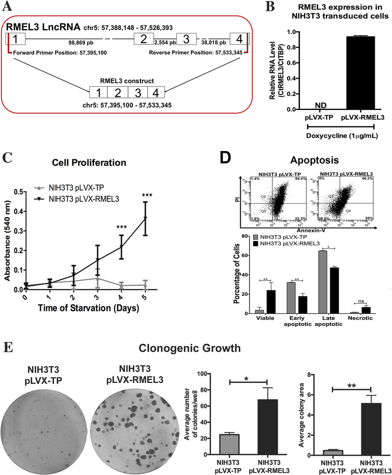
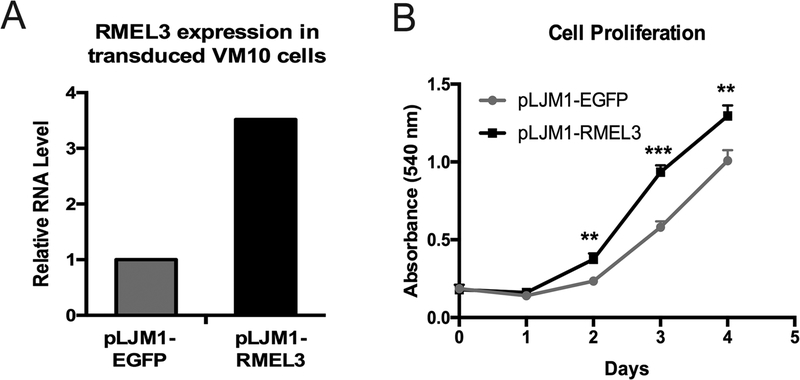
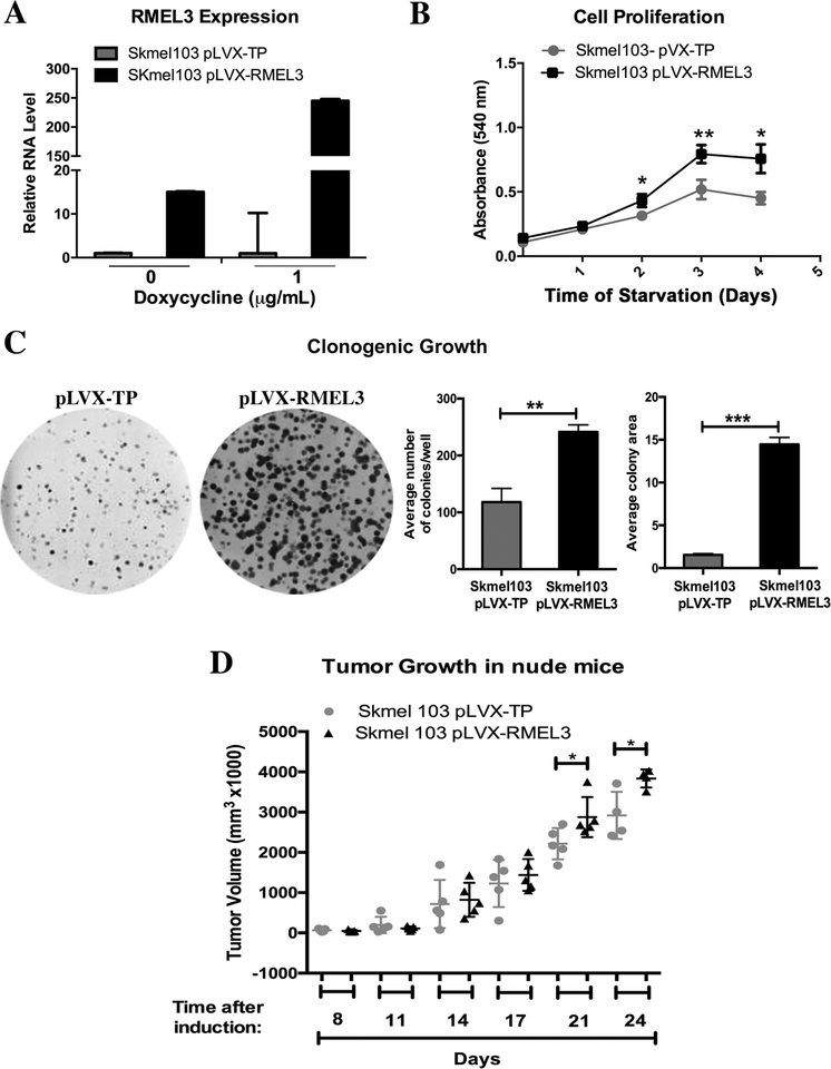
RMEL3 is a recently identified lncRNA associated with BRAFV600E mutation and melanoma cell survival. Here, we demonstrate strong and moderate RMEL3 upregulation in BRAF and NRAS mutant melanoma cells, respectively, compared to melanocytes. High expression is also more frequent in cutaneous than in acral/mucosal melanomas, and analysis of an ICGC melanoma dataset showed that mutations in RMEL3 locus are preponderantly C > T substitutions at dipyrimidine sites including CC > TT, typical of UV signature. RMEL3 mutation does not correlate with RMEL3 levels, but does with poor patient survival, in TCGA melanoma dataset. Accordingly, RMEL3 lncRNA levels were significantly reduced in BRAFV600E melanoma cells upon treatment with BRAF or MEK inhibitors, supporting the notion that BRAF-MEK-ERK pathway plays a role to activate RMEL3 gene transcription. RMEL3 overexpression, in immortalized fibroblasts and melanoma cells, increased proliferation and survival under serum starvation, clonogenic ability, and xenografted melanoma tumor growth. Although future studies will be needed to elucidate the mechanistic activities of RMEL3, our data demonstrate that its overexpression bypasses the need of mitogen activation to sustain proliferation/survival of non-transformed cells and suggest an oncogenic role for RMEL3.

Keywords: BRAFV600E; CTD-2023N9.1; ENSG00000250961.1; LncGPBP1-1:1; MAPK; chr5:57395060-57533424 (GRCh38/hg38); melanoma; mitogen; serum response.


Figures

Figure 1.
Figure 1.. High RMEL3 expression predominates in BRAFV600E followed by NRAS mutant melanoma cell lines, as well as in nodular/ superficial spreading in comparison with acral/mucosal melanomas.
(A) RT-qPCR analysis of RMEL3 RNA levels in melanocytes and melanoma cell lines of indicated genotype regarding major câncer drivers (BRAF, NRAS, and NF1). (B) RT-qPCR analysis of RMEL3 expression in melanoma subtypes: acral/mucosal and nodular/superficial spreading cutaneous melanoma, for which patient specimens were obtained from the Barretos Cancer Hospital, São Paulo state, Brazil. In A and B, relative expression was calculated according to 2−ΔΔCT method usingTBP (Tata-box binding protein) as endogenous control, and the mean levels of RMEL3 RNA in different foreskin human melanocyte primary cultures were used as reference in both A and B, totaling the use of four different melanocyte cultures derived from different donors. Error bars represent SEM of 3 independent experiments. ***p < 0.005. Asterisks indicate statistically significant differences between groups based on Fisher’s exact test. (C) Frequency of base substitution mutations in the RMEL3 locus from a total of 579 base substitutions reported by ICGC in a set of 129 melanoma samples. (D) RMEL3 expression levels (TPM) grouped according to the occurrence (RMEL3 mutant) or not (RMEL3 wt) of somatic mutation in the RMEL3 gene detected in the TCGA melanoma dataset of 450 samples. (E) Survival curves for melanoma patients of the TCGA database carrying (RMEL3 mutant) or not (RMEL3 wt) mutations in the RMEL3 gene. Statistical analyses used chi-squared test in D and E
Figure 2.
Figure 2.. RMEL3 lncRNA levels decrease in BRAFV600E mutant melanoma cells after treatment with BRAF and MEK inhibitors.
(A - D) RT-qPCR expression analyses of (A, B) RMEL3 lncRNA and (C, D) FOXD3 (positive control) in melanoma cell lines treated with the BRAF inhibitor vemurafenib (PLX4032) (1 μM, 6 h or 10 μM, 48 h) and respective controls (vehicle), as indicated. (E) Western blot analysis for phosphorylated ERK (pERK) and total ERK (tERK) in total protein lysates from melanoma cells treated with BRAF inhibitor (1 μM, 6 h or 10 μM, 48 h) or vehicle. (F) RT-qPCR expression analysis of RMEL3 in UACC-62 melanoma cell line treated with the MEK inhibitor PD98059 (25 μM) or vehicle for 48 hr, and corresponding Western blot analysis for phosphorylated ERK (pERK) and total ERK (tERK) in total protein lysates of treated and control melanoma cells. For all RT-qPCR analyses, relative expression was calculated according to 2−ΔΔCT method using TBP (Tata-box binding protein) as endogenous control and the normalized Ct of the sample treated with the vehicle alone as reference. Error bars represent SEM of 3 independent experiments. *p < 0.05; **p < 0.005; ***p < 0.0005; ****p < 0.00005. Asterisks indicate statistically significant differences between groups based on unpaired parametric Student’s t test
Figure 3.
Figure 3.. Ectopic expression of human RMEL3 protects NIH3T3 murine fibroblasts from serum withdrawal-induced growth arrest/ apoptosis.
(A) Schematic representation of RMEL3 locus (above), its transcript (center), and the region cloned into expression vectors (below). (B) Efficiency of exogenous RMEL3 induction in cells transduced with pLVX-RMEL3 or pLVX-TP (as control) after treatment with doxycycline (1 μM, 24 h), analyzed by RT-qPCR. ND (not-detected). Relative expression was calculated according to ΔCT using TBP (Tatabox binding protein) as endogenous control. (C-E) Functional assays using NIH3T3 cells stably transduced with pLVX-RMEL3 or pLVX-TP (as control) in the presence of doxycycline (1 μM). (C) Proliferation rates. During the time course of the assay, cells were maintained under serum starvation (in medium supplemented with 0.5% FBS). After the indicated time points, cells were stained with crystal violet and cell density was quantified according to the absorbance in an ELISA microplate reader. ***p < 0.0005 (D) Apoptosis rate. Flow cytometry-based detection of annexin-V- and propidium iodide (PI)-stained cells. Dot plot from one assay representative of three replicates, and below, a summary graphics of three independent replicates. Cells were cultured in FBS-free medium for 48 h, and afterward, culture medium was supplemented with 0.5% FBS and cells were cultured for additional 48 h, when they were assayed. *p < 0.05; **p < 0.005. (E) Clonogenic ability. Cells were seeded in 60-mm-diameter plates and allowed to grow for 9 days, when they were fixed with paraformaldehyde and stained with crystal violet to reveal the colonies. *p < 0.05; **p < 0.005. Error bars represent SEM of 3 independent experiments for B-E. Asterisks indicate statistically significant differences between groups based on unpaired parametric Student’s t test
Figure 4.
Figure 4.. Overexpression of RMEL3 increases proliferation of a BRAFV600E mutant, RMEL3-low expresser, melanoma cell line.
(A) Efficiency of stable expression of RMEL3 RNA in VM10 melanoma cells transduced with pLJM1-RMEL3, analyzed by RT-qPCR. Relative expression was calculated according to 2−ΔΔCT method using TBP (Tata-box binding protein) as endogenous control and the normalized Ct of cells transduced with control vector (pLJM1-EGFP) as reference. (B) Proliferation of VM10 cells transduced with pLJM1-RMEL3 or pLJM1-EGFP as control. Cells were cultured in medium supplemented with 10% FBS, and after the indicated time points, they were stained with crystal violet for quantification of cell density according to the absorbance in an ELISA microplate plate reader. Error bars represent SEM of 3 independent experiments. Asterisks indicate statistically significant differences between groups based on unpaired parametric Student’s t test. Day 2 (**p < 0.0034); Day 3 (***p < 0.0004); Day 4 (**p < 0.0065)
Figure 5.
Figure 5.. Overexpression of RMEL3 in an NRAS mutant melanoma cell line increases proliferation rates, clonogenic ability, and tumor growth.
(A) Efficiency of RMEL3 transgene expression in SKMEL103 cells stably transduced with pLVX-RMEL3 or pLVX-TP (empty vector), not treated or treated with doxycycline (1 μg/mL, 24 h), as analyzed by RT-qPCR. The relative expression was calculated according to 2−ΔΔCT method using TBP (Tata-box binding protein) as endogenous control and the normalized Ct of the pLVX-TP-transduced cells as reference. (B) Proliferation of SKMEL103 cells transduced with pLVX-RMEL3 or pLVX-TP (as control), cultured in FBS-free medium in the presence of doxycycline (1 μg/mL). After the indicated time points, cells were stained with crystal violet and cell density was quantified according to the absorbance in an ELISA microplate reader. *p < 0.05; **p < 0.005. (C) Clonogenic ability of SKMEL103 cells transduced with pLVX-RMEL3 or pLVX-TP (as control). Cells were seeded in 60-mm-diameter plates with DMEM Complete Medium containing doxycycline (1 μg/mL). After 14 days, they were fixed with paraformaldehyde and stained with crystal violet. **p < 0.0014, ***p < 0.0001. Error bars represent SEM of 3 independent experiments for A-C. (D) Tumor growth. Nude mice were injected subcutaneously with 106 SKMEL103 cells, in the right flank with pLVX-RMEL3-transduced cells and in the left flank with pLVX-TP-transduced control cells. After injections, mice were maintained with diet ad libitum and drinking water was supplemented with 1 mg/ml doxycycline and changed every 2 days. *p < 0.05. This experiment was done once. Asterisks indicate statistically significant differences between groups based on unpaired parametric Student’s t test

References

    1. Abel EV, Aplin AE (2010). FOXD3 is a mutant B-RAF-regulated inhibitor of G1-S progression in melanoma cells. Cancer Res, 70(7): 2891–2900. - PMC - PubMed
    1. Abel EV, Basile KJ, Kugel CH III, Witkiewicz AK, Le K, Amaravadi RK, Karakousis GC, Xu X, Xu W, Schuchter LM, Lee JB, Ertel A, Fortina P, Aplin AE (2013). Melanoma adaptsto RAF/MEK inhibitorsthrough FOXD3-mediated upregulationof ERBB3. J Clin Invest, 123(5):2155–2168. - PMC - PubMed
    1. Ascierto PA, Marincola FM, Atkins MB (2015). What’s new in melanoma? Combination!. J Transl Med, 13:213. - PMC - PubMed
    1. Basile KJ, Abel EV, Aplin AE (2012). Adaptive upregulation of FOXD3 and resistance to PLX4032/4720-induced cell death in mutant B-RAF melanoma cells. Oncogene, 31: 2471–2479. - PMC - PubMed
    1. Bhatia P, Friedlander P, Zakaria EA, Kandil E (2015). Impact of BRAF mutation status in the prognosis of cutaneous melanoma: an area of ongoing research. Ann Transl Med, 3(2):24. - PMC - PubMed

Publication types

MeSH terms