. 2018 Dec;24(12):2210-2218.
doi: 10.3201/eid2412.180702.

Novel Type of Chronic Wasting Disease Detected in Moose (Alces alces), Norway

Novel Type of Chronic Wasting Disease Detected in Moose (Alces alces), Norway

Laura Pirisinu et al. Emerg Infect Dis. 2018 Dec.

Abstract

Chronic wasting disease (CWD) persists in cervid populations of North America and in 2016 was detected for the first time in Europe in a wild reindeer in Norway. We report the detection of CWD in 3 moose (Alces alces) in Norway, identified through a large scale surveillance program. The cases occurred in 13-14-year-old female moose, and we detected an abnormal form of prion protein (PrPSc) in the brain but not in lymphoid tissues. Immunohistochemistry revealed that the moose shared the same neuropathologic phenotype, characterized by mostly intraneuronal deposition of PrPSc. This pattern differed from that observed in reindeer and has not been previously reported in CWD-infected cervids. Moreover, Western blot revealed a PrPSc type distinguishable from previous CWD cases and from known ruminant prion diseases in Europe, with the possible exception of sheep CH1641. These findings suggest that these cases in moose represent a novel type of CWD.

Keywords: Alces alces; CWD; Nor16CWD; Norway; TSE; cervid; chronic wasting disease; moose; prions; reindeer; transmissible spongiform encephalopathy; zoonoses.


Figures

Figure 1
Figure 1
Immunohistochemical detection of disease-associated prion protein in brain sections at the level of the obex in cervids with chronic wasting disease, Norway. A–E) Reindeer; F–J) moose. mAbs used were 12B2 (A, F), 9A2 (B, G), L42 (C, H), SAF 84 (D, I), and F99/97.6 (E, J). Staining obtained in the reindeer tissues is similar regardless of mAbs used (A–E). Conversely, for moose tissues, the staining was primarily observed intraneuronally with L42, SAF84, and F99/97.6 (H–J) but was not observed using the more N-terminal mAbs 12B2 and 9A2 (F, G). Scale bars indicate 40 µm. mAbs, monoclonal antibodies.
Figure 2
Figure 2
Western blot analysis of PrPres in brains of chronic wasting disease–affected cervids from Norway and Canada. A) Western blot analysis PrPres in brains of moose and reindeer from Norway. Membrane was probed with L42 monoclonal antibodies. Molecular weights (kDa) are indicated on the right. Tissue equivalent loaded per lane was 1 mg. B) Western blot analysis of PrPres from moose isolates from Norway (lanes 6–7) compared with PrPres from chronic wasting disease–affected elk or wapiti (lanes 1–3), white-tailed deer (lane 4), and moose (lane 5) from Canada. Membranes were probed with L42 (upper) and 12B2 (lower) monoclonal antibodies. A scrapie sheep sample from Italy was added as control (lane 8). Molecular weights (kDa) are indicated on the right of each blot. Tissue equivalents loaded per lane were 1 mg for Canadian isolates, 2 mg for Norwegian isolates, and 0.15 mg for scrapie sheep control. PrPres, protease-resistant core of abnormal form of prion protein.
Figure 3
Figure 3
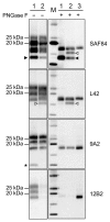
Characterization of PrPres fragments from moose (Alces alces) in Europe by epitope mapping. Mapping with mAbs spanning the whole prion protein enabled the analysis of PrPres in moose samples before (PNGase F–) and after (PNGase F+) deglycosylation, based on presence or absence of the epitopes and apparent molecular weight. Lanes 1, moose no. 1; lanes 2, moose no. 3; lane M, protein standards; lane 3, sheep scrapie sample. Solid arrowheads indicate C-terminal fragment of ≈13 kDa fragment (present in both samples and detected with SAF84 mAbs). Open arrowheads indicate C-terminal fragment of ≈16 kDa fragment in moose no. 2 with SAF84 and L42 mAbs. Asterisk indicates the internal fragment detected in moose no. 1 with 9A2 mAbs. Molecular weights are indicated on the left. In the blots on the right, protein standards are shown in lane M (10, 15, 20, 25, and 37 kDa). The mAbs used are indicated on the right. mAbs, monoclonal antibodies; PrPres, protease-resistant core of abnormal form of prion protein.
Figure 4
Figure 4
Bar graph of antibody-signal ratios (y-axis) showing discrimination of the ovine, bovine, moose, and reindeer samples (x-axis) analyzed in a study characterizing chronic wasting disease in moose (Alces alces), Norway. Numbers indicate sample type: 1, scrapie; 2, CH1641; 3, CH1641-like; 4, classical bovine spongiform encephalopathy (BSE); 5, H-type atypical BSE; 6, L-type atypical BSE; 7, moose no. 1; 8, moose no. 2; 9, moose no. 3; 10, reindeer. The antibody ratio is the L42/12B2 ratio of the chemiluminescence signal relative to the L42/12B2 ratio of the control scrapie loaded in each blot. Bars represent median values of >3 independent determinations; error bars represent the range of observed values. Bars start at y = 2, which is the cutoff value of the antibody ratio for the discrimination of low molecular weight samples (i.e., suspected bovine spongiform encephalopathy cases) from scrapie, according to discriminatory Western blot.
Figure 5
Figure 5
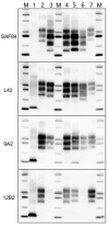
Comparison of protease-resistant PrPres from moose (Alces alces) with chronic wasting disease and from sheep with scrapie, Europe. Representative blots show epitope mapping analysis of PrPres (lane 4, CH1641; lane 5, moose no. 1; lane 6, moose no. 2) in comparison with different ovine transmissible spongiform encephalopathy isolates (lane 1, atypical/Nor98; lane 2, classical scrapie; and lane 3, CH1641). A chronic wasting disease isolate from Canada was loaded as control (lane 7). The antibodies used are indicated on the left. Protein standards are shown in lane M (10, 15, 20, 25, 37, and 50 kDa). The small amount of PrPres with intact 12B2 epitope in moose no.1 had a molecular weight higher than that observed with more C-terminal monoclonal antibodies (18.7 +0.3 kDa measured with 12B2 vs. 17.2 +0.1 kDa measured with L42). Even if the increase of the apparent molecular weight might be a known behavior when proteinase K cleavage occurs near the epitope, we noted that, in the case of moose no. 1, the 12B2-positive PrPres had a molecular weight higher than scrapie (18.1 +0.1 kDa measured with 12B2) and CH1641-like sample (18.1 +0.4 kDa when detected with 12B2). PrPres, protease-resistant core of abnormal form of prion protein.
Figure 6
Figure 6
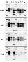
Comparison of protease-resistant core of abnormal form of prion protein from moose (Alces alces) in Europe with chronic wasting disease and from cattle with BSE. Representative blots show epitope mapping analysis of protease-resistant core of abnormal form of prion protein in moose (lane 5, moose no. 1; lane 6, moose no. 2) in comparison with different BSE isolates (lane 2, classical BSE; lane 3, H-type BSE; and lane 4, L-type BSE). A sheep scrapie isolate was loaded as control (lane 1). The antibodies are indicated on the left. Protein standards are shown in lane M (10, 15, 20, 25, 37, and 50 kDa). BSE, bovine spongiform encephalopathy.

References

    1. Prusiner SB. Molecular biology of prion diseases. Science. 1991;252:1515–22. 10.1126/science.1675487 - DOI - PubMed
    1. Benestad SL, Telling GC. Chronic wasting disease: an evolving prion disease of cervids. Handb Clin Neurol. 2018;153:135–51. 10.1016/B978-0-444-63945-5.00008-8 - DOI - PubMed
    1. EFSA Panel on Biological Hazards. Scientific opinion on chronic wasting disease (CWD) in cervids. EFSA J. 2017;15:4667.
    1. Kim TY, Shon HJ, Joo YS, Mun UK, Kang KS, Lee YS. Additional cases of Chronic Wasting Disease in imported deer in Korea. J Vet Med Sci. 2005;67:753–9. 10.1292/jvms.67.753 - DOI - PubMed
    1. Miller MW, Swanson HM, Wolfe LL, Quartarone FG, Huwer SL, Southwick CH, et al. Lions and prions and deer demise. PLoS One. 2008;3:e4019. 10.1371/journal.pone.0004019 - DOI - PMC - PubMed

Publication types