Case Reports
. 2018 Dec;24(12):2241-2250.
doi: 10.3201/eid2412.180937.

Rat Hepatitis E Virus as Cause of Persistent Hepatitis after Liver Transplant

Case Reports

Rat Hepatitis E Virus as Cause of Persistent Hepatitis after Liver Transplant

Siddharth Sridhar et al. Emerg Infect Dis. 2018 Dec.

Abstract

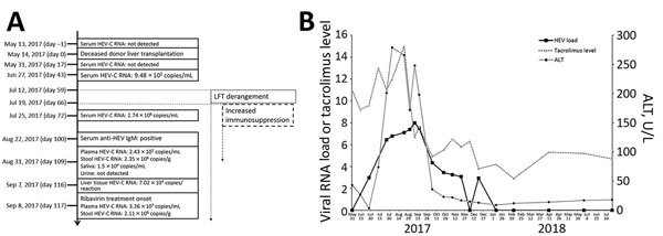
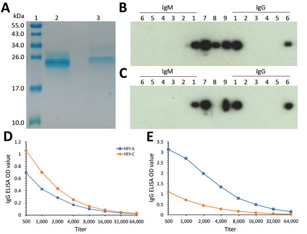
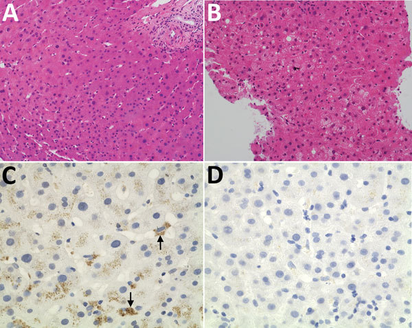
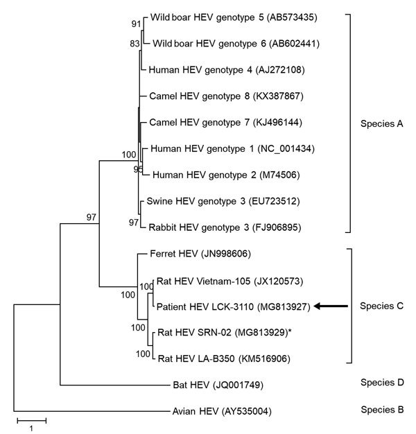
All hepatitis E virus (HEV) variants reported to infect humans belong to the species Orthohepevirus A (HEV-A). The zoonotic potential of the species Orthohepevirus C (HEV-C), which circulates in rats and is highly divergent from HEV-A, is unknown. We report a liver transplant recipient with hepatitis caused by HEV-C infection. We detected HEV-C RNA in multiple clinical samples and HEV-C antigen in the liver. The complete genome of the HEV-C isolate had 93.7% nt similarity to an HEV-C strain from Vietnam. The patient had preexisting HEV antibodies, which were not protective against HEV-C infection. Ribavirin was an effective treatment, resulting in resolution of hepatitis and clearance of HEV-C viremia. Testing for this zoonotic virus should be performed for immunocompromised and immunocompetent patients with unexplained hepatitis because routine hepatitis E diagnostic tests may miss HEV-C infection. HEV-C is also a potential threat to the blood product supply.

Keywords: HEV; Hong Kong; chronic hepatitis; hepatitis; hepatitis E; immunocompromised; liver transplant; rat hepatitis E virus; rodents; viruses; zoonoses.


Figures

Figure 1
Figure 1
Natural course of HEV-C infection in a 56-year-old man at Queen Mary Hospital, Hong Kong. A) Timeline of major clinical events. All days are post transplant. B) Kinetics of liver function tests, tacrolimus levels (μg/L), and plasma HEV-C RNA load (log10 copies/mL) with relation to ribavirin therapy. ALT, alanine aminotransferase; HEV-C, Orthohepevirus C; LFT, liver function test.
Figure 2
Figure 2
Serologic testing for HEV infection at Queen Mary Hospital, Hong Kong. A) Sodium dodecyl sulfate polyacrylamide gel electrophoresis gel showing purified HEV-A and HEV-C 239-aa recombinant proteins used in Western blot and ELISA. Lane 1, molecular weight marker; lane 2, HEV-A protein; lane 3, HEV-C protein. B–C) IgM and IgG Western blot using HEV-A protein (B) and HEV-C protein (C). Lane 1, patient serum (posttransplant day 100); lanes 2–5, individual platelet donor serum; lane 6, organ donor serum; lane 7, murine polyclonal serum against HEV-C; lane 8, specific monoclonal antibody against HEV-A; lane 9, cross-reactive monoclonal antibody against HEV-A and HEV-C. D, E) HEV-A and HEV-C ELISA IgG titers of patient pretransplant (D) and posttransplant serum (E) using an OD of 0.3 as assay cutoff as described in the Technical Appendix. HEV, hepatitis E virus; HEV-A, Orthohepevirus A; HEV-C, Orthohepevirus C; OD, optical density.
Figure 3
Figure 3
Histologic and immunohistochemical staining of liver tissue from a 56-year-old man at Queen Mary Hospital, Hong Kong. A, B) Liver tissue sections (original magnification ×200) stained with hematoxylin and eosin obtained at day 0 (A), showing normal hepatocyte architecture, and day 98 (B) after transplant showing progressive increase in hepatocyte ballooning and degenerative changes. C, D) Liver tissue section stained with cross-reactive monoclonal antibody (original magnification ×400); arrows show perinuclear antigen staining (C) and negative control with bovine serum albumin (D).
Figure 4
Figure 4
Phylogenetic analysis using complete open reading frame 2 nucleotide sequences of LCK-3110 and other hepatitis E virus strains. The tree was constructed using maximum-likelihood method with bootstrap values calculated from 1,000 trees. Only bootstrap values >70% are shown. GenBank accession numbers are provided. Arrows indicate strain LCK-3110; asterisk indicates strain SRN-02 detected in a street rodent in the epidemiologic investigation. Scale bar indicates mucleotide substitutions per site.
Figure 5
Figure 5
Isolation of HEV-C from 56-year-old male patient’s feces in cell culture, Queen Mary Hospital, Hong Kong. A) HEV-C RNA loads in culture S and day-7 CL of A549, Huh-7, and Caco-2 cell lines after inoculation by patient’s filtered fecal suspension. Mean of 3 replicates; error bars indicate SEM. B) Uninfected A549 cell monolayer stained with anti–HEV-C polyclonal antiserum. C) Infected A549 cell monolayer stained with anti–HEV-C polyclonal antiserum. Original magnification ×400. CL, cell lysate; HEV, hepatitis E virus; S, supernatant.

References

    1. Rein DB, Stevens GA, Theaker J, Wittenborn JS, Wiersma ST. The global burden of hepatitis E virus genotypes 1 and 2 in 2005. Hepatology. 2012;55:988–97. 10.1002/hep.25505 - DOI - PubMed
    1. Lee GH, Tan BH, Teo EC, Lim SG, Dan YY, Wee A, et al. Chronic infection with camelid hepatitis E virus in a liver transplant recipient who regularly consumes camel meat and milk. Gastroenterology. 2016;150:355–7.e3. 10.1053/j.gastro.2015.10.048 - DOI - PubMed
    1. Abravanel F, Lhomme S, El Costa H, Schvartz B, Peron JM, Kamar N, et al. Rabbit hepatitis E virus infections in humans, France. Emerg Infect Dis. 2017;23:1191–3. 10.3201/eid2307.170318 - DOI - PMC - PubMed
    1. Li TC, Chijiwa K, Sera N, Ishibashi T, Etoh Y, Shinohara Y, et al. Hepatitis E virus transmission from wild boar meat. Emerg Infect Dis. 2005;11:1958–60. 10.3201/eid1112.051041 - DOI - PMC - PubMed
    1. Tei S, Kitajima N, Takahashi K, Mishiro S. Zoonotic transmission of hepatitis E virus from deer to human beings. Lancet. 2003;362:371–3. 10.1016/S0140-6736(03)14025-1 - DOI - PubMed

Publication types

MeSH terms

Substances