

doi: 10.1289/EHP2268.
The Brain before Birth: Using fMRI to Explore the Secrets of Fetal Neurodevelopment
- PMID: 30457876
- PMCID: PMC6371691
- DOI: 10.1289/EHP2268
Item in Clipboard
The Brain before Birth: Using fMRI to Explore the Secrets of Fetal Neurodevelopment
Environ Health Perspect.
2018 Nov.
No abstract available
Figures
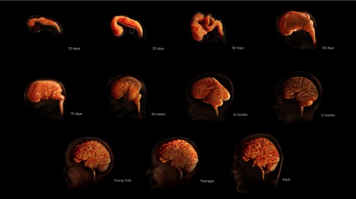
The process that will ultimately give rise to the connectome begins about 25 days after conception, when the neural tube begins to form. By the end of the embryonic period (gestational week 10), the basics of the neural system are established. All the structures continue to develop throughout the fetal period and early childhood. By 6 years of age, the brain has reached 90% of its adult volume. By age 25, it typically is fully developed. Image: © TheVisualMD/Science Source.

The connectome is shaped by internal and external stimuli throughout the course of life. In the fetus and young child, certain chemical exposures and situational factors (such as maternal stress and low socioeconomic status) are risk factors for neurodevelopmental problems. However, positive influences, such as parental engagement, may help to build resilience and mitigate any negative impacts. Image: © Daniel Atkin/Alamy Stock Photo.
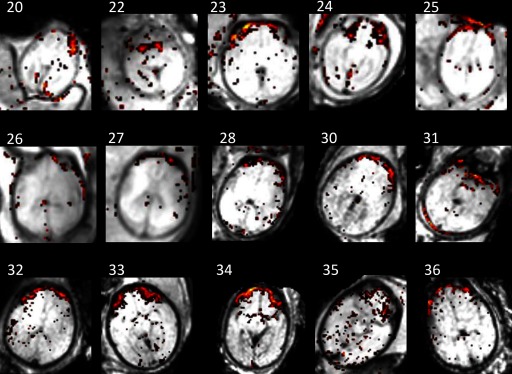
In 2012, Veronika Schöpf et al. captured functional images of fetal brains at gestational weeks 20–36 (the numbers in the figure above indicate gestational week). The team was the first to show that resting-state networks can be detected in utero. This imaging was a major advance over the use of task-based fMRI because, as Schöpf put it, “You could never know what a fetus was up to, whether it was performing a task or at rest.” Image: Schöpf et al. (2012).
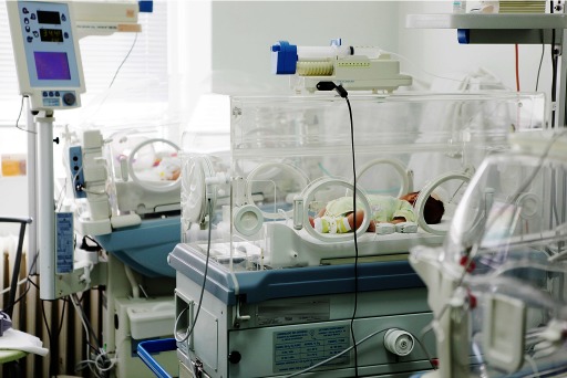
Some developmental abnormalities result from disease processes that started in the womb, but others may arise from the very act of being born prematurely and the stress of subsequent medical interventions. Annemarie Stroustrup et al. are investigating whether the NICU environment contributes to the latter category. If it does, that’s one negative influence that could be changed to a positive—or at least improved. Image: © Nenov/Getty Images.
References
-
- Seung S. 2013. Connectome: How the Brain’s Wiring Makes Us Who We Are. New York, NY: Mariner Books.
Publication types
MeSH terms
LinkOut - more resources
Full Text Sources
Medical
