

Artificial MicroRNA-Mediated Inhibition of Japanese Encephalitis Virus Replication in Neuronal Cells
- PMID: 30457923
- PMCID: PMC6277082
- DOI: 10.1089/nat.2018.0743
Artificial MicroRNA-Mediated Inhibition of Japanese Encephalitis Virus Replication in Neuronal Cells
Abstract
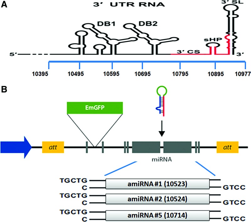
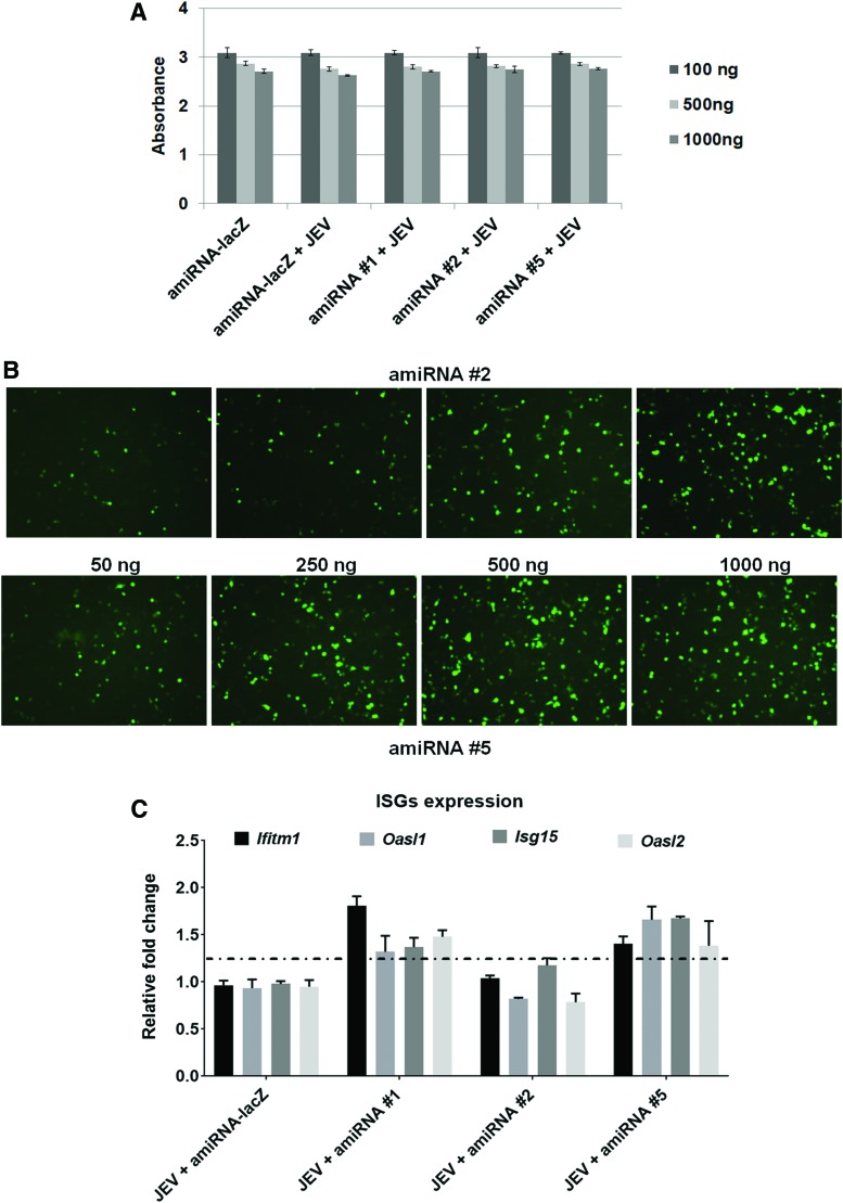
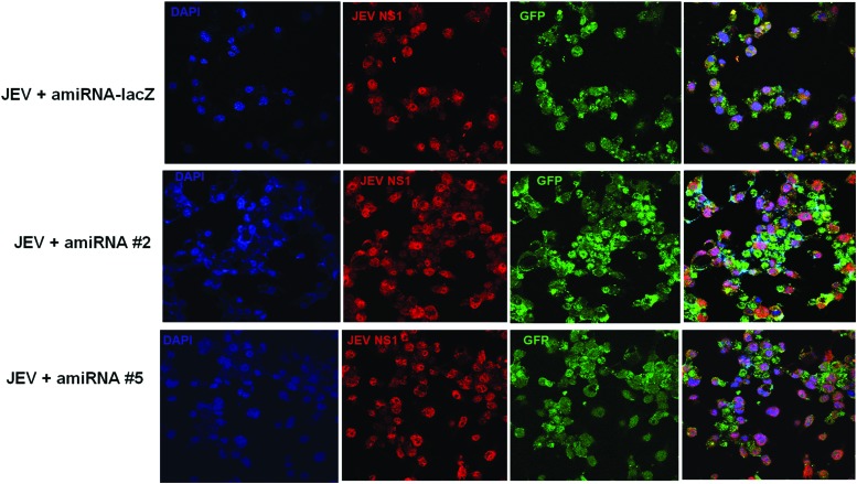
Artificial microRNA (amiRNA)-mediated inhibition of viral replication has recently gained importance as a strategy for antiviral therapy. In this study, we evaluated the benefit of using the amiRNA vector against Japanese encephalitis virus (JEV). We designed three single amiRNA sequences against the consensus sequence of 3' untranslated region (3'UTR) of JEV and tested their efficacy against cell culture-grown JEV Vellore strain (P20778) in neuronal cells. The binding ability of three amiRNAs on 3'UTR region was tested in vitro in HEK293T cells using a JEV 3'UTR tagged with luciferase reporter vector. Transient transfection of amiRNAs was nontoxic to cells as evident from the MTT assay and caused minimal induction in interferon-stimulated gene expression. Furthermore, our result suggested that transient expression of two amiRNAs (amiRNA #1 and amiRNA #2) significantly reduced intracellular viral RNA and nonstructural 1 (NS1) protein, as well as diminished infectious viral particle release up to 95% in the culture supernatant as evident from viral plaque reduction assay. Overall, our results indicated that RNA interference based on amiRNAs targeting viral conserved regions at 3'UTR was a useful approach for improvements of nucleic acid inhibitors against JEV.
Keywords: 3′UTR; JEV; artificial microRNA; replication.
Conflict of interest statement
No competing financial interests exist.
Figures
Similar articles
-
Susceptibility of naïve and differentiated PC12 cells to Japanese encephalitis virus infection.IUBMB Life. 2017 Feb;69(2):79-87. doi: 10.1002/iub.1595. Epub 2017 Jan 23. IUBMB Life. 2017. PMID: 28111888
-
Effective inhibition of different Japanese encephalitis virus genotypes by RNA interference targeting two conserved viral gene sequences in vitro and in vivo.Virus Genes. 2018 Dec;54(6):746-755. doi: 10.1007/s11262-018-1602-z. Epub 2018 Sep 18. Virus Genes. 2018. PMID: 30229544
-
Japanese Encephalitis Virus exploits the microRNA-432 to regulate the expression of Suppressor of Cytokine Signaling (SOCS) 5.Sci Rep. 2016 Jun 10;6:27685. doi: 10.1038/srep27685. Sci Rep. 2016. PMID: 27282499 Free PMC article.
-
Broad-spectrum antiviral activity of RNA interference against four genotypes of Japanese encephalitis virus based on single microRNA polycistrons.PLoS One. 2011;6(10):e26304. doi: 10.1371/journal.pone.0026304. Epub 2011 Oct 18. PLoS One. 2011. PMID: 22028851 Free PMC article.
-
A Review of miRNA Regulation in Japanese Encephalitis (JEV) Virus Infection.Curr Pharm Biotechnol. 2024;25(5):521-533. doi: 10.2174/0113892010241606231003102047. Curr Pharm Biotechnol. 2024. PMID: 37888811 Review.
Cited by
-
Interactions of host miRNAs in the flavivirus 3´UTR genome: From bioinformatics predictions to practical approaches.Front Cell Infect Microbiol. 2022 Oct 13;12:976843. doi: 10.3389/fcimb.2022.976843. eCollection 2022. Front Cell Infect Microbiol. 2022. PMID: 36310869 Free PMC article. Review.
-
Molecular Mechanism and Role of Japanese Encephalitis Virus Infection in Central Nervous System-Mediated Diseases.Viruses. 2022 Nov 30;14(12):2686. doi: 10.3390/v14122686. Viruses. 2022. PMID: 36560690 Free PMC article. Review.
-
A Comprehensive Review of the Development and Therapeutic Use of Antivirals in Flavivirus Infection.Viruses. 2025 Jan 8;17(1):74. doi: 10.3390/v17010074. Viruses. 2025. PMID: 39861863 Free PMC article. Review.
-
Bone marrow-derived extracellular vesicles modulate the abundance of infiltrating immune cells in the brain and exert an antiviral effect against the Japanese encephalitis virus.FASEB Bioadv. 2022 Oct 28;4(12):798-815. doi: 10.1096/fba.2022-00071. eCollection 2022 Dec. FASEB Bioadv. 2022. PMID: 36479206 Free PMC article.
-
An RBM10 and NF-κB interacting host lncRNA promotes JEV replication and neuronal cell death.J Virol. 2023 Dec 21;97(12):e0118323. doi: 10.1128/jvi.01183-23. Epub 2023 Nov 22. J Virol. 2023. PMID: 37991381 Free PMC article.
References
Publication types
MeSH terms
Substances
LinkOut - more resources
Full Text Sources