. 2019 Jan 2;129(1):150-162.
doi: 10.1172/JCI121372. Epub 2018 Nov 19.

TNF/TNFR axis promotes pyrin inflammasome activation and distinctly modulates pyrin inflammasomopathy

Affiliations

TNF/TNFR axis promotes pyrin inflammasome activation and distinctly modulates pyrin inflammasomopathy

Deepika Sharma et al. J Clin Invest. .

Abstract

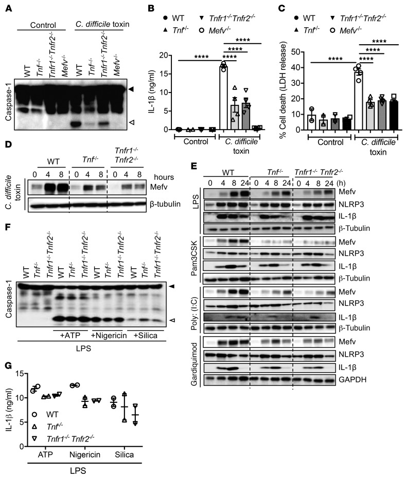
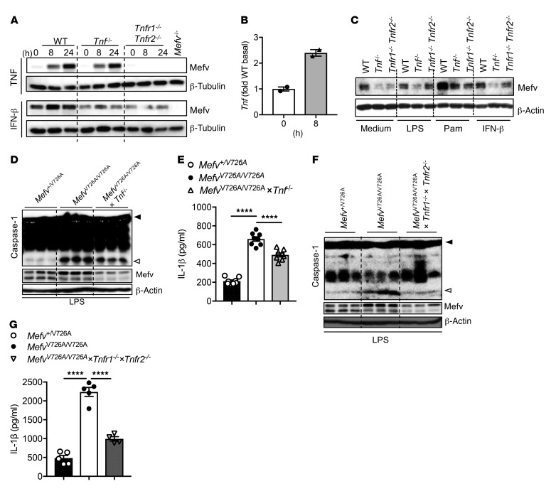
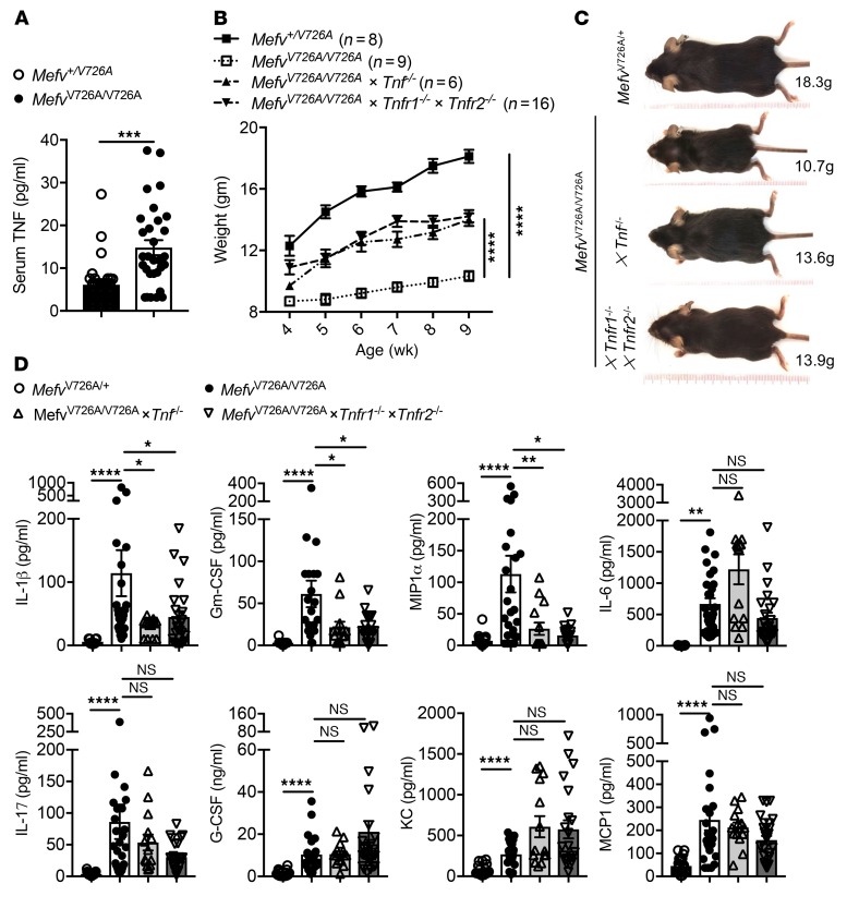
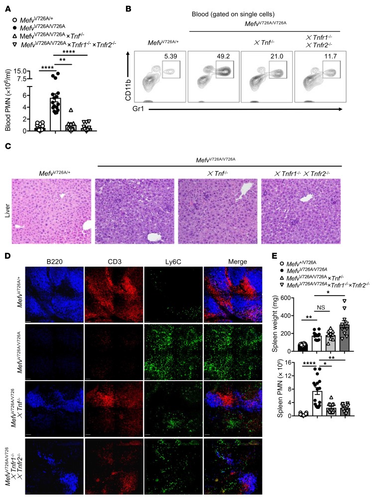
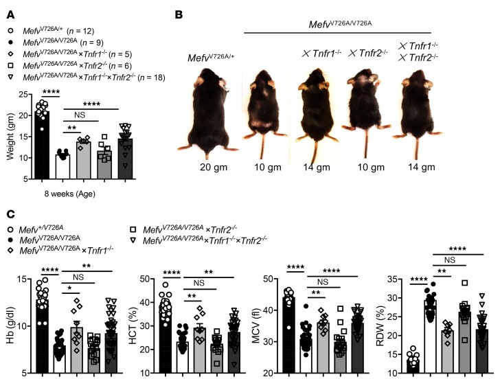
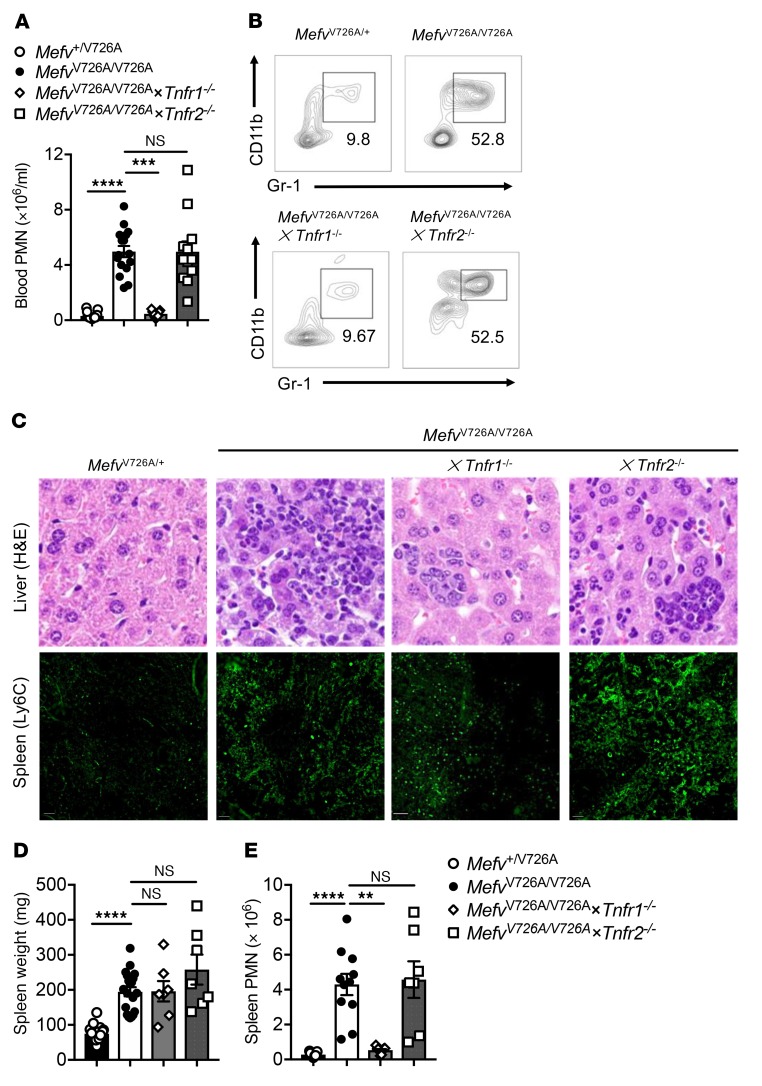
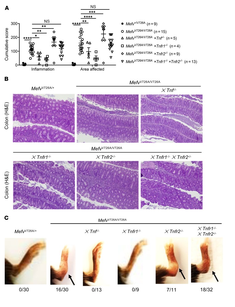
Pyrin is an inflammasome sensor that promotes caspase-1-mediated pyroptotic cell death and maturation of proinflammatory cytokines IL-1β and IL-18. Familial Mediterranean fever (FMF), an autoinflammatory disorder, is associated with mutations in the gene encoding pyrin (MEFV). FMF-knockin (FMF-KI) mice that express chimeric pyrin protein with FMF mutation (MefvV726A/V726A) exhibit an autoinflammatory disorder mediated by autoactivation of the pyrin inflammasome. Increase in the levels of TNF are observed in FMF-KI mice, and many features of FMF overlap with the autoinflammatory disorder associated with TNF receptor signaling. In this study, we assessed the contribution of TNF signaling to pyrin inflammasome activation and its consequent role in distinct FMF pathologies. TNF signaling promoted the expression of pyrin in response to multiple stimuli and was required for inflammasome activation in response to canonical pyrin stimuli and in myeloid cells from FMF-KI mice. TNF signaling promoted systemic wasting, anemia, and neutrophilia in the FMF-KI mice. Further, TNF-induced pathology was induced specifically through the TNFR1 receptor, while TNFR2-mediated signaling was distinctly protective in colitis and ankle joint inflammation. Overall, our data show that TNF is a critical modulator of pyrin expression, inflammasome activation, and pyrin-inflammasomopathy. Further, specific blockade of TNFR1 or activation of TNFR2 could provide substantial protection against FMF pathologies.

Keywords: Cellular immune response; Immunology; Inflammation.


Conflict of interest statement

Conflict of interest: The authors have declared that no conflict of interest exists.

Figures

Figure 1
Figure 1. TNF signaling promotes pyrin expression and inflammasome activation.
(A) Caspase-1 processing, (B) IL-1β release, and (C) LDH release in BMDMs in response to C. difficile stimuli for 24 hours. (D) Pyrin expression in BMDMs in response to C. difficile stimuli. (E) Pyrin, NLRP3, and IL-1β expression in BMDMs following TLR stimuli LPS (TLR4), Pam3CSK (TLR1/TLR2), poly(I:C) (TLR3), and gardiquimod (TLR7). (F) Caspase-1 processing and (G) IL-1β release BMDMs in response to LPS priming for 4 hours followed by ATP, nigericin. or silica for 30 minutes, 2 hours, or 12 hours, respectively. (B, C, G) Data are presented as mean ± SEM and are representative of at least 3 independent experiments. ****P < 0.0001 compared with WT, 1-way ANOVA followed by Fischer’s LSD test.
Figure 2
Figure 2. Pyrin activation observed in MefvV726A/V726A monocytes is promoted by TNF signaling.
Pyrin expression in (A) BMDMs and (C) peritoneal cells following TLR and cytokine stimuli (TNF, IFN-β) over 24 hours. (B) Tnf expression in WT BMDMs stimulated with IFN-β. (D and F) Caspase-1 processing, pyrin expression, and (E and G) IL-1β release in monocytes in response to LPS (200 ng/ml) stimuli for 24 hours. (B, E, G) Data are presented as mean ± SEM and are representative of at least 3 independent repeats. ****P < 0.0001 compared with MefvV726A/V726A, 1-way ANOVA followed by Fischer’s LSD test.
Figure 3
Figure 3. TNF signaling promotes runting and systemic inflammation in MefvV726A/V726A mice.
(A) Level of TNF in serum of indicated mice at 8 to 10 weeks of age. (B) Body weights of indicated number of female mice and (C) representative whole-body image of mice at 8 weeks of age. (D) Cytokine levels in serum samples. (A, B, D) Data are presented as mean ± SEM with (A and D) n = 14–30 for each genotype. *P < 0.05; **P < 0.01; ***P < 0.001; ****P < 0.0001 compared with MefvV726A/V726A, Student’s t test (A), 2-way ANOVA (B), and Kruskal-Wallis test followed by Dunn’s post test (D).
Figure 4
Figure 4. TNF signaling promotes neutrophilia and granulopoiesis in MefvV726A/V726A mice.
(A) Number of neutrophils (PMN) in blood (/ml) and (B) representative contour plot depicting proportion of PMNs (CD11b+Gr1+) in blood of indicated mice. Cells were gated on single cells; the percentage of cells identified as neutrophils is noted on each plot. (C) Representative H&E-stained sections of liver tissues. Original magnification, ×20. (D) Cellular composition in spleens of MefvV726A/V726A mice. B220, CD3, and Ly6C represent B cells, T cells, and monocytes/granulocytes, respectively. Scale bars: 50 μM. (E) Weight and total number of granulocytes in the spleens of indicated mice. (A and E) Data are presented as mean ± SEM with n = 8–16 for each genotype. *P < 0.05; **P < 0.01; ****P < 0.0001 compared with MefvV726A/V726A mice, using Kruskal-Wallis test followed by Dunn’s post test.
Figure 5
Figure 5. TNFR1 signaling promotes runting and anemia in MefvV726A/V726A mice.
(A) Body weights of indicated numbers of female mice and (B) representative whole-body images of mice at 8 weeks of age. (C) Features of anemia, including Hb, HCT, MCV, and RDW levels in the blood of indicated mice. n = 9–39 per genotype. (A and C) Data are presented as mean ± SEM. *P < 0.05; **P < 0.01; ****P < 0.0001 compared with MefvV726A/V726A using (A and C) 1-way ANOVA or Kruskal-Wallis followed by Fisher’s LSD or Dunn’s post test.
Figure 6
Figure 6. TNFR1 signaling promotes neutrophilia and granulopoiesis in MefvV726A/V726A mice.
(A) Number of neutrophils (PMN) in blood (/ml) and (B) representative dot plot depicting proportion of PMNs (CD11b+Gr1+) in blood of indicated mice. Cells were gated on single cells; percentage of cells identified as neutrophils is noted on each plot. (C) Representative H&E-stained sections of liver tissues and immunofluorescence for myeloid cells (Ly6C) in the spleen. Scale bars: 50 μM. Original magnification, ×40. (D) Weight and (E) total number of granulocytes in the spleens of indicated mice. (A, D, E) n =6–16 per genotype. ***P < 0.01; **P < 0.01; ****P < 0.0001 compared with MefvV726A/V726A by Kruskal-Wallis followed by Dunn’s post test.
Figure 7
Figure 7. Serosal inflammation in MefvV726A/V726A mice is distinctly mediated by TNFR signaling.
(A) Histological analysis of colon tissue in mice. (B) Representative images of H&E staining of proximal colon. Original magnification, ×10. (C) Photograph of mice highlighting ankle joint inflammation in strains of MefvV726A/V726A mice. The numbers below each image represent the proportion of mice in which joint inflammation was visibly discernible. *P < 0.05; **P < 0.01; ***P < 0.001; ****P < 0.0001 compared with MefvV726A/V726A, 2-way ANOVA followed by Fischer’s LSD test.

References

    1. Chae JJ, Aksentijevich I, Kastner DL. Advances in the understanding of familial Mediterranean fever and possibilities for targeted therapy. Br J Haematol. 2009;146(5):467–478. doi: 10.1111/j.1365-2141.2009.07733.x. - DOI - PMC - PubMed
    1. [No authors listed] Ancient missense mutations in a new member of the RoRet gene family are likely to cause familial Mediterranean fever. The International FMF Consortium. Cell. 1997;90(4):797–807. doi: 10.1016/S0092-8674(00)80539-5. - DOI - PubMed
    1. French FMF Consortium. A candidate gene for familial Mediterranean fever. Nat Genet. 1997;17(1):25–31. doi: 10.1038/ng0997-25. - DOI - PubMed
    1. Papin S, et al. The SPRY domain of Pyrin, mutated in familial Mediterranean fever patients, interacts with inflammasome components and inhibits proIL-1beta processing. Cell Death Differ. 2007;14(8):1457–1466. doi: 10.1038/sj.cdd.4402142. - DOI - PubMed
    1. Chae JJ, et al. Gain-of-function Pyrin mutations induce NLRP3 protein-independent interleukin-1β activation and severe autoinflammation in mice. Immunity. 2011;34(5):755–768. doi: 10.1016/j.immuni.2011.02.020. - DOI - PMC - PubMed

Publication types

MeSH terms