. 2018 Nov 20;13(11):e0207593.
doi: 10.1371/journal.pone.0207593. eCollection 2018.

The effect of human placental chorionic villi derived mesenchymal stem cell on triple-negative breast cancer hallmarks

Affiliations

The effect of human placental chorionic villi derived mesenchymal stem cell on triple-negative breast cancer hallmarks

Alaa T Alshareeda et al. PLoS One. .

Abstract

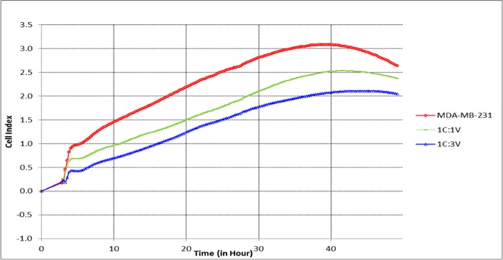
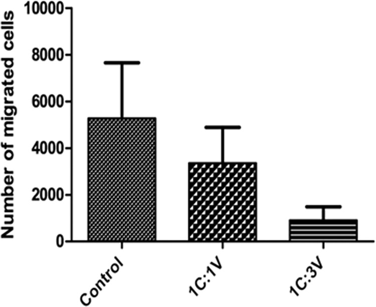
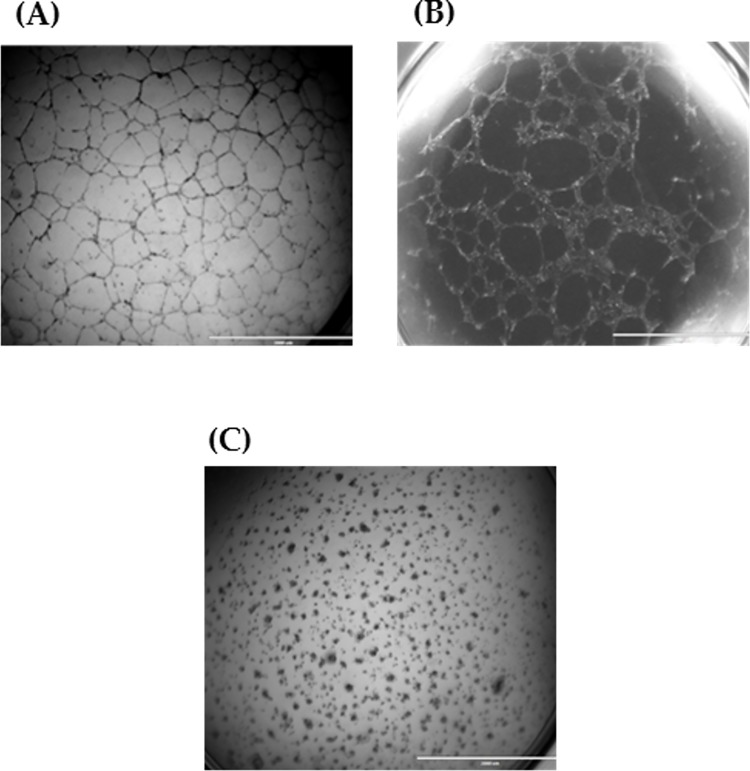
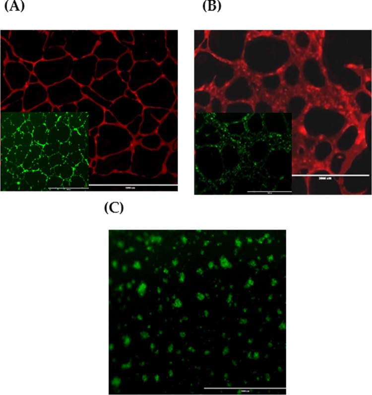
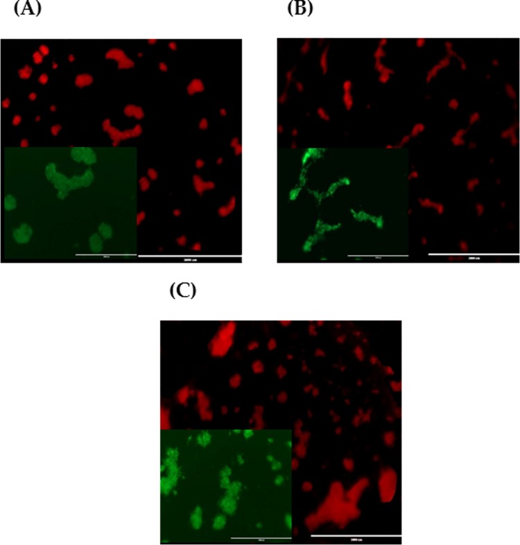
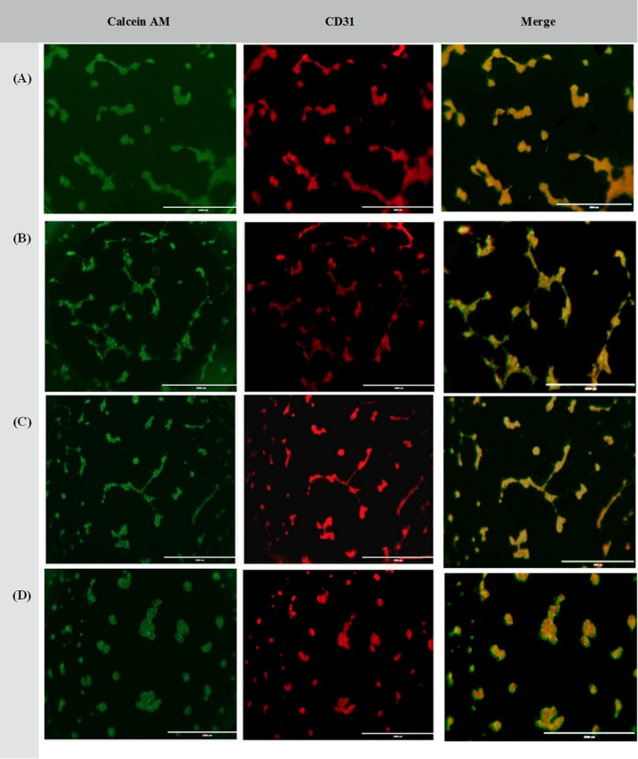
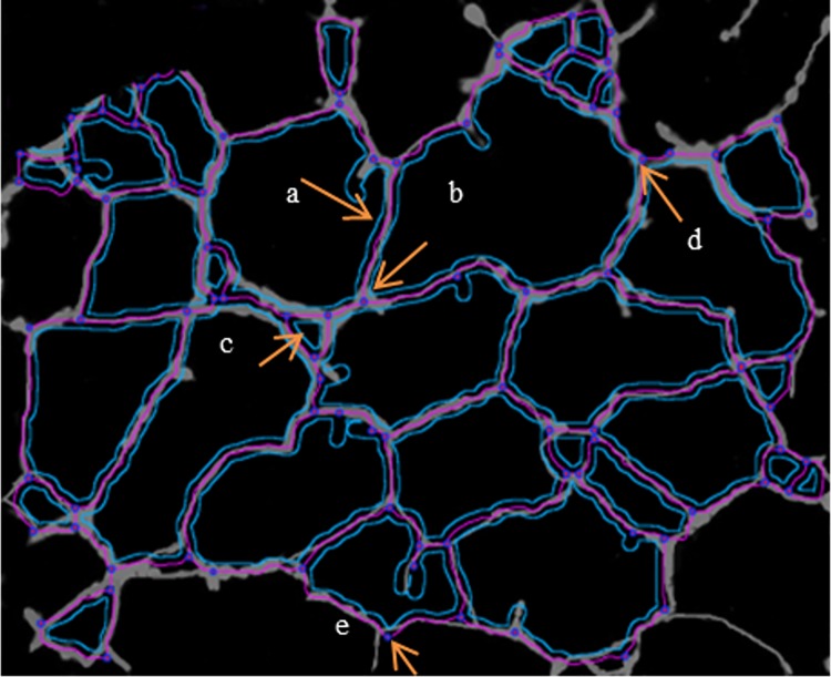
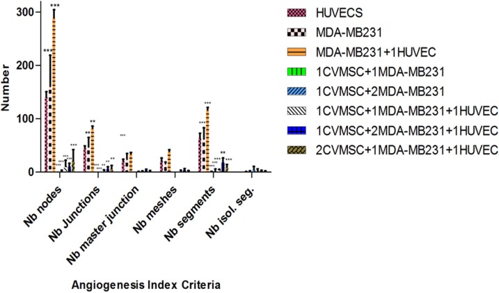
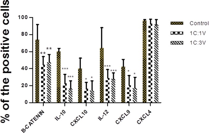
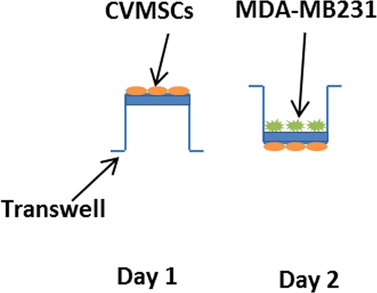
Mesenchymal stem cells (MSCs) can influence the tumour microenvironment (TEM) and play a major role in tumourigenesis. Triple-negative [Ostrogen receptor (ER-), Progesterone receptor (PgR-), and HER2/neu receptor (HER2-)] breast cancer (TNBC) is an aggressive class of BC characterized by poor prognosis and lacks the benefit of routinely available targeted therapies. This study aims to investigate the effect of human placental chorionic villi derived MSCs (CVMSCs) on the behavior of TNBC in vitro. This was done by assaying different cancer hallmarks including proliferation, migration and angiogenesis. Cell proliferation rate of TNBC cell line (MDA-MB231) was monitored in real time using the xCELLigence system. Whereas, Boyden chamber migration assay was used to measure MDA-MB231 motility and invasiveness toward CVMSCs. Finally, a three-dimensional (3D) model using a co-culture system of CVMSCs with MDA-MB231 with or without the addition of human umbilical vein endothelial cells (HUVECs) was created to assess tumour angiogenesis in vitro. CVMSCs were able to significantly reduce the proliferative and migratory capacity of MDA-MB231 cells. Co-culturing of MDA-MB231 with CVMSCs, not only inhibited the tube formation ability of HUVECs but also reduced the expression of the BC characteristic cytokines; IL-10, IL-12, CXCL9 and CXCL10 of CVMSCs. These results support the hypothesis that CVMSCs can influence the behavior of TNBC cells and provides a basic for a potential therapeutic approach in a pre-clinical settings. The data from this study also highlight the complexity of the in vitro cancer angiogenesis model settings and regulations.


Conflict of interest statement

The authors have declared that no competing interests exist.

Figures

Fig 1
Fig 1. Validation of MSCs markers by flow cytometry.
MSCs found positive for the following markers CD144, CD90, CD105, and CD166. In addition, MSCs were negative for HLA-DR, and CD-14 markers. These results are typical for MSCs validation assay.
Fig 2
Fig 2. Nestin validation to confirm MSCs ability to differentiate into neurons.
MSCs were differentiated into neurons through neuronal specific media. Top raw shows differentiated MSCs which are nestin, a neural positive marker (indicated by the green dye). Meddle raw shows positive control U87 (Neural cell line) which are also nestin positive, green dye. Bottom raw shows negative stain for nestin on undifferentiated MSCs (Indicated by the absence of color).
Fig 3
Fig 3. The effect of CVMSCs on MDA-MB231 proliferation.
The effect of CVMSCs on MDA-MB231 cells proliferation was assessed in vitro over 50 hour. C is MDA-MB231, and V is CVMSC. MDA-MB231 cells were pretreated with CVMSCs through a transwell membrane. The reduction in the proliferative capacity of MDA-MB231 induced by CVMSCs was statistically significant (P<0.0001) compared to the control; untreated MDA-MB231). Each experiment was done in quadruplicate.
Fig 4
Fig 4. The effect of CVMSCs on MDA-MB231 cell migration using Boyden chamber migration assay.
Where the control is C (MDA-MB231), and V is human placental chorionic villi derived MSC. The reduction in the migration response induced by CVMSCs was statistically significant (P = 0.037). Each experiment was repeated 3 times.
Fig 5
Fig 5. Tube formation of individual cell population.
(A) HUVECs; (B) MDA-MB231 and; (C) CVMSCs on Matrigel with VEGF for 24h incubation. Tubes started to form following 14 hour of incubation. 2x magnification.
Fig 6
Fig 6. CD31 and Calcein AM staining of individual cell population.
(A) HUVECs; (B) MDA-MB231 and; (C) CVMSCs. Images show both CD31 (red) and Calcein AM staining (green) for HUVECs and MDA-MB231. Whereas CVMSCs, show no expression of CD31. (2x magnification for CD31 and 10x magnification for Calcein AM). 2x magnification for (C).
Fig 7
Fig 7. Tube formation of CVMSCs and MDA-MB231 co-culture at different ratios.
(A) Co-culture 1 CVMSCs and 1 MDA-MB231; (B) Co-culture 1 CVMSCs and 2 MDA-MB231 and; (C) Co-culture 3 CVMSCs and 1 MDA-MB231 on Matrigel with VEGF for 24h incubation. Images show CD31 (red) and Calcein AM staining (green) staining. 10x magnification for Calcein AM and 2x magnification for CD31.
Fig 8
Fig 8. Tube formation of CVMSCs, MDA-MB231 and HUVECs co-culture at different ratios.
(A) Co-culture of 1 HUVECs: 1CVMSCs and 1 MDA-MB231; (B) Co-culture of 1 HUVECs: 1 CVMSCs and 2MDA-MB231 and; (C) Co-culture of 1 HUVECs: 2 CVMSCs and 1 MDA-MB231 and; (D) Co-culture of 1 HUVECs: 3 CVMSCs and 1 MDA-MB231 on Matrigel with VEGF for 24h incubation. Images show CD31 (red) and Calcein AM (green) staining or both overlay at 10x magnification.
Fig 9
Fig 9. The detection of constitutive elements of the network—Summary.
A tree composed from master segment (a); associated by master junction (b); delimiting the meshes (c); junction (note that this pointed junction is composed by several nodes) (d); node identified as three neighbors (e).
Fig 10
Fig 10. Statistical computation of angiogenic parameters using angiogenesis analyzer macro connected image J software.
The bars show a comparison between untreated MDA-MB231 and treated MDA-MB231 with different ration of CVMSCs in the presence of HUVECs or not. Nb = number. All the groups were analyzed by Two-way ANOVA and each group was compared to the positive group (HUVECs). HUVEC = human vascular endothelial cells, CVMSC = of human placental chorionic villi derived MSCs.
Fig 11
Fig 11. Expression of cytokines and chemokines in pre-treated CVMSCs with MDA-MB231 through a transwell membrane.
Two-way ANOVA analyzed the two treated groups; 1:1 and 1:3 and each group was compared to the control. The control is CVMSCs without any treatment. C is MDA-MB231, V is CVMSCs. CVMSCs = human placental chorionic villi derived MSCs.
Fig 12
Fig 12. A direct contact between CVMSCs and MDA-MB231 through a transwell membrane.
CVMSCs were incubated overnight on the underside of the transwell. After 24 h the transwell was flipped over and MDA-MB231 cells were added on the upper chamber of the transwell and incubated for 48hr.

References

    1. Augello A, Kurth TB, De Bari C. Mesenchymal stem cells: a perspective from in vitro cultures to in vivo migration and niches. European cells & materials. 2010;20:121–33. Epub 2011/01/21. . - PubMed
    1. Krampera M, Pizzolo G, Aprili G, Franchini M. Mesenchymal stem cells for bone, cartilage, tendon and skeletal muscle repair. Bone. 2006;39(4):678–83. Epub 2006/06/13. 10.1016/j.bone.2006.04.020 . - DOI - PubMed
    1. Tanaka M, Miyajima A. Identification and isolation of adult liver stem/progenitor cells. Methods Mol Biol. 2012;826:25–32. Epub 2011/12/15. 10.1007/978-1-61779-468-1_3 . - DOI - PubMed
    1. Sakaguchi Y, Sekiya I, Yagishita K, Muneta T. Comparison of human stem cells derived from various mesenchymal tissues: superiority of synovium as a cell source. Arthritis and rheumatism. 2005;52(8):2521–9. Epub 2005/07/30. 10.1002/art.21212 . - DOI - PubMed
    1. Roubelakis MG, Pappa KI, Bitsika V, Zagoura D, Vlahou A, Papadaki HA, et al. Molecular and proteomic characterization of human mesenchymal stem cells derived from amniotic fluid: comparison to bone marrow mesenchymal stem cells. Stem cells and development. 2007;16(6):931–52. Epub 2007/12/01. 10.1089/scd.2007.0036 . - DOI - PubMed

Publication types