

Spectral features of nuclear DNA in human sperm assessed by Raman Microspectroscopy: Effects of UV-irradiation and hydration
- PMID: 30458032
- PMCID: PMC6245842
- DOI: 10.1371/journal.pone.0207786
Spectral features of nuclear DNA in human sperm assessed by Raman Microspectroscopy: Effects of UV-irradiation and hydration
Abstract
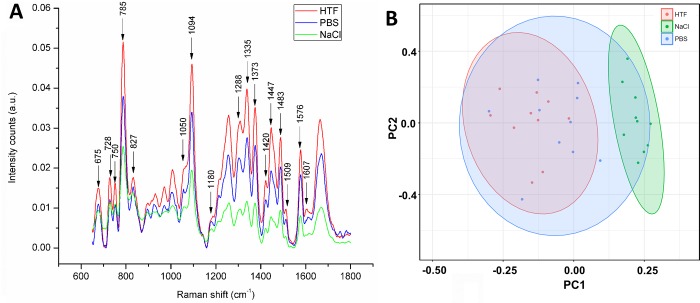
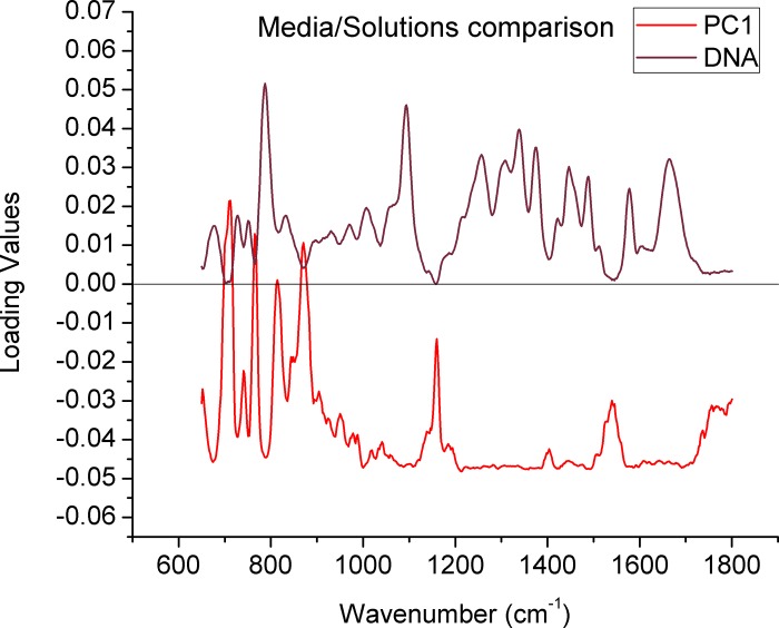
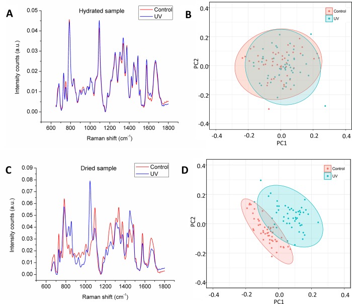
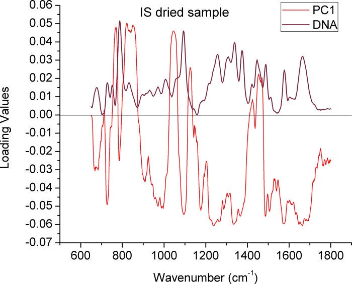
Raman Microspectroscopy represents an innovative tool for the assessment of sperm biochemical features otherwise undetectable by routine semen analysis. Previously, it was shown that induced DNA damage can be detected in smeared sperm by this technique. This novel readout may be of value for clinical settings especially if it can be transferred to living cells. Yet, starting with living sperms this study was carried-out using a variety of conditions to disclose the Raman features of sperm nuclei under different hydration conditions and UV exposure. Human sperm were immobilized and Raman spectra were obtained from individual sperm as repeated measurements. To create conditions with controlled DNA damage, sperm samples were exposed to ultraviolet light. Several media were used to evaluate their effect on Raman spectra in aqueous conditions. To substantiate differences between the experimental conditions, the spectra were analyzed by Principal Component Analysis. We observed that spectra of sperm nuclei obtained in different solutions showed a qualitatively unchanged spectral pattern showing the principal signals related to DNA. Evaluating the effect of ultraviolet light generated the finding that spectra representing DNA damage were only observed in dry conditions but not in aqueous medium. Thus, Raman microspectroscopy was successfully applied for sperm analysis in different conditions, among them in live spermatozoa in aqueous solution during the initial measurement, revealing the principle use of this technique. However, implementation of Raman spectroscopy as a technique for clinical sperm analysis and selection may be especially relevant when DNA evaluation can be established using live sperm.
Conflict of interest statement
The authors have declared that no competing interests exist.
Figures

References
-
- Sánchez V, Wistuba J, Mallidis C (2013) Semen analysis: update on clinical value, current needs and future perspectives. Reproduction 146: 249–258. - PubMed
-
- Wang C, Swerdloff RS (2014) Limitations of semen analysis as a test of male fertility and anticipated needs from newer tests. Fertility and Sterility 102: 1502–1507. 10.1016/j.fertnstert.2014.10.021 - DOI - PMC - PubMed
-
- Barratt CLR, Mansell SA (2013) Andrology is desperate for a new assay–Let us make sure we get it right this time…. Middle East Fertility Society Journal 18: 82–83.
-
- Tüttelmann F, Nieschlag E (2010) Classification of Andrological Disorders In: Nieschlag E, Behre HM, Nieschlag S, editors. Andrology: Male Reproductive Health and Dysfunction. Berlin, Heidelberg: Springer Berlin Heidelberg; pp. 87–92.
-
- Kliesch S (2014) Diagnosis of Male Infertility: Diagnostic Work-up of the Infertile Man. European Urology Supplements 13: 73–82.
Publication types
MeSH terms
Substances
LinkOut - more resources
Full Text Sources
Other Literature Sources
