Review
. 2018 Nov 19;28(22):R1313-R1324.
doi: 10.1016/j.cub.2018.09.018. Epub 2018 Nov 19.

Sex Chromosome Effects on Male-Female Differences in Mammals

Affiliations
Review

Sex Chromosome Effects on Male-Female Differences in Mammals

Daniel M Snell et al. Curr Biol. .

Abstract

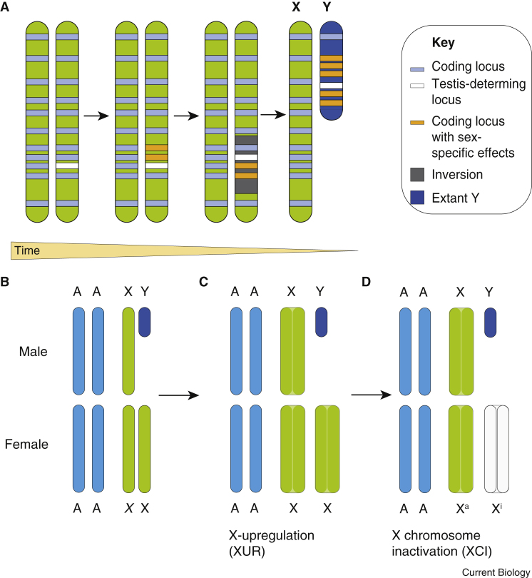
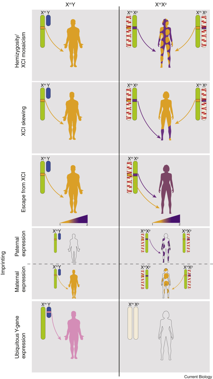
Fundamental differences exist between males and females, encompassing anatomy, physiology, behaviour, and genetics. Such differences undoubtedly play a part in the well documented, yet poorly understood, disparity in disease susceptibility between the sexes. Although traditionally attributed to gonadal sex hormone effects, recent work has begun to shed more light on the contribution of genetics - and in particular the sex chromosomes - to these sexual dimorphisms. Here, we explore the accumulating evidence for a significant genetic component to mammalian sexual dimorphism through the paradigm of sex chromosome evolution. The differences between the extant X and Y chromosomes, at both a sequence and regulatory level, arose across 166 million years. A functional result of these differences is cell autonomous sexual dimorphism. By understanding the process that changed a pair of homologous ancestral autosomes into the extant mammalian X and Y, we believe it easier to consider the mechanisms that may contribute to hormone-independent male-female differences. We highlight key roles for genes with homologues present on both sex chromosomes, where the X-linked copy escapes X chromosome inactivation. Finally, we summarise current experimental paradigms and suggest areas for developments to further increase our understanding of cell autonomous sexual dimorphism in the context of health and disease.


Figures

Figure 1
Figure 1
The evolution of the mammalian sex chromosomes and dosage compensation mechanisms. (A) A testis-determining locus (proto-SRY, white) was acquired on an autosome around 148–166 million years ago. Sexually antagonistic alleles (orange) then evolved at nearby loci, selected for in males due to their tight linkage to SRY. Recombination suppression between the proto-X and -Y chromosomes likely followed on from chromosomal inversions (grey), which were subsequently only carried by males. Over evolutionary time, the lack of sexual recombination led to the appearance of repetitive DNA sequences and short-term expansion. In the longer term, large deletions took place. The outcome of this process is the small, relatively gene poor Y chromosome observed in most eutherian mammals today. Concurrent with this process, X upregulation (XUR) evolved to balance X gene dosage between the single X chromosome and the autosomes in males: this is depicted as the doubled surface area of the X chromosomes in (C) compared to (B). However, XUR was passed on to XX offspring, resulting in X:autosome dosage disparity between males and females. X chromosome inactivation (XCI) then evolved to repress one of the two X chromosomes in XX cells (D). This is depicted as the loss of colour of the X chromosome. Abbreviations: Xa, active X chromosome; Xi, inactive X chromosome.
Figure 2
Figure 2
Possible mechanisms underlying male–female genetic sexual dimorphism in eutherian mammals. The organism-wide expression of an individual gene allele is represented by block colour, with XY males in the left-hand column and XX females in the right-hand column. (A) A single allele of an X-linked gene is expressed in all cells in the male, whereas due to X chromosome inactivation (XCI), the same allele is only expressed in 50% of cells in the female. (B) XCI skewing can result in a change to the percentage of cells expressing any given X allele in females. (C) As both alleles of XCI escapee genes are expressed in females, the relative expression is increased compared to males. (D) Imprinting resulting in Xp allelic expression would be absent in males due to the absence of Xp, and would be present in 50% of cells in females. Imprinting resulting in Xm allelic expression would be present in all cells in males and 50% of cells in females. (E) Ubiquitously expressed Y-linked genes are only present in males. Abbreviations: Xm, maternally derived X chromosome; Xp, paternally derived X chromosome. Gene expression is depicted in arbitrary units, taking 1 as normal expression for a single chromosome.
Figure 3
Figure 3
Genes escaping XCI are implicated in a range of sexually dimorphic diseases. A small number of X-linked genes show ubiquitous expression, have extant Y-linked homologues, and escape XCI. These genes have roles in the regulation of gene expression and are implicated in male–female sexual dimorphism in a wide range of diseases. Specific organs affected are indicated by the gene-related colours, i.e. KDM5C in orange.
Figure 4
Figure 4
The Four Core Genotypes (FCG) model. In this Punnett square, the maternal genotype is depicted on the left-hand side and the paternal genotype is depicted at the top. A gamete from each parent carries a single sex chromosome, which come together to create the two possible offspring genotypes, XX (green column) and XY (blue column). Furthermore, the father carries Sry as a transgene, the inheritance of which determines the gonadal sex of the offspring: female above the dashed line (XX, XY), and male below the dashed line (XXSry, XYSry). The FCG model can therefore be used to separate sex chromosome effects (XX, green; XY, blue) from gonadal sex hormone effects (ovarian hormones above dotted line, and testicular hormones below dotted line) in mouse.

References

    1. Randall J.C., Winkler T.W., Kutalik Z., Berndt S.I., Jackson A.U., Monda K.L., Kilpeläinen T.O., Esko T., Mägi R., Li S. Sex-stratified genome-wide association studies including 270,000 individuals show sexual dimorphism in genetic loci for anthropometric traits. PLoS Genet. 2013;9:e1003500. - PMC - PubMed
    1. Plavcan J.M. Sexual dimorphism in primate evolution. Am. J. Phys. Anthropol. 2002;116:25–53. - PubMed
    1. Arnold A.P. Sex chromosomes and brain gender. Nat. Rev. Neurosci. 2004;5:701–708. - PubMed
    1. Burgoyne P.S., Thornhill A.R., Boudrean S.K., Darling S.M., Bishop C.E., Evans E.P., Capel B., Mittwoch U. The genetic basis of XX-XY differences present before gonadal sex differentiation in the mouse [and Discussion] Philos. Trans. R. Soc. Lond. B Biol. Sci. 1995;350:253–261. - PubMed
    1. Cui W., Ma C.-X., Tang Y., Chang V., Rao P.V., Ariet M., Resnick M.B., Roth J. Sex differences in birth defects: A study of opposite-sex twins. Birth Defect Res. A Clin. Mol. Teratol. 2005;73:876–880. - PubMed

Publication types

LinkOut - more resources