Pharmacological stress impairs working memory performance and attenuates dorsolateral prefrontal cortex glutamate modulation
- PMID: 30458306
- PMCID: PMC6491044
- DOI: 10.1016/j.neuroimage.2018.11.017
Pharmacological stress impairs working memory performance and attenuates dorsolateral prefrontal cortex glutamate modulation
Abstract
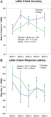
Working memory processes are associated with the dorsolateral prefrontal cortex (dlPFC). Prior research using proton functional magnetic resonance spectroscopy (1H fMRS) observed significant dlPFC glutamate modulation during letter 2-back performance, indicative of working memory-driven increase in excitatory neural activity. Acute stress has been shown to impair working memory performance. Herein, we quantified dlPFC glutamate modulation during working memory under placebo (oral lactose) and acute stress conditions (oral yohimbine 54 mg + hydrocortisone 10 mg). Using a double-blind, randomized crossover design, participants (N = 19) completed a letter 2-back task during left dlPFC 1H fMRS acquisition (Brodmann areas 45/46; 4.5 cm3). An automated fitting procedure integrated with LCModel was used to quantify glutamate levels. Working memory-induced glutamate modulation was calculated as percentage change in glutamate levels from passive visual fixation to 2-back levels. Results indicated acute stress significantly attenuated working memory-induced glutamate modulation and impaired 2-back response accuracy, relative to placebo levels. Follow-up analyses indicated 2-back performance significantly modulated glutamate levels relative to passive visual fixation during placebo but not acute stress. Biomarkers, including blood pressure and saliva cortisol, confirmed that yohimbine + hydrocortisone dosing elicited a significant physiological stress response. These findings support a priori hypotheses and demonstrate that acute stress impairs dlPFC function and excitatory activity. This study highlights a neurobiological mechanism through which acute stress may contribute to psychiatric dysfunction and derail treatment progress. Future research is needed to isolate noradrenaline vs. cortisol effects and evaluate anti-stress medications and/or behavioral interventions.
Keywords: (1)H fMRS; Dorsolateral prefrontal cortex; Glutamate; Noradrenaline; Stress; Working memory.
Copyright © 2018 Elsevier Inc. All rights reserved.
Conflict of interest statement
Conflict of Interest
All authors declare no conflict of interest with respect to the conduct or content of this work.
Figures


Similar articles
-
A neurobiological correlate of stress-induced nicotine-seeking behavior among cigarette smokers.Addict Biol. 2020 Jul;25(4):e12819. doi: 10.1111/adb.12819. Epub 2019 Aug 16. Addict Biol. 2020. PMID: 31418989 Free PMC article.
-
Working Memory Modulates Glutamate Levels in the Dorsolateral Prefrontal Cortex during 1H fMRS.Front Psychiatry. 2018 Mar 6;9:66. doi: 10.3389/fpsyt.2018.00066. eCollection 2018. Front Psychiatry. 2018. PMID: 29559930 Free PMC article.
-
Transcranial Stimulation of the Dorsolateral Prefrontal Cortex Prevents Stress-Induced Working Memory Deficits.J Neurosci. 2016 Jan 27;36(4):1429-37. doi: 10.1523/JNEUROSCI.3687-15.2016. J Neurosci. 2016. PMID: 26818528 Free PMC article. Clinical Trial.
-
Stress and Inflammation Target Dorsolateral Prefrontal Cortex Function: Neural Mechanisms Underlying Weakened Cognitive Control.Biol Psychiatry. 2025 Feb 15;97(4):359-371. doi: 10.1016/j.biopsych.2024.06.016. Epub 2024 Jun 27. Biol Psychiatry. 2025. PMID: 38944141 Review.
-
Unusual Molecular Regulation of Dorsolateral Prefrontal Cortex Layer III Synapses Increases Vulnerability to Genetic and Environmental Insults in Schizophrenia.Biol Psychiatry. 2022 Sep 15;92(6):480-490. doi: 10.1016/j.biopsych.2022.02.003. Epub 2022 Feb 12. Biol Psychiatry. 2022. PMID: 35305820 Free PMC article. Review.
Cited by
-
Treatment evaluation of Kami Guibi-tang on participants with amnestic mild cognitive impairment using magnetic resonance imaging on brain metabolites, gamma-aminobutyric acid, and cerebral blood flow.J Appl Clin Med Phys. 2021 Nov;22(11):151-164. doi: 10.1002/acm2.13443. Epub 2021 Oct 11. J Appl Clin Med Phys. 2021. PMID: 34633758 Free PMC article.
-
Function and biochemistry of the dorsolateral prefrontal cortex during placebo analgesia: how the certainty of prior experiences shapes endogenous pain relief.Cereb Cortex. 2023 Aug 23;33(17):9822-9834. doi: 10.1093/cercor/bhad247. Cereb Cortex. 2023. PMID: 37415068 Free PMC article.
-
A neurobiological correlate of stress-induced nicotine-seeking behavior among cigarette smokers.Addict Biol. 2020 Jul;25(4):e12819. doi: 10.1111/adb.12819. Epub 2019 Aug 16. Addict Biol. 2020. PMID: 31418989 Free PMC article.
-
Long-term effects of glucocorticoid excess on the brain.J Neuroendocrinol. 2022 Aug;34(8):e13142. doi: 10.1111/jne.13142. Epub 2022 Aug 18. J Neuroendocrinol. 2022. PMID: 35980208 Free PMC article. Review.
-
Functional MRS studies of GABA and glutamate/Glx - A systematic review and meta-analysis.Neurosci Biobehav Rev. 2023 Jan;144:104940. doi: 10.1016/j.neubiorev.2022.104940. Epub 2022 Nov 2. Neurosci Biobehav Rev. 2023. PMID: 36332780 Free PMC article.
References
-
- Apšvalka D, Gadie A, Clemence M, Mullins PG, 2015. Event-related dynamics of glutamate and BOLD effects measured using functional magnetic resonance spectroscopy (fMRS) at 3T in a repetition suppression paradigm. NeuroImage 118, 292–300. - PubMed
-
- Arnsten AF, Mathew R, Ubriani R, Taylor JR, Li B-M, 1999. α−1 noradrenergic receptor stimulation impairs prefrontal cortical cognitive function. Biological Psychiatry 45, 26–31. - PubMed
-
- Birnbaum S, Gobeske KT, Auerbach J, Taylor JR, Arnsten AF, 1999. A role for norepinephrine in stress-induced cognitive deficits: α−1-adrenoceptor mediation in the prefrontal cortex. Biological Psychiatry 46, 1266–1274. - PubMed
Publication types
MeSH terms
Substances
Grants and funding
LinkOut - more resources
Full Text Sources
Medical

