. 2018 Dec 3;47(5):564-575.e5.
doi: 10.1016/j.devcel.2018.09.024. Epub 2018 Oct 25.

A Hippo-like Signaling Pathway Controls Tracheal Morphogenesis in Drosophila melanogaster

Affiliations

A Hippo-like Signaling Pathway Controls Tracheal Morphogenesis in Drosophila melanogaster

Carole L C Poon et al. Dev Cell. .

Abstract

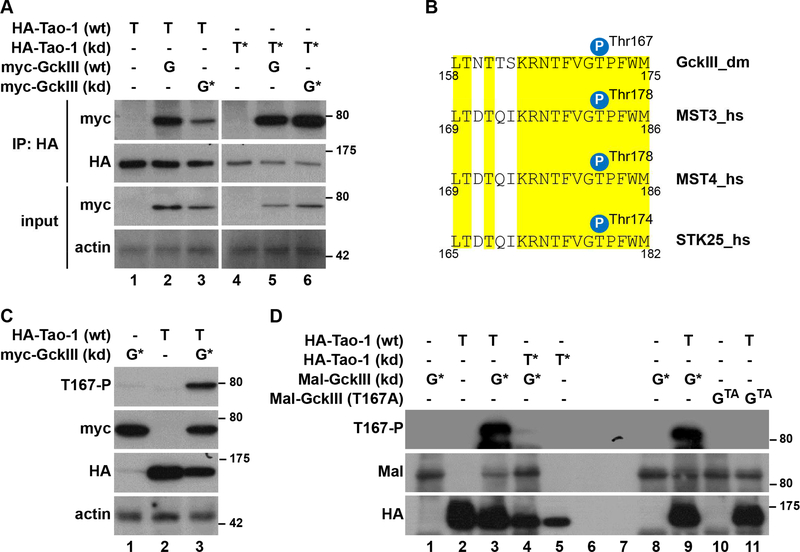
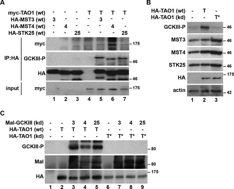
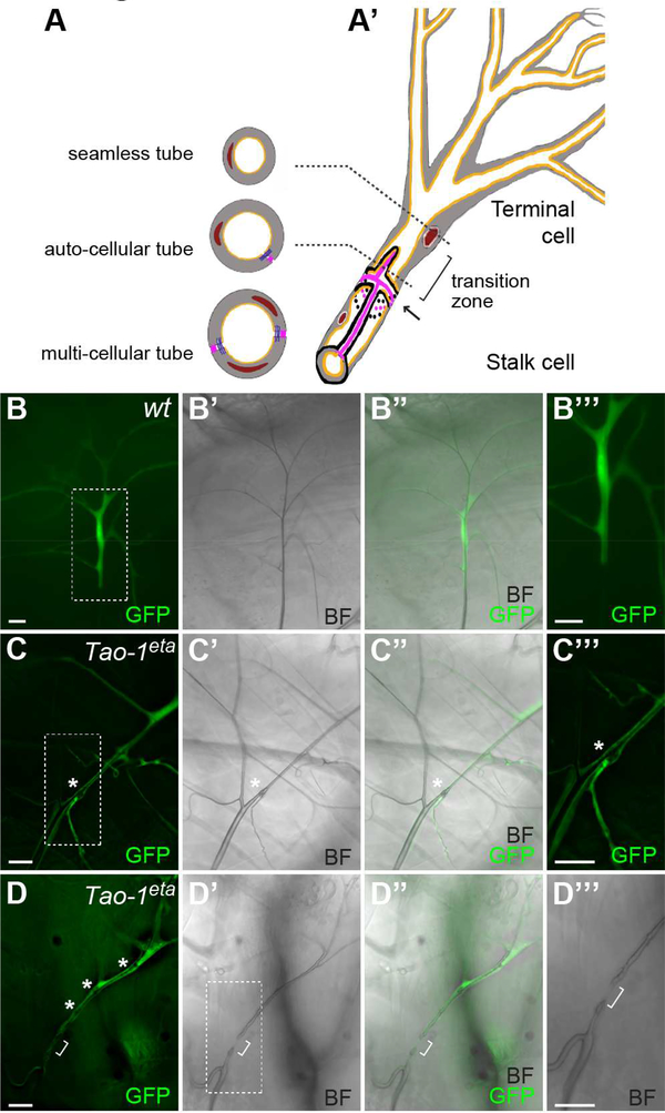
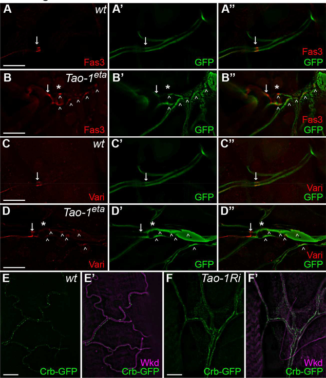
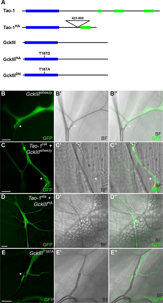
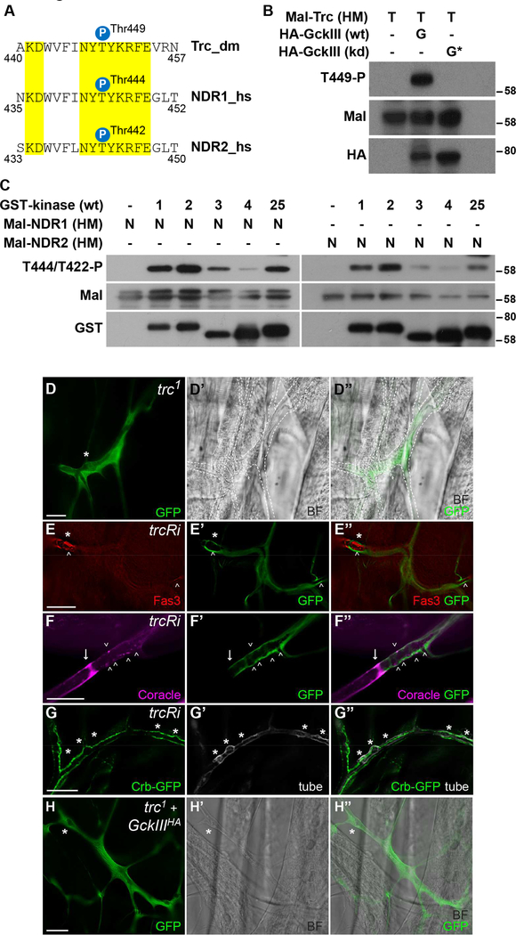
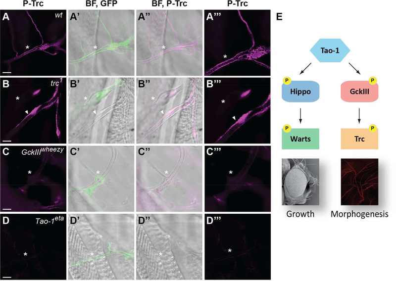
Hippo-like pathways are ancient signaling modules first identified in yeasts. The best-defined metazoan module forms the core of the Hippo pathway, which regulates organ size and cell fate. Hippo-like kinase modules consist of a Sterile 20-like kinase, an NDR kinase, and non-catalytic protein scaffolds. In the Hippo pathway, the upstream kinase Hippo can be activated by another kinase, Tao-1. Here, we delineate a related Hippo-like signaling module that Tao-1 regulates to control tracheal morphogenesis in Drosophila melanogaster. Tao-1 activates the Sterile 20-like kinase GckIII by phosphorylating its activation loop, a mode of regulation that is conserved in humans. Tao-1 and GckIII act upstream of the NDR kinase Tricornered to ensure proper tube formation in trachea. Our study reveals that Tao-1 activates two related kinase modules to control both growth and morphogenesis. The Hippo-like signaling pathway we have delineated has a potential role in the human vascular disease cerebral cavernous malformation.

Keywords: Hippo-like pathways; kinases; morphogenesis; signaling; trachea; tubular networks.


Conflict of interest statement

DECLARATION OF INTERESTS

The authors declare no competing interests.

Figures

Figure 1.
Figure 1.. D. melanogaster Tao-1 kinase binds and phosphorylates GckIII kinase on its activation loop.
(A) Lysates of D. melanogaster S2R+ cells expressing wild-type (wt) or kinase dead (kd) HA-Tao-1 together with wt or kd myc-GckIII were subjected to immunoprecipitation (IP) using anti-HA antibodies. Immunoprecipitates and input lysates were analysed by immunoblotting with the indicated antibodies. (B) Alignment of the conserved T-loop of D. melanogaster (dm) and human (hs) GCKIII kinase sequences. Identical residues are highlighted in yellow. The positions of the regulatory T-loop phosphorylation sites are indicated. The T-loop phosphorylation site of GCKIII kinases is conserved in flies and humans. (C) S2R+ cells were transfected with the indicated plasmids and immunoblotted using anti-T167-P (top), anti-myc (top middle), anti-HA (bottom middle) and anti-actin (bottom) antibodies. (D) Lysates of S2R+ cells transiently expressing HA-Tao-1 variants were subjected to immunoprecipitation with anti-HA antibodies. Immunopurified proteins were then used in kinase assays with or without or with the indicated recombinant versions of Mal-GckIII. Following kinase reactions, the samples were immunoblotted with the indicated antibodies. Importantly, the antiT167-P antibody specifically detected Tao1-mediated phosphorylation of GckIII on Thr167 (compare lanes 3, 9 and 11). Relative molecular masses are shown in kDa for each blot. See also Table S1.
Figure 2.
Figure 2.. TAO1 kinase binds and phosphorylates GCKIII kinases on their activation loop in human cells.
(A) Lysates of human HEK293 cells expressing HA-GCKIII kinases (MST3, MST4 or STK25) together with myc-TAO1 were subjected to immunoprecipitation using anti-HA antibodies, before immunoblotting with the indicated antibodies. (B) HEK293 cells expressing wt or kd human TAO1 were lysed and immunoblotted with the indicated antibodies. (C) Lysates of HEK293 cells expressing human wild-type (wt) or kinase dead (kd) HA-TAO1 were subjected to immunoprecipitation with anti-HA antibodies. Immunopurified kinases were used in kinase assays with or without the indicated recombinant wt or kd Mal-GCKIII kinases. Following kinase reactions, samples were examined by immunoblotting with the indicated antibodies. Importantly, TAO1-mediated activation loop phosphorylation of human GCKIII kinases was only observed when wild-type TAO1 was incubated with kinase-dead GCKIII kinases. Relative molecular masses are shown in kDa for each blot.
Figure 3.
Figure 3.. Tao-1 regulates development of D. melanogaster trachea.
(A-A’) Schematic illustration of D. melanogaster trachea, which are comprised of three different tube architectures: seamless tubes, auto-cellular tubes and multi-cellular tubes (cross sections represented in (A)). (A’) A depiction of part of a terminal cell (a seamless tube) that joins to an autocellular stalk cell at the transition zone (marked by bracket), which is located between an intracellular junction (marked by arrow) and the nucleus. Nucleus is brown; cytoplasm is grey; septate junctions are pink; adherens junctions are purple; apical lumens are orange (adapted from (Song et al., 2013)). (B-D”’) Wild-type (B-B”’) or Tao-1eta (C-D”’) terminal cell clones, marked by GFP (green). Boxed areas in (B), (C) and (D’) are shown as close up images in (B”’), (C”’) and (D”’). Brightfield (BF) images are shown in (B’), (C’), (D’) and (D”’) and the merged images (B”), (C”) and (D”). (B-B”’) Gas-filled tubes that pierce the cytoplasm are evident in the wild-type terminal cell. The tube extends beyond the clone through the unmarked neighbouring stalk cell. (C-C”’) Loss of Tao-1 results in prominent dilations (*) in the transition zone of 73% of terminal cells scored (D-D”’) A Tao-1eta terminal cell clone that displays a “gap” defect, in which a portion of the terminal cell tube is absent within the transition zone (bracket). The cell also displays several small tube dilations (marked by *). Scale bar = 20 μm in (A-D”’). See also Figures S1-S3 and Table S2.
Figure 4.
Figure 4.. Tao-1 prevents ectopic accumulation of septate junction proteins and Crumbs in trachea.
(A-B”) Wild-type (A-A”) or Tao-1eta (B-B”) terminal cell clones, marked by GFP (green) and stained for antibodies specific for Fas3 (red). The arrow indicates the intercellular junction. Ectopic Fas3 is present in the transition zone dilation (indicated by *) of Tao-1eta terminal cell clones and in more distal positions of the cell (indicated by arrowheads, B). (C-D”) Wild-type (C-C”) or Tao-1eta (D-D”) terminal cell clones, marked by GFP (green) and stained for antibodies specific for Varicose (Vari, red). The arrow indicates the intercellular junction. Ectopic Vari is present in the transition zone dilation (indicated by *) of Tao-1eta terminal cell clones and more distally as well (arrowheads, D). (E-E’) A wild-type terminal cell with Crumbs-GFP (green) and stained with antibodies specific for Wkd (magenta) to mark the tracheal tubes. Crumbs decorates the lumenal membrane in well spaced foci. (F-F’) A terminal cell expressing Tao-1 RNAi with btl-Gal4. Crumbs-GFP levels were elevated and appeared to almost continuously line the lumenal membrane.
Figure 5.
Figure 5.. GckIII functions epistatically to Tao-1 in trachea
(A) Schematic of Tao-1 and GckIII wild-type and mutant transgenes utilised for genetic epistasis experiments. Blue boxes indicate a kinase domain, and green boxes indicate coiled-coil domains. Wild type Tao-1 contains a kinase domain at the N-terminus and 3 coiled coil domains at the C-terminus. Hyperactive Tao-1 (Tao-1HA) contains a deletion in the central region (amino acids 423–900). Wild type GckIII contains a kinase domain at the N terminus; hyperactive GckIII (GckIIIHA) contains a point mutation where T is mutated to D to mimic phosphorylation at 167 in the activation loop of the kinase domain; and dominant negative GckIII (GckIIIDN) contains a point mutation where T is mutated to A to prevent phosphorylation at 167 in the activation loop of the kinase domain. (B-E) Terminal cell clones of the indicated genotypes are marked by GFP (green) with accompanying brightfield images (BF). Transition zone tube dilations are marked by an asterisk (*). (B-B”) GckIIIwheezy terminal cell clones, marked by GFP (green). Transition zone tube dilations (*) are observed in 100% of GckIIIwheezy mutant clones scored. (C-C”) A GckIIIwheezy terminal cell clone (marked by GFP) expressing a hyperactive Tao-1 (Tao-1HA) transgene. Expression of hyperactive Tao-1 failed to rescue transition zone dilations in GckIIIwheezy mutant cells, and transition zone tube dilations were observed in 100% terminal cells scored. (D-D”) A Tao-1eta terminal cell clone (marked by GFP) expressing a hyperactive GckIII (GckIIIHA) transgene. Expression of hyperactive GckIIIHA in Tao-1eta mutant clones caused transition zone tube dilations in 51% terminal cells scored. (E-E”) Expression of a GckIII transgene bearing the point mutation T167A in the activation loop (GckIIIT167A) results in transition zone tube dilations in 66% of terminal cells scored. Scale bars = 20 μm. See also Figure S4 and Table S2.
Figure 6.
Figure 6.. The Tricornered kinase is phosphorylated on its hydrophobic motif by GckIII, and regulates tracheal development.
(A) Alignment of the hydrophobic motif of D. melanogaster (dm) and human (hs) NDR kinase sequences. Identical residues are highlighted in yellow. The positions of the regulatory hydrophobic motif phosphorylation sites are indicated. The hydrophobic motif phosphorylation site of Trc/NDR kinases is conserved from flies to humans. (B) Lysates of S2R+ cells transiently expressing wild-type (wt) or kinase-dead (kd) HA-GckIII were immunoprecipitated with anti-HA antibodies and incubated with a recombinant Mal-tagged hydrophobic motif fragment of Trc [Mal-Trc(HM)] in kinase assays. Subsequently, samples were examined by immunoblotting using the indicated antibodies. Relative molecular masses are shown in kDa for each blot. (C) Recombinant GST-tagged wild-type full-length MST kinases (MST [1], MST2 [2], MST3 [3], MST4 [4] or STK25 [25], with respective abbreviations in square brackets) were incubated with recombinant Mal-tagged hydrophobic motif fragments of human NDR1 or NDR2 [Mal-NDR1/2 (HM)]. Following kinase reactions, the samples were analysed by Western blotting using the indicated antibodies. Relative molecular masses are shown in kDa for each blot. (D-D”) A trc1 terminal cell clone marked by GFP (green). Brightfield (BF) images are shown in (D’) and the merged image (D”). The * indicates a prominent transition zone dilation. The terminal cell also exhibits a gas-filling defect and so is outlined with a white dashed line in (C’ and C”). (E-E”) Terminal cell expressing a trc-RNAi (trcRi) transgene using btl-GAL4 (marked by GFP) and stained for Fas3 (red). Large dilations (*) in the transition zone were readily detected, and ectopic Fas3 is present in the transition zone dilation (arrowheads) of Tao-1eta terminal cell clones and more distally as well (arrowheads). (F-F”) Terminal cells marked with GFP (green) expressing a trc-RNAi (trcRi) transgene using drm-GAL4 and stained for Coracle (magenta) displayed ectopic localization of Coracle in the transition zone (arrowheads) and distally (data not shown). Arrows mark intercellular junction. (G-G”) Terminal cell (tubes visualised with UV) expressing a trc-RNAi (trcRi) transgene using drm-GAL4 with Crumbs-GFP almost continuously lined the lumenal membrane. (H-H”) A trc1 terminal cell clone [marked by GFP (green)] expressing a hyperactive GckIII transgene. A brightfield (BF) image is shown in (H’) and the merged image (H”). The * indicates a transition zone tube dilation; expression of hyperactive GckIII failed to rescue transition zone dilations in trc1 cells. Scale bar = 20 μm in (D-H). See also Figure S5 and Table S2.
Figure 7.
Figure 7.. Tricornered activity in trachea requires Tao-1 and GckIII.
(A-D) Third instar larval trachea stained with antibodies that recognise phosphorylation of the activation loop of Trc (P-Trc). GFP positive terminal cells are shown of the following genotypes: (A-A”’) wild-type; (B-B”’) trc1; (C-C”’) GckIIIwheezy; (D-D”’) Tao-1eta. P-Trc is in magenta (A-D). Clones are marked by GFP (green) in panels merged with brightfield images (A’, B’, C’, D’). P-Trc (magenta) is merged with brightfield images (A”, B”, C”, D”). Close-up images of P-Trc (magenta) are shown in (A”’, B”’, C”’, D”’). GFP positive terminal cells are indicated by arrowheads. P-Trc is evident in puncta that line the apical membrane of wild-type and control tracheal lumens. P-Trc staining is reduced or absent in trachea that are mutant for trc, GckIII or Tao-1 in (B–D. In (B-B”’) the asterisk indicates a control GFP negative terminal cell which stains positive for P-Trc and serves as an internal control. Scale bars = 10 μm. (E) Tao-1 regulates two Hippo-like signalling modules. In addition to regulating Hippo-Warts signalling in organ growth, we have discovered that Tao-1 kinase regulates a related Hippo-like pathway consisting of GckIII and Trc in morphogenesis.

References

    1. AVRUCH J, ZHOU D, FITAMANT J, BARDEESY N, MOU F & BARRUFET LR (2012). Protein kinases of the Hippo pathway: regulation and substrates. Semin Cell Dev Biol, 23, 770–84. - PMC - PubMed
    1. BACHMANN A, DRAGA M, GRAWE F & KNUST E (2008). On the role of the MAGUK proteins encoded by Drosophila varicose during embryonic and postembryonic development. BMC Dev Biol, 8, 55. - PMC - PubMed
    1. BAER MM, BILSTEIN A & LEPTIN M (2007). A clonal genetic screen for mutants causing defects in larval tracheal morphogenesis in Drosophila. Genetics, 176, 2279–91. - PMC - PubMed
    1. BARDIN AJ & AMON A (2001). Men and sin: what’s the difference? Nat Rev Mol Cell Biol, 2, 815–26. - PubMed
    1. BEITEL GJ & KRASNOW MA (2000). Genetic control of epithelial tube size in the Drosophila tracheal system. Development, 127, 3271–82. - PubMed

Publication types