. 2018 Nov 20;9(6):e01524-18.
doi: 10.1128/mBio.01524-18.

Molecular Basis for the Evolution of Species-Specific Hemoglobin Capture by Staphylococcus aureus

Affiliations

Molecular Basis for the Evolution of Species-Specific Hemoglobin Capture by Staphylococcus aureus

Jacob E Choby et al. mBio. .

Abstract

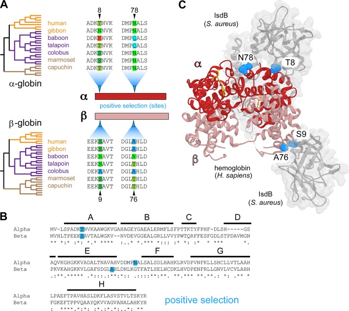
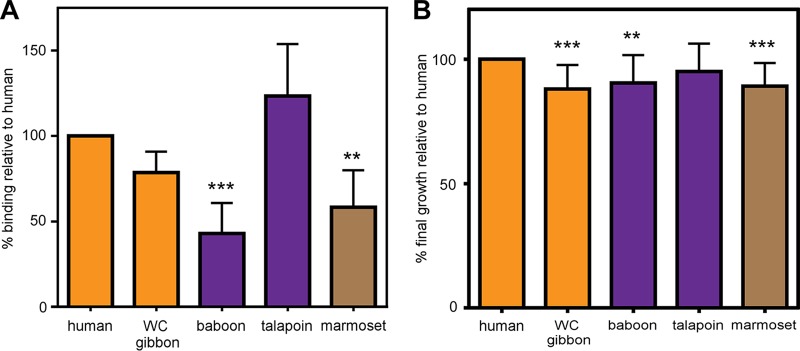
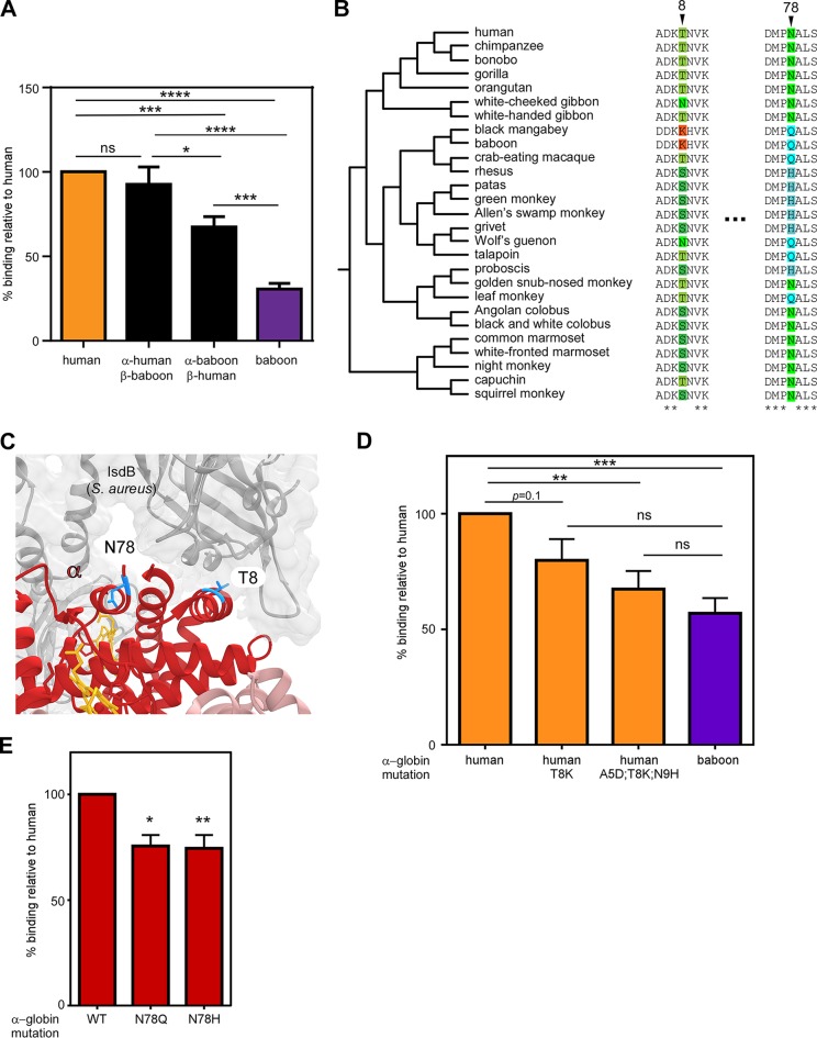
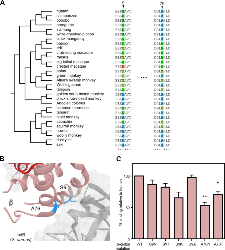
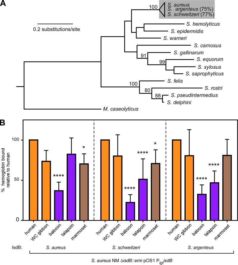
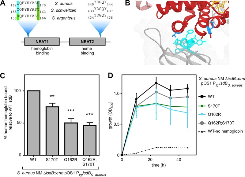
Metals are a limiting resource for pathogenic bacteria and must be scavenged from host proteins. Hemoglobin provides the most abundant source of iron in the human body and is required by several pathogens to cause invasive disease. However, the consequences of hemoglobin evolution for bacterial nutrient acquisition remain unclear. Here we show that the α- and β-globin genes exhibit strikingly parallel signatures of adaptive evolution across simian primates. Rapidly evolving sites in hemoglobin correspond to binding interfaces of IsdB, a bacterial hemoglobin receptor harbored by pathogenic Staphylococcus aureus Using an evolution-guided experimental approach, we demonstrate that the divergence between primates and staphylococcal isolates governs hemoglobin recognition and bacterial growth. The reintroduction of putative adaptive mutations in α- or β-globin proteins was sufficient to impair S. aureus binding, providing a mechanism for the evolution of disease resistance. These findings suggest that bacterial hemoprotein capture has driven repeated evolutionary conflicts with hemoglobin during primate descent.IMPORTANCE During infection, bacteria must steal metals, including iron, from the host tissue. Therefore, pathogenic bacteria have evolved metal acquisition systems to overcome the elaborate processes mammals use to withhold metal from pathogens. Staphylococcus aureus uses IsdB, a hemoglobin receptor, to thieve iron-containing heme from hemoglobin within human blood. We find evidence that primate hemoglobin has undergone rapid evolution at protein surfaces contacted by IsdB. Additionally, variation in the hemoglobin sequences among primates, or variation in IsdB of related staphylococci, reduces bacterial hemoglobin capture. Together, these data suggest that S. aureus has evolved to recognize human hemoglobin in the face of rapid evolution at the IsdB binding interface, consistent with repeated evolutionary conflicts in the battle for iron during host-pathogen interactions.

Keywords: Staphylococcus aureus; evolution; heme transport; hemoglobin; host-pathogen interactions; iron acquisition.


Figures

FIG 1
FIG 1
Parallel signatures of positive selection among primate hemoglobins at the bacterial IsdB binding interface. (A) Species phylogenies for the 27 α-globin and 30 β-globin orthologs analyzed (left). Alignments from representative species across hominoid (orange), Old World monkey (purple), and New World monkeys (brown) are shown, the full phylogenetic trees are displayed in Fig. 3 and 4. Amino acid sites under positive selection were identified in α-globin and β-globin (blue arrows) using PAML with both species and gene trees (posterior probabilities, >0.95). (B) Amino acid alignment of human α-globin and β-globin proteins. The position of conserved helices A to H are shown, and identity between globins is noted as conserved (*), highly similar (:), and similar (.). (C) The residues of α-globin (red) and β-globin (salmon) under positive selection (blue spheres) at the interface of hemoglobin capture by Staphylococcus aureus IsdB (gray) (PDB 5VMM).
FIG 2
FIG 2
Primate hemoglobin variation dictates S. aureus binding and heme iron acquisition. (A) S. aureus binding of recombinant hemoglobin of various primate species. An iron-starved S. aureus wild type was incubated with purified recombinant hemoglobin from representative species across hominoid (orange), Old World monkey (purple), and New World monkeys (brown). Hemoglobin bound to the surface of S. aureus was eluted and analyzed by SDS-PAGE; relative hemoglobin abundance was measured by densitometry analysis (Image J) and compared to human hemoglobin for each replicate. (B) Growth of S. aureus in iron-depleted medium with 2.5 µg/ml of purified recombinant hemoglobin as the sole iron source. Shown is the final growth yield of S. aureus after 48 h. Growth of each replicate is compared to growth using human hemoglobin. Panel A shows the means from two independent experiments in biological triplicates, panel B shows the means from three independent experiments with 2 to 3 biological replicates, ± SEM; **, P < 0.005; ***, P < 0.0005 by two-way analyses of variance (ANOVA) with Sidak’s correction for multiple comparisons comparing transformed (percent value) data.
FIG 3
FIG 3
Species-specific diversity in α-globin restricts heme scavenging by S. aureus. (A). An iron-starved S. aureus wild type was incubated with purified recombinant hemoglobin, and bound hemoglobin was quantified. (B) Species phylogenies and sequence alignments surrounding positions exhibiting signatures of positive selection in α-globin. (C) Residues 8 and 78 of human α-globin (red) interact closely with IsdB (gray) (PDB 5VMM). (D) An iron-starved S. aureus wild type was incubated with purified recombinant hemoglobin, including mutagenized human hemoglobin, and bound hemoglobin was quantified. (E) An iron-starved S. aureus wild type was incubated with purified recombinant hemoglobin, including mutagenized human hemoglobin, and bound hemoglobin was quantified. Panel A shows the means from 3 independent experiments with 2 to 3 biological replicates, panel D shows the means from 6 independent experiments with 2 to 3 biological replicates, and panel E shows the means from 2 independent experiments with 3 biological replicates ± SEM; ns, no significance; *, P < 0.05; **, P < 0.005; ***, P < 0.0005 by two-way ANOVA with Sidak’s correction for multiple comparisons comparing transformed (percent value) data.
FIG 4
FIG 4
β-Globin divergence contributes to S. aureus hemoglobin binding. (A) Species phylogenies and sequence alignments surrounding positions exhibiting signatures of positive selection in β-globin. (B) Residues 9 and 76 of human β-globin (salmon) interact closely with IsdB (gray) (PDB 5VMM). (C) An iron-starved S. aureus wild type was incubated with purified recombinant human hemoglobin or variants of human hemoglobin encoding variants in β-globin, and bound hemoglobin was quantified. The means from four independent experiments with 3 biological replicates ± SEM are shown; *, P < 0.05; **, P < 0.005 by two-way ANOVA with Sidak’s correction for multiple comparisons comparing transformed (percent value) data.
FIG 5
FIG 5
IsdB diversity among related staphylococcal strains impacts primate-specific hemoglobin capture. (A) Maximum likelihood phylogeny of the DNA gyrase A protein from representative staphylococci generated using PhyML. M. caseolyticus was included as an outgroup. The similarity of IsdB in S. argenteus and S. schweitzeri relative to S. aureus is shown on the right. Bootstrap values above 80 are indicated. (B) S. aureus lacking native isdB but harboring constitutively expressed plasmid-borne isdB variants were incubated with purified recombinant hemoglobin and from hominoid (orange), Old World monkey (purple), and New World monkeys (brown) and bound hemoglobin was quantified. The means from three independent experiments with 3 biological replicates ± SEM are shown; *, P < 0.05; ****, P < 0.0001 by two-way ANOVA with Sidak’s correction for multiple comparisons, comparing transformed (percent value) data.
FIG 6
FIG 6
IsdB NEAT1 domain diversity among staphylococci modulates human hemoglobin recognition. (A) An alignment of the NEAT1 subdomain critical for hemoglobin binding shows variation among staphylococcal IsdB, while no variation was observed for the NEAT2 subdomain required for heme binding. (B) The Q162 to S170 subdomain of NEAT1 (cyan) is proximal to helices containing T8 and N78 of α-globin (red). (C) S. aureus lacking native isdB but harboring constitutively expressed plasmid-borne S. aureus isdB variants was incubated with purified recombinant human hemoglobin, and bound hemoglobin was quantified. (D) The growth of S. aureus lacking native isdB but harboring constitutively expressed plasmid-borne S. aureus isdB variants using hemoglobin as the sole iron source was monitored over time. Panel C shows, the means from three independent experiments with 3 biological replicates ± SEM; **, P < 0.005; ***, P < 0.0005 by two-way ANOVA with Sidak’s correction for multiple comparisons, comparing transformed (percent value) data. Panel D shows the results of two independent experiments with six biological replicates each ± standard deviations.

Similar articles

Cited by

References

    1. Palmer LD, Skaar EP. 2016. Transition metals and virulence in bacteria. Annu Rev Genet 50:67–91. doi:10.1146/annurev-genet-120215-035146. - DOI - PMC - PubMed
    1. Weinberg ED. 1975. Nutritional immunity: host's attempt to withhold iron from microbial invaders. JAMA 231:39–41. doi:10.1001/jama.1975.03240130021018. - DOI - PubMed
    1. Schade AL, Caroline L. 1944. Raw hen egg white and the role of iron in growth inhibition of Shigella dysenteriae, Staphylococcus aureus, Escherichia coli and Saccharomyces cerevisiae. Science 100:14–15. doi:10.1126/science.100.2584.14. - DOI - PubMed
    1. Cassat JE, Skaar EP. 2013. Iron in infection and immunity. Cell Host Microbe 13:509–519. doi:10.1016/j.chom.2013.04.010. - DOI - PMC - PubMed
    1. Choby JE, Skaar EP. 2016. Heme synthesis and acquisition in bacterial pathogens. J Mol Biol 428:3408–3428. doi:10.1016/j.jmb.2016.03.018. - DOI - PMC - PubMed

Publication types

LinkOut - more resources