. 2018 Nov 20;9(6):e02087-18.
doi: 10.1128/mBio.02087-18.

Cryptococcus neoformans Cda1 and Its Chitin Deacetylase Activity Are Required for Fungal Pathogenesis

Affiliations

Cryptococcus neoformans Cda1 and Its Chitin Deacetylase Activity Are Required for Fungal Pathogenesis

Rajendra Upadhya et al. mBio. .

Abstract

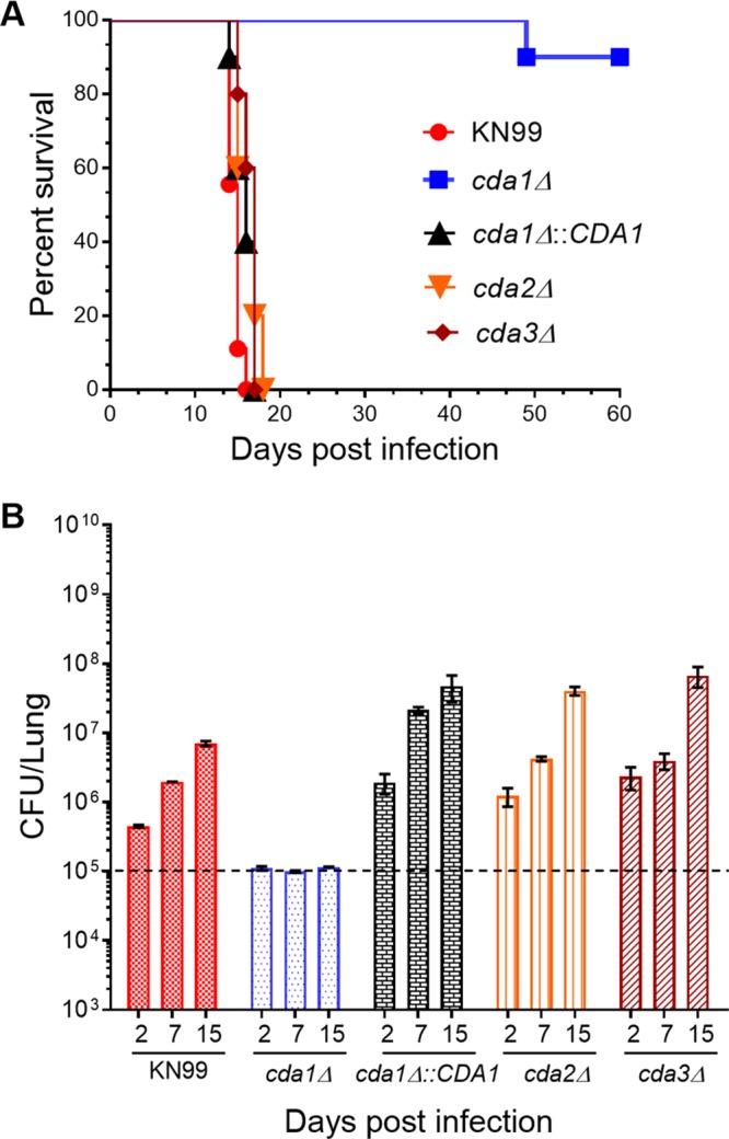
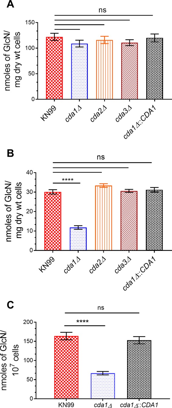
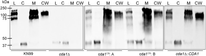
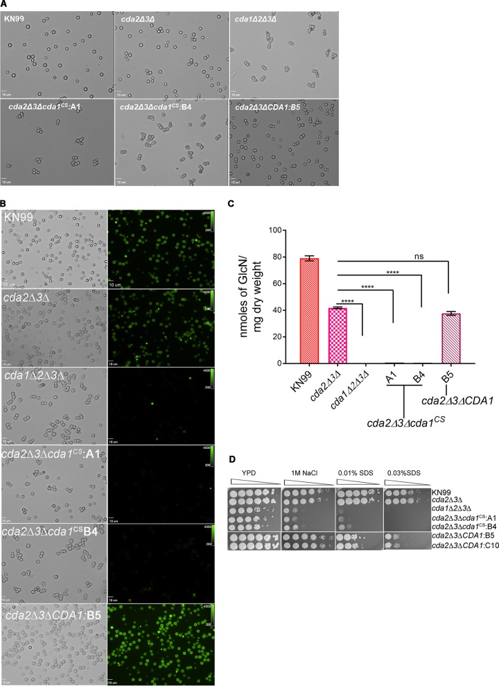
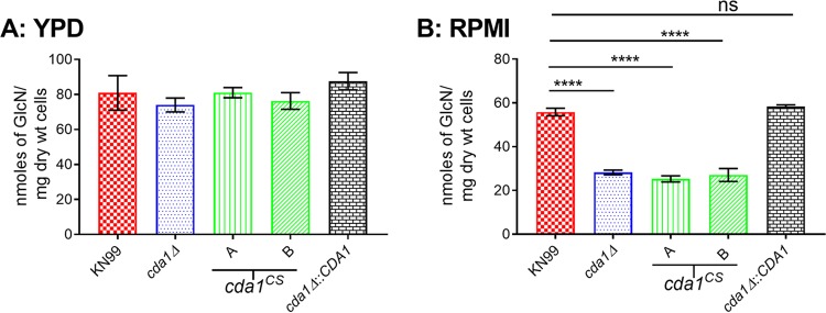
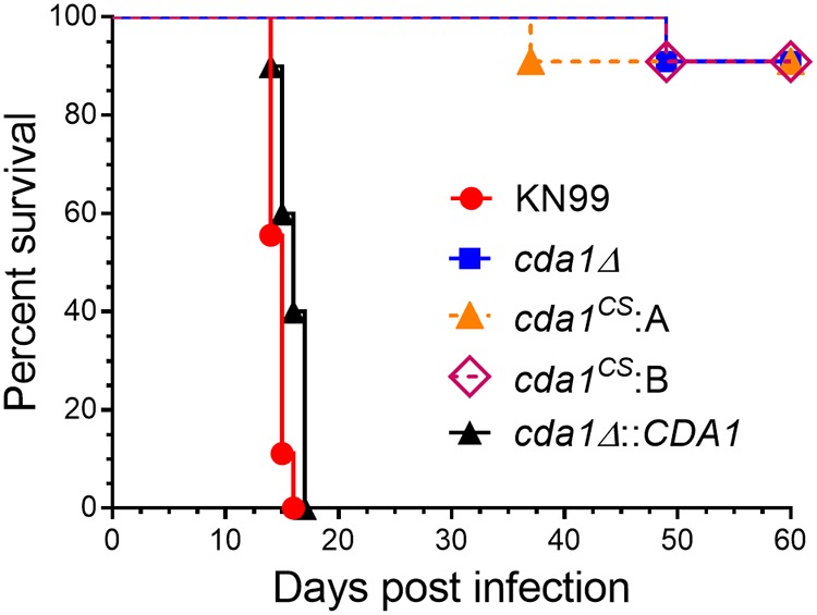
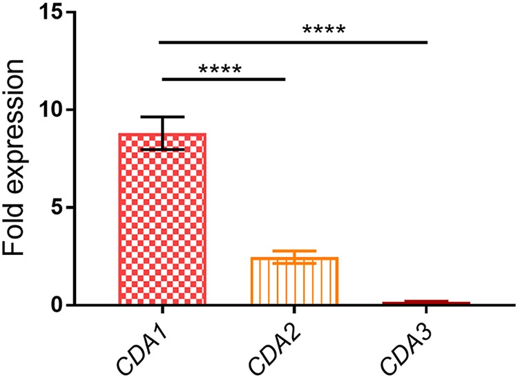
Chitin is an essential component of the cell wall of Cryptococcus neoformans conferring structural rigidity and integrity under diverse environmental conditions. Chitin deacetylase genes encode the enyzmes (chitin deacetylases [Cdas]) that deacetylate chitin, converting it to chitosan. The functional role of chitosan in the fungal cell wall is not well defined, but it is an important virulence determinant of C. neoformans Mutant strains deficient in chitosan are completely avirulent in a mouse pulmonary infection model. C. neoformans carries genes that encode three Cdas (Cda1, Cda2, and Cda3) that appear to be functionally redundant in cells grown under vegetative conditions. Here we report that C. neoformans Cda1 is the principal Cda responsible for fungal pathogenesis. Point mutations were introduced in the active site of Cda1 to generate strains in which the enzyme activity of Cda1 was abolished without perturbing either its stability or localization. When used to infect CBA/J mice, Cda1 mutant strains produced less chitosan and were attenuated for virulence. We further demonstrate that C. neoformans Cda genes are transcribed differently during a murine infection from what has been measured in vitroIMPORTANCECryptococcus neoformans is unique among fungal pathogens that cause disease in a mammalian host, as it secretes a polysaccharide capsule that hinders recognition by the host to facilitate its survival and proliferation. Even though it causes serious infections in immunocompromised hosts, reports of infection in hosts that are immunocompetent are on the rise. The cell wall of a fungal pathogen, its synthesis, composition, and pathways of remodelling are attractive therapeutic targets for the development of fungicides. Chitosan, a polysaccharide in the cell wall of C. neoformans is one such target, as it is critical for pathogenesis and absent in the host. The results we present shed light on the importance of one of the chitin deacetylases that synthesize chitosan during infection and further implicates chitosan as being a critical factor for the pathogenesis of C. neoformans.

Keywords: catalytic site; cell wall localization; chitin deacetylase; chitosan; fungal virulence; metal binding site.


Figures

FIG 1
FIG 1
C. neoformans cda1Δ strain displays severely attenuated virulence. (A) CBA/J mice (4 to 6 weeks old, female) were infected intranasally with 105 CFU of each strain. Survival of the animals was recorded as mortality of mice for 60 days postinfection. Mice that lost 20% of their body weight were considered ill and sacrificed. Data are representative of two independent experiments with 10 animals for each strain. Virulence was determined using Mantel-Cox curve comparison with statistical significance determined by the log rank test (P < 0.0001 comparing KN99 with the cda1Δ strain). (B) Fungal burden in the lungs of mice infected with indicated strains at different days after infection. Data are the average of two experiments with four mice per group at each time point. The dashed line indicates the CFU of the initial infection for each strain.
FIG 2
FIG 2
C. neoformans Cda1 plays a major role in the synthesis of chitosan under host infection conditions. (A) Chitosan levels of strains grown in YPD. The indicated strains were grown in YPD for 48 h. The amount of chitosan in the cell wall of the strains was quantified by the MBTH assay. Data are the averages for three biological experiments with two technical replicates and are expressed as nanomoles of glucosamine per milligram (dry weight) of yeast cells. (B) Chitosan levels of strains grown in RPMI containing 10% FBS and 5% CO2 at 37°C for 5 days. The indicated strains were grown in YPD for 48 h. Yeast cells were harvested, washed with PBS, and inoculated at 500 cells/μl in RPMI containing 10% FBS and incubated for 5 days at 37°C in the presence of 5% CO2. At the end of incubation, chitosan was measured by the MBTH assay and expressed as nanomoles of glucosamine per milligram (dry weight) of cells. Data represent the averages for three biological experiments with two technical replicates. (C) Chitosan levels of strains growing in the murine lung. Mice (CBA/J; four mice per group) were intranasally inoculated with 107 CFU of each strain. On day 7 PI, lungs were excised and homogenized, and the lung tissue was removed by alkaline extraction, leaving the fungal cells to be harvested, counted, and subjected to the MBTH assay. Data were expressed as nanomoles of glucosamine per 107 cells. Significant differences between the groups were compared by one-way ANOVA, followed by Bonferroni’s multiple-comparison test (P < 0.0001 comparing KN99 with any other strain; ns, not significant).
FIG 3
FIG 3
Identification of potential active site residues of C. neoformans Cda1 by sequence alignment. Multiple-sequence alignment of polysaccharide deacetylases. The sequences of C. neoformans Cdas (Cn Cda1, Cn Cda2, and Cn Cda3 [CNAG_05799, CNAG_01230, and CNAG_01239, respectively]) were aligned by the T-coffee program to the peptidoglycan N-deacetylase of S. pneumoniae (Sp PgdA) (GenBank accession number EDK82277.1) and Cda of C. lindemuthianum (Cc Cda) (GenBank accession number AAT68493.1) (60, 61). Identical amino acids are in black boxes, and similar amino acids are in gray boxes. The conserved residues corresponding to catalytic site and metal binding pocket are colored red and blue, respectively.
FIG 4
FIG 4
Effects of changes in amino acids to the catalytic site and metal binding site of Cda1 on protein stability. (A) Western blot analysis of the whole-cell lysates of the wild-type strain and the corresponding Cda1 mutant strains. Equal amounts of protein quantified by BCA protein assay were separated on a 12% Stain-Free Tris-glycine gels. Proteins were transferred to a nitrocellulose membrane and probed with a C. neoformans Cda1-specific polyclonal antibody. (B) Total proteins detected by the photo-activation of the stain-free gel used in the immunoblot shown in panel A. (C) Immunoblot analysis of the whole-cell lysates of catalytic and metal binding site mutants generated in cda2Δ3Δ background. (D) Visualization of total proteins present in each lane of the blot in panel C using stain-free technology.
FIG 5
FIG 5
Disruption of C. neoformans Cda1 catalytic site did not interfere with its cellular localization. The indicated strains were streaked onto YPD agar and incubated for 4 days at 30°C. Cells were collected from the plates, washed with PBS, and subjected to subcellular fractionation. Equal quantities of protein as determined by the BCA method were separated on 10% SDS gel and analyzed by Western blotting using a C. neoformans Cda1-specific polyclonal antibody. Lanes: L, total cell lysate; C, cytosol fraction; M, cell membrane fraction; CW, cell wall fraction.
FIG 6
FIG 6
Point mutations in the catalytic site of Cda1 expressed in a cda2Δ3Δ strain results in chitosan deficiency and phenotypes similar to those of the chitosan-deficient cda1Δ2Δ3Δ strain. (A) Cda1 catalytic point mutant strains generated in the cda2Δ3Δ background display a budding defect when grown in YPD medium similar to the budding defect observed for a chitosan-deficient cda1Δ2Δ3Δ strain. The wild-type and mutant strains were grown in YPD for 36 h, collected, washed, and visualized with an Olympus BX61 microscope, and photographs were taken at 100× magnification. (B) The wild-type, corresponding Cda deletion, and mutant strains were grown as described above in panel A and stained with Eosin Y to detect cell wall chitosan. Bright-field and fluorescence isothiocyanate panels are shown. Photographs were taken at 100× magnification. (C) Quantitative determination of cell wall chitosan in wild-type and various Cda deletion and mutant strains by the MBTH assay. Cells were grown in YPD for 48 h, collected, washed, and used for the assay. Data represent the averages for three biological and two technical replicates. Significant differences between the groups were compared by one-way ANOVA, followed by Dunnett’s multiple-comparison test (****, P < 0.0001 comparing the chitosan amount for each strain to that for the cda2Δ3Δ strain). (D) Wild-type and various C. neoformans Cda deletion and catalytic point mutant strains were grown in YPD for 36 h. Cells were harvested, washed, and diluted to an OD650 of 1.0. Four microliters of 10-fold serially diluted cell suspension was spotted on YPD agar alone and YPD agar containing cell wall stress reagents NaCl and SDS. The plates were incubated for 4 days at 30°C and photographed.
FIG 7
FIG 7
C. neoformans Cda1 catalytic point mutant strains fail to produce chitosan when grown under host-mimicking conditions. (A) Yeast strains were grown in YPD at 30°C for 72 h. Cells were collected, washed, and used for the measurement of cell wall chitosan by the MBTH assay. (B) The indicated strains were initially grown in YPD for 36 h, then collected, washed, and inoculated at a cell density of 500 cells/μl in RPMI medium containing 10% FBS, and incubated for 5 days at 37°C and 5% CO2. At the end of incubation, cells were collected, washed, and used for chitosan measurement by the MBTH assay. Data are the averages for three biological experiments with each experiment having two technical replicates. Significant differences between the groups were compared by one-way ANOVA, followed by Bonferroni’s multiple-comparison test (****, P < 0.0001 comparing chitosan levels of strain KN99 to each other strain).
FIG 8
FIG 8
Chitin deacetylase activity of C. neoformans Cda1 is critical for fungal pathogenesis. Mice (female CBA/J; 4 to 6 weeks old) were inoculated with 105 CFU of the indicated yeast strains by intranasal instillation. Mice were monitored for up to 60 days PI for survival by monitoring their body weight. Animals that lost 20% of their weight during inoculation were considered moribund and were euthanized. Data for each strain (n = 10 mice) are the combined results of two experiments, each with five animals. Survival curves were compared by log rank (Mantel-Cox) test (P < 0.0001 comparing strain KN99 to cda1Δ or cda1CS isolate A or cda1CS isolate B).
FIG 9
FIG 9
C. neoformans CDA1 is transcriptionally upregulated during host infection. Mice were inoculated with 107 CFU of KN99 cells. At day 7 PI, the lungs were excised and homogenized, and fungal cells were harvested and used for the isolation of total RNA. Total RNA (0.5 µg) was used for the synthesis of cDNA, which was subsequently subjected to quantitative real-time PCR using CDA1-specific primers. C. neoformans 18S rRNA transcript levels were used as a reference gene. Transcript levels of the respective genes in the cells used as inoculum served as control. Data are the averages for two independent experiments each with three animals per group. Fold expression was calculated for each gene comparing the extent of its upregulation in the lung samples to YPD-grown samples. Significant differences in the expression levels between genes were compared by ordinary one-way ANOVA, followed by Dunnett’s multiple-comparison test. (****, P < 0.0001 comparing fold expression of CDA1, CDA2, and CDA3 in the lung samples to their respective levels in the inoculum sample grown in YPD).

Similar articles

Cited by

References

    1. Rajasingham R, Smith RM, Park BJ, Jarvis JN, Govender NP, Chiller TM, Denning DW, Loyse A, Boulware DR. 2017. Global burden of disease of HIV-associated cryptococcal meningitis: an updated analysis. Lancet Infect Dis 17:873–881. doi:10.1016/S1473-3099(17)30243-8. - DOI - PMC - PubMed
    1. Litvintseva AP, Carbone I, Rossouw J, Thakur R, Govender NP, Mitchell TG. 2011. Evidence that the human pathogenic fungus Cryptococcus neoformans var. grubii may have evolved in Africa. PLoS One 6:e19688. doi:10.1371/journal.pone.0019688. - DOI - PMC - PubMed
    1. Litvintseva AP, Thakur R, Reller LB, Mitchell TG. 2005. Prevalence of clinical isolates of Cryptococcus gattii serotype C among patients with AIDS in Sub-Saharan Africa. J Infect Dis 192:888–892. doi:10.1086/432486. - DOI - PubMed
    1. Hiremath SS, Chowdhary A, Kowshik T, Randhawa HS, Sun S, Xu J. 2008. Long-distance dispersal and recombination in environmental populations of Cryptococcus neoformans var. grubii from India. Microbiology 154:1513–1524. doi:10.1099/mic.0.2007/015594-0. - DOI - PubMed
    1. Idnurm A, Bahn YS, Nielsen K, Lin X, Fraser JA, Heitman J. 2005. Deciphering the model pathogenic fungus Cryptococcus neoformans. Nat Rev Microbiol 3:753–764. doi:10.1038/nrmicro1245. - DOI - PubMed

Publication types

LinkOut - more resources