. 2018 Nov 20;9(6):e02095-18.
doi: 10.1128/mBio.02095-18.

Prion Seeds Distribute throughout the Eyes of Sporadic Creutzfeldt-Jakob Disease Patients

Affiliations

Prion Seeds Distribute throughout the Eyes of Sporadic Creutzfeldt-Jakob Disease Patients

Christina D Orrù et al. mBio. .

Abstract

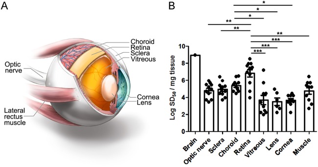
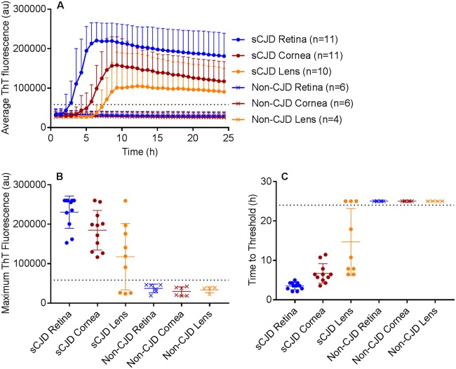
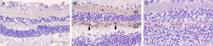
Sporadic Creutzfeldt-Jakob disease (sCJD) is the most common prion disease in humans and has been iatrogenically transmitted through corneal graft transplantation. Approximately 40% of sCJD patients develop visual or oculomotor symptoms and may seek ophthalmological consultation. Here we used the highly sensitive real-time quaking-induced conversion (RT-QuIC) assay to measure postmortem prion seeding activities in cornea, lens, ocular fluid, retina, choroid, sclera, optic nerve, and extraocular muscle in the largest series of sCJD patient eyes studied by any assay to date. We detected prion seeding activity in 100% of sCJD eyes, representing three common sCJD subtypes, with levels varying by up to 4 log-fold among individuals. The retina consistently showed the highest seed levels, which in some cases were only slightly lower than brain. Within the retina, prion deposits were detected by immunohistochemistry (IHC) in the retinal outer plexiform layer in most sCJD cases, and in some eyes the inner plexiform layer, consistent with synaptic prion deposition. Prions were not detected by IHC in any other eye region. With RT-QuIC, prion seed levels generally declined in eye tissues with increased distance from the brain, and yet all corneas had prion seeds detectable. Prion seeds were also present in the optic nerve, extraocular muscle, choroid, lens, vitreous, and sclera. Collectively, these results reveal that sCJD patients accumulate prion seeds throughout the eye, indicating the potential diagnostic utility as well as a possible biohazard.IMPORTANCE Cases of iatrogenic prion disease have been reported from corneal transplants, yet the distribution and levels of prions throughout the eye remain unknown. This study probes the occurrence, level, and distribution of prions in the eyes of patients with sporadic Creutzfeldt-Jakob disease (sCJD). We tested the largest series of prion-infected eyes reported to date using an ultrasensitive technique to establish the prion seed levels in eight regions of the eye. All 11 cases had detectable prion seeds in the eye, and in some cases, the seed levels in the retina approached those in brain. In most cases, prion deposits could also be seen by immunohistochemical staining of retinal tissue; other ocular tissues were negative. Our results have implications for estimating the risk for iatrogenic transmission of sCJD as well as for the development of antemortem diagnostic tests for prion diseases.

Keywords: Creutzfeldt-Jakob disease; RT-QuIC; eye; prion.


Figures

FIG 1
FIG 1
Comparison of prion seed levels in brain (temporal cortex) and ocular compartments from posterior to anterior eye as measured by RT-QuIC analysis. (A) Image showing the ocular tissues tested by RT-QuIC. (B) The seeding dose 50 per mg of tissue (logSD50) is shown. The average prion seed level in eye was highest in the retina, and lowest in the vitreous and lens. One vitreous sample, 3 lens samples, and 1 muscle sample were negative and are not shown. Retinal prion seed levels were significantly higher than most other ocular tissues, including optic nerve, sclera, lens, cornea, vitreous, and extraocular muscle. The prion seed level in a representative sCJD brain sample (termporal cortex) is shown for comparison. For retina, optic nerve, sclera, cornea, vitreous, and extraocular muscle, n = 11; for choroid, n = 10; and for lens, n = 9. *, P < 0.05; **, P < 0.01; ***, P < 0.001, one-way ANOVA with Tukey’s multiple comparison test. Graphics in panel A by Ryan Kissinger.
FIG 2
FIG 2
RT-QuIC of retina, cornea, and lens from sCJD and non-sCJD patients. (A) The average prion seeding amplification kinetics are shown for retina (blue), cornea (red), and lens (orange) from sCJD (circles) and non-CJD (X’s) patients. The dotted line indicates the ThT fluorescence threshold for a positive result (see Materials and Methods). (B) The maximum ThT fluorescence reached within 24 hours is shown. The thin lines represent mean and standard deviation, whereas the dotted line indicates the ThT fluorescence threshold for a positive result. (C) The time to reach the threshold for positivity is shown for each sample. The dotted line indicates the end of the 24-hour experiment. If a sample did not reach the threshold within 24 h, it was marked as 25 h to indicate a negative result. n = 4 (lens) or 6 (retina, cornea) non-CJD control cases were analyzed.
FIG 3
FIG 3
PrPSc deposits in retinal plexiform layers. (A) Retina from a non-CJD control patient. (B) Retina from an sCJD patient having abundant labeling of PrPSc deposits (patient 5, VV2). Deposits consisted of an admixture of diffuse and focal PrPSc (arrowheads) in the outer plexiform layer (opl) as well as diffuse aggregates (*) in the inner plexiform layer (ipl). (C) Retina from an sCJD patient, subtype MM1 (patient 1), showing PrPSc in the opl, but not the ipl. Scale bar = 50 μm.

Similar articles

Cited by

References

    1. Prusiner SB. 1998. Prions. Proc Natl Acad Sci U S A 95:13363–13383. doi:10.1073/pnas.95.23.13363. - DOI - PMC - PubMed
    1. DeArmond SJ, Prusiner SB. 1995. Etiology and pathogenesis of prion diseases. Am J Pathol 146:785–811. - PMC - PubMed
    1. Collinge J. 2016. Mammalian prions and their wider relevance in neurodegenerative diseases. Nature 539:217–226. doi:10.1038/nature20415. - DOI - PubMed
    1. Takada LT, Geschwind MD. 2013. Prion diseases. Semin Neurol 33:348–356. doi:10.1055/s-0033-1359314. - DOI - PubMed
    1. Ironside JW, Ritchie DL, Head MW. 2017. Prion diseases. Handb Clin Neurol 145:393–403. doi:10.1016/B978-0-12-802395-2.00028-6. - DOI - PubMed

Publication types

Supplementary concepts