. 2019 Feb;18(2):320-337.
doi: 10.1074/mcp.RA118.001044. Epub 2018 Nov 20.

Human Stress-inducible Hsp70 Has a High Propensity to Form ATP-dependent Antiparallel Dimers That Are Differentially Regulated by Cochaperone Binding

Affiliations

Human Stress-inducible Hsp70 Has a High Propensity to Form ATP-dependent Antiparallel Dimers That Are Differentially Regulated by Cochaperone Binding

Filip Trcka et al. Mol Cell Proteomics. 2019 Feb.

Abstract

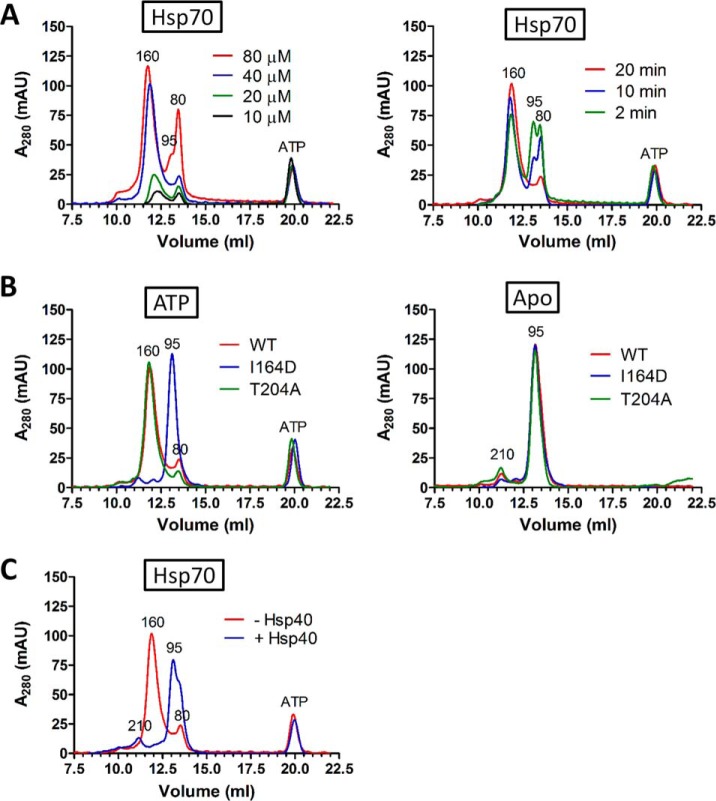
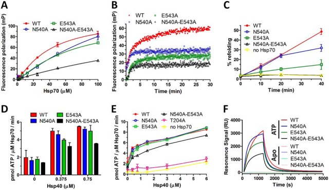
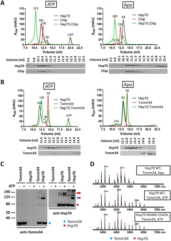
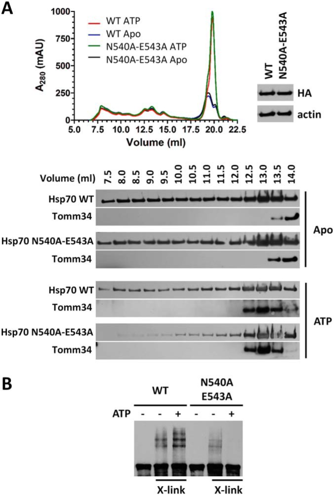
Eukaryotic protein homeostasis (proteostasis) is largely dependent on the action of highly conserved Hsp70 molecular chaperones. Recent evidence indicates that, apart from conserved molecular allostery, Hsp70 proteins have retained and adapted the ability to assemble as functionally relevant ATP-bound dimers throughout evolution. Here, we have compared the ATP-dependent dimerization of DnaK, human stress-inducible Hsp70, Hsc70 and BiP Hsp70 proteins, showing that their dimerization propensities differ, with stress-inducible Hsp70 being predominantly dimeric in the presence of ATP. Structural analyses using hydrogen/deuterium exchange mass spectrometry, native electrospray ionization mass spectrometry and small-angle X-ray scattering revealed that stress-inducible Hsp70 assembles in solution as an antiparallel dimer with the intermolecular interface closely resembling the ATP-bound dimer interfaces captured in DnaK and BiP crystal structures. ATP-dependent dimerization of stress-inducible Hsp70 is necessary for its efficient interaction with Hsp40, as shown by experiments with dimerization-deficient mutants. Moreover, dimerization of ATP-bound Hsp70 is required for its participation in high molecular weight protein complexes detected ex vivo, supporting its functional role in vivo As human cytosolic Hsp70 can interact with tetratricopeptide repeat (TPR) domain containing cochaperones, we tested the interaction of Hsp70 ATP-dependent dimers with Chip and Tomm34 cochaperones. Although Chip associates with intact Hsp70 dimers to form a larger complex, binding of Tomm34 disrupts the Hsp70 dimer and this event plays an important role in Hsp70 activity regulation. In summary, this study provides structural evidence of robust ATP-dependent antiparallel dimerization of human inducible Hsp70 protein and suggests a novel role of TPR domain cochaperones in multichaperone complexes involving Hsp70 ATP-bound dimers.

Keywords: Allostery; Chaperone; Cochaperone; Mass Spectrometry; Protein Conformation; Protein Structure; Protein-Protein Interactions; Structural Biology.


Figures

None
Graphical abstract
Fig. 1.
Fig. 1.
Homologs of Hsp70 differ in their propensity to dimerize in the presence of ATP. A, Bacterially purified DnaK, Hsp70, Hsc70 and BiP proteins (40 μm) were pre-incubated with or without ATP (0.2 mm, 20 mins, 21 °C) before separation by analytical SEC. Apparent MW of eluting peaks is indicated (see Experimental procedures). B, Hsp70 homologs (40 μm) were pre-incubated with or without ATP for 20 mins before addition of chemical cross-linker (10 molar excess of DSA). The cross-linked complexes were separated by LDS-PAGE (pre-cast gradient (4–12%) gel, NuPage). The molecular weight standard is indicated. C, Native ESI-MS spectra of Hsp70 and Hsc70 proteins (20 μm) were acquired after their pre-incubation without (Apo) or with ATP (0.2 mm). The charged states corresponding to monomers and dimers are labeled with single and double dots, respectively.
Fig. 2.
Fig. 2.
ATP-induced dimerization of Hsp70 requires its intact NBD-SBDβ docked conformation. A, Hsp70 was mixed at different concentrations (10, 20, 40, 80 μm; 20 min incubation) or for different time intervals (2, 10, 20 min) with ATP (0.2 mm) at 21 °C before separation by SEC. B, Hsp70 and its mutant forms (I164D, T204A) (40 μm) were incubated without (Apo) or with ATP (0.2 mm, 20 min) at 21 °C before SEC. C, Hsp70 (40 μm) was incubated with ATP (0.2 mm, 21 °C) for 20 min. Hsp40 (10 μm) was added or not for 5 min and samples were separated by SEC. Apparent MW of eluting peaks is indicated (see Experimental procedures).
Fig. 3.
Fig. 3.
Conserved N540 and E543 residues are required for stable ATP-induced dimerization of Hsp70. A, Crystal structure of DnaK ATP-bound dimer (PDB code 4jne) with N537 and E540 residues highlighted in red (N540, E543 numbering in human inducible Hsp70). The NBD domain is depicted in yellow, SBDβ in marine and SBDα in green. B, Hsp70 and N540A, E543A and N540A-E543A (40 μm) were incubated without (Apo) or with ATP (0.2 mm, 20 min) at 21 °C before SEC. C, Hsp70 WT, N540A, E543A and N540A-E543A proteins (40 μm) were pre-incubated with or without ATP for 20 min before addition of chemical cross-linker (10 molar excess of DSA). The cross-linked complexes were separated by LDS-PAGE (pre-cast gradient (4–12%) gel, NuPage). The molecular weight standard is indicated. D, Native ESI-MS spectra of Hsp70 WT, N540A, E543A and N540A-E543A proteins (20 μm) were acquired after their pre-incubation without (Apo) or with ATP. The charged states corresponding to monomers and dimers are labeled with single and double dots, respectively. E, Deuteration level (exchanged deuterons, D) differences of Hsp70 WT, N540A, E543A and N540A-E543A peptides in ATP-bound and nucleotide-free state (Apo) after 1200 s (for other incubation times see supplemental Fig. S2B) incubation in deuterated buffer. Numbers at the left indicate the Hsp70 peptide fragments; schematic representation at the left shows Hsp70 domain constitution; L, interdomain linker; T, C-terminal tail. The asterisks indicate peptides covering allosterically important regions of Hsp70 molecule. Dashed lines in the graph indicate significance level of 0.35 Da (see Experimental procedures).
Fig. 4.
Fig. 4.
ATP-bound Hsp70 monomers assemble as antiparallel dimers in solution as determined by small-angle X-ray scattering. A, χ2-ranked fitting of Hsp70 dimeric crystal structures (PDB codes with indicated chains) and homology-based Hsp70 dimer model (see Experimental Procedures) to the SAXS experimental data obtained for Hsp70-T204A mutant at two concentrations. Fitting to the experimental scattering curves was performed using CRYSOL ATSAS v.2.8.3. B, CRYSOL fit of Hsp70 ATP dimer atomic model. Simulated scattering data of dimeric Hsp70 atomic model (red) is fitted to experimentally obtained solution scattering showing overall good fit with χ2 = 1.1. C, Atomic model of Hsp70 ATP-dependent dimer, developed as described in Experimental Procedures. NBD is highlighted in yellow, SBDβ in marine and SBDα in green. N- and C-terminal unstructured regions of the model are omitted from the figure. The image was created in PyMOL.
Fig. 5.
Fig. 5.
Hsp70 ATP-dependent dimers assemble through NBD-NBD and NBD-SBDα interfaces. A, Deuteration level (exchanged deuterons, D) differences between Hsp70 WT and N540A, E543A and N540A-E543A peptides in nucleotide-free state (Apo) measured after 120 s (for other incubation times see supplemental Fig. S3B) incubation in deuterated buffer. The graph is labeled as in Fig. 3E. Red dots indicate peptides highlighted in part B and C of this figure. B, Crystal structure of human SBD (PDB code 4po2). SBDα (lid) helices and the lid-SBDβ positioning ionic/polar contacts are indicated. SBDβ is labeled in marine and SBDα in green. Regions covered by peptides highlighted by red dots in A are red. C, Deuteration kinetics of indicated peptides from Apo and ATP-bound Hsp70 proteins. D, Deuteration level (exchanged deuterons, D) differences between Hsp70 WT and N540A, E543A and N540A-E543A in ATP-bound state measured after 20 s (for other incubation times see supplemental Fig. S3B) incubation in deuterated buffer. The graph is labeled similarly as in Fig. 3E. Red dots indicate peptides highlighted (red/pink) on the atomic structures in E, E. 48–68, 268–282, 294–302, 519–529 and 530–542 peptides highlighted in D were projected onto the homology-based Hsp70 ATP-bound dimer atomic model (see Experimental Procedures) and the corresponding peptides (see supplemental Fig. S4C) in DnaK were projected onto the crystal structure of DnaK ATP-bound dimer (PDB code 4jne). The peptides are colored in red or pink in the respective protomer. The atomic structures are shown in both ribbon and surface representations. Surface representation was also manually separated for clarity (bottom structures). NBD is highlighted in yellow, SBDβ in marine and SBDα in green. N- and C-terminal unstructured regions of the model are omitted from the figure. The image was created in PyMOL.
Fig. 6.
Fig. 6.
E543A and N540A-E543A mutants have severely impaired substrate binding/refolding activities and lower interaction with Hsp40. A, Equilibrium binding curves of F-NRLLLTG peptide binding to Hsp70 WT and mutants under nucleotide-free conditions. Fluorescence polarization was determined at 30 nm peptide and increasing Hsp70 concentrations. Error bars represent S.D.; n = 3 independent experiments. B, Kinetics of F-NRLLLTG interaction with WT and mutant Hsp70s under nucleotide- free conditions determined by fluorescence polarization. Protein and peptide concentrations were 50 μm and 30 nm, respectively. C, Firefly luciferase was chemically denatured, mixed with Hsp70 WT or mutants (1 μm), Hsp40 (2 μm), Bag-1 (0.5 μm) and ATP (2 mm) and recovered luminescence was measured. Error bars represent S.D.; n = 3 independent experiments. D, ATPase activity of Hsp70 WT and mutants at lower Hsp40 concentrations, for full results see E. E, ATPase activity of Hsp70 WT and mutants (2 μm) was tested at various Hsp40 concentrations in malachite green assay. Error bars represent S.D.; n = 3 independent experiments. F, SPR measurement of Hsp70 WT and mutants binding to Hsp40 under nucleotide-free conditions (Apo) or in the presence of ATP.
Fig. 7.
Fig. 7.
ATP-dependent dimer of Hsp70 associates with Chip and Tomm34 TPR domain cochaperones through different mechanisms. A and B, Hsp70 WT and Chip or Tomm34 were mixed (both at 40 μm) and pre-incubated with or without ATP (0.2 mm, 20 min, 21 °C) before separation by analytical SEC. Apparent MW of eluting peaks is indicated (see Experimental Procedures). Proteins in the separated fractions were analyzed by SDS-PAGE and Coomassie staining. C, Hsp70, Tomm34 or both (40 μm each) were mixed in the presence or absence of ATP (0.2 mm) for 20 min at 21 °C. Glutaraldehyde was added (0.5 mm final concentration) for 10 mins and the reactions stopped with Tris, pH 8 (80 mm final concentration). Samples were diluted in 2× CSB loading buffer, separated by SDS-PAGE, blotted and probed by Tomm34 or Hsp70 antibodies. Blue and red dots, respectively, indicate positions of Tomm34 and Hsp70 monomers, homodimers and heterodimers. D, Native ESI-MS spectra of Hsp70 WT or N540A-E543A mutant mixtures with Tomm34 (20 μm) were acquired after pre-incubation without (Apo) or with ATP. The charged states corresponding to Tomm34/Hsp70 monomers and heterodimers are labeled with single and double dots, respectively.
Fig. 8.
Fig. 8.
ATP-dependent dimerization of Hsp70 is required for its participation in high molecular weight complexes detected ex vivo. A, Desalted lysates from HEK293 cells overexpressing HA-tagged Hsp70 WT or N540A-E543A mutant to the same level (evaluated by Western blotting using anti-HA antibody) were incubated with/without 2 mm ATP for 20 min at 21 °C before SEC. The presence of Hsp70 and Tomm34 proteins in the separated fractions was tested by Hsp70/Tomm34 antibodies. B, Desalted HEK293 cell lysates with overexpressed HA-tagged Hsp70 WT or N540A-E543A were incubated with/without 2 mm ATP for 20 min at 21 °C before mixing with glutaraldehyde (0.5 mm final concentration) for 10 min at 21 °C. The reaction was stopped by adding Tris, pH 8 (80 mm final concentration), samples were supplemented with 2× CSB loading buffer and analyzed by SDS-PAGE and Western blotting with Hsp70 antibody.

References

    1. Calamini B., and Morimoto R. I. (2012) Protein homeostasis as a therapeutic target for diseases of protein conformation. Curr. Topics Med. Chem. 12, 2623–2640 - PMC - PubMed
    1. Young J. C., Agashe V. R., Siegers K., and Hartl F. U. (2004) Pathways of chaperone-mediated protein folding in the cytosol. Nat. Rev. Mol. Cell Biol. 5, 781–791 - PubMed
    1. Young J. C. (2010) Mechanisms of the Hsp70 chaperone system. Biochim. Biol. Cell. 88, 291–300 - PMC - PubMed
    1. Boorstein W. R., Ziegelhoffer T., and Craig E. A. (1994) Molecular evolution of the HSP70 multigene family. J. Mol. Evolution 38, 1–17 - PubMed
    1. Radons J. (2016) The human HSP70 family of chaperones: where do we stand? Cell Stress Chaperones 21, 379–404 - PMC - PubMed

Publication types

MeSH terms

Substances

LinkOut - more resources