. 2019 Jan 9;39(2):271-280.
doi: 10.1523/JNEUROSCI.1188-18.2018. Epub 2018 Nov 20.

Auditory Thalamostriatal and Corticostriatal Pathways Convey Complementary Information about Sound Features

Affiliations

Auditory Thalamostriatal and Corticostriatal Pathways Convey Complementary Information about Sound Features

Nicholas D Ponvert et al. J Neurosci. .

Abstract

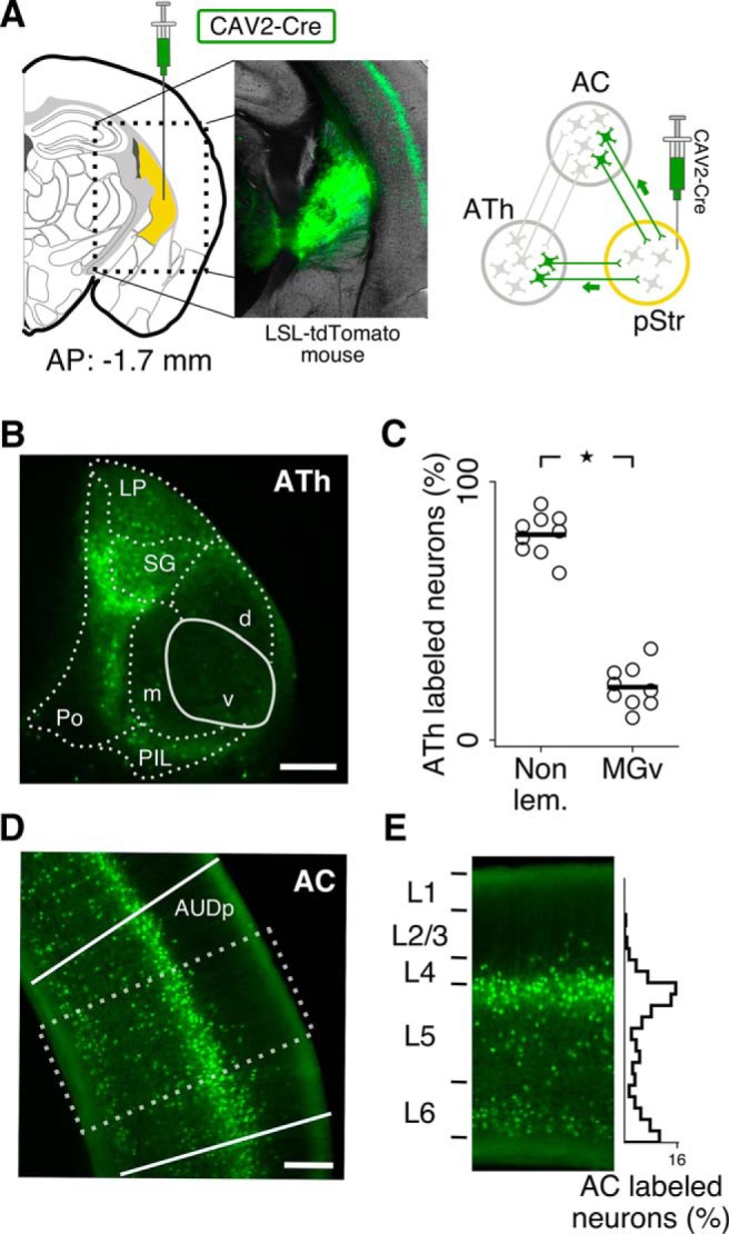
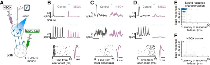
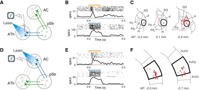
Multiple parallel neural pathways link sound-related signals to behavioral responses. For instance, the striatum, a brain structure involved in action selection and reward-related learning, receives neuronal projections from both the auditory thalamus and auditory cortex. It is not clear whether sound information that reaches the striatum through these two pathways is redundant or complementary. We used an optogenetic approach in awake mice of both sexes to identify thalamostriatal and corticostriatal neurons during extracellular recordings, and characterized neural responses evoked by sounds of different frequencies and amplitude modulation rates. We found that neurons in both pathways encode sound frequency with similar fidelity, but display different coding strategies for amplitude modulated noise. Whereas corticostriatal neurons provide a more accurate representation of amplitude modulation rate in their overall firing rate, thalamostriatal neurons convey information about the precise timing of acoustic events. These results demonstrate that auditory thalamus and auditory cortex neurons provide complementary information to the striatum, and suggest that these pathways could be differentially recruited depending on the requirements of a sound-driven behavior.SIGNIFICANCE STATEMENT Sensory signals from the cerebral cortex and the thalamus converge onto the striatum, a nucleus implicated in reward-related learning. It is not clear whether these two sensory inputs convey redundant or complementary information. By characterizing the sound-evoked responses of thalamostriatal and corticostriatal neurons, our work demonstrates that these neural pathways convey complementary information about the temporal features of sounds. This work opens new avenues for investigating how these pathways could be selectively recruited depending on task demands, and provides a framework for studying convergence of cortical and thalamic information onto the striatum in other sensory systems.

Keywords: amplitude modulation; auditory cortex; auditory thalamus; neural coding; pathway-specific; striatum.


Figures

Figure 1.
Figure 1.
Neurons in auditory thalamus (ATh) and auditory cortex (AC) project to the posterior striatum (pStr). A, A retrograde virus (CAV2-Cre) was injected in the posterior striatum (yellow) of LSL-tdTomato mice, shown here in a coronal brain slice. The virus is picked up by the axon terminals and transported to the cell bodies, causing excision of the loxP-flanked STOP cassette and subsequent expression of tdTomato in neurons that project to the injection location. B, Expression of tdTomato in ATh neurons (AP: −3.16 mm. v, d, and m: Ventral, dorsal, and medial nuclei of the medial geniculate body, respectively; LP, lateral posterior nucleus; SG, suprageniculate nucleus; PO, posterior thalamic nucleus; PIL, posterior intralaminar thalamic nucleus. Scale bar, 200 μm. C, Most thalamic striatal-projecting neurons were located in non-lemniscal thalamic nuclei: d, m, SG, LP, PO, and PIL. Each point represents the fraction of labeled neurons for one of three interleaved sets of histological sections from each animal (3 mice, stars represent p < 0.05). D, Expression of tdTomato in primary auditory cortex (AUDp) neurons. Dotted rectangle represents cortical region shown in E. Scale bar, 200 μm. E, Striatal-projecting neurons in AC were present across deep cortical layers with high density in the superficial portion of layer 5. Results were consistent across all cortical slices analyzed.
Figure 2.
Figure 2.
Identification of striatal-projecting neurons during extracellular recording. A, CAV2-Cre injected in the posterior striatum (pStr) of LSL-ChR2 mice travels retrogradely and causes expression of ChR2 in striatal-projecting neurons. ChR2-expressing neurons were identified by their responses to laser light during extracellular recording. To validate approaches for distinguishing between ChR2-expressing neurons and neurons that respond to light because of synaptic excitation, we measured changes in light-evoked responses after blocking synaptic transmission with NBQX. B, Top row, Response of an example cortical neuron to a 100 ms pulse of blue laser light before and after application of NBQX. Middle row, This neuron responds reliably to a 5 Hz train of 10 ms laser light pulses before and after NBQX. Bottom row, This neuron responds within 10 ms of light onset (left). Spike shape was consistent before and after NBQX injection (right). C, Example cortical neuron that responds to light (top) but cannot follow a train of laser pulses (middle). The light-evoked responses of this neuron disappear after NBQX application. D, Example cortical neuron that responds to a train of laser pulses (middle) but has a long latency response (bottom). The light-evoked responses of this neuron disappear after NBQX application. E, Neurons collected during sound response characterization recordings were classified as striatal-projecting if they displayed fast, reliable responses to light (blue points). Laser-responsive neurons that did not meet these criteria were excluded (gray points). Dotted gray line represents a threshold requiring neurons to reach 1/2 of their maximum laser-evoked firing rate within 10 ms. F, Neurons collected during NBQX control experiments that continued to respond after application of NBQX display fast, reliable responses to light (blue points). Laser-responsive neurons which failed to respond after application of NBQX did not meet the criteria used to operationally define striatal-projecting neurons.
Figure 3.
Figure 3.
Striatal-projecting neurons in auditory thalamus and auditory cortex display reliable sound responses. We recorded from ChR2-tagged striatal-projecting neurons in auditory thalamus (ATh; A) or auditory cortex (AC; D). B, Example sound-evoked response and laser-evoked response from a ChR2-tagged neuron in auditory thalamus (yellow bar: 100 ms burst of 60 dB SPL white noise; blue bar: 100 ms pulse of 445 nm laser light). C, Coronal sections showing locations of recorded tagged neurons in ATh. Each recording location was projected onto the closest example section. Thicker lines represent the border of MGv. E, Same as B for an example ChR2-tagged neuron in auditory cortex. F, Coronal sections showing locations of recorded tagged neurons in auditory cortex. Each recording location was projected onto the closest example section. Thicker lines represent the border of the primary auditory field (AUDp). AUDd and AUDv, Dorsal and ventral fields, respectively.
Figure 4.
Figure 4.
Auditory thalamostriatal and corticostriatal neurons encode sound frequency with similar fidelity. A, Example frequency-intensity tuning curve from a striatal-projecting neuron in auditory thalamus (ATh). Tuning curves were generated by recording neural responses to 100 ms pure tone pips at 16 frequencies (2–40 kHz) and 12 intensities (15–70 dB SPL in 5 dB steps). B, Example frequency-intensity tuning curve from a striatal-projecting neuron in auditory cortex. C, No statistically significant difference in tuning bandwidth at BW10 was observed between tuned auditory thalamostriatal (n = 24) and auditory corticostriatal (AC; n = 27) neurons. Str, Striatal. D, Intensity threshold for responses to pure tones was lower in auditory corticostriatal neurons than thalamostriatal neurons. E, Auditory thalamostriatal neurons display shorter response latencies to pure tones than corticostriatal neurons. F, No statistically significant difference was observed in the ratio of onset spike rate to sustained spike rate between thalamostriatal and corticostriatal neurons. G, Thalamostriatal neurons display more monotonic responses to sound level than corticostriatal neurons. Stars represent p < 0.05. Black bars indicate the median value of each group.
Figure 5.
Figure 5.
Thalamostriatal and corticostriatal neurons convey different information about temporal features of sounds. A, B, Example responses of two auditory thalamostriatal (ATh) neurons to AM white noise at different modulation rates. Average firing rates vary little as a function of AM rate. Dotted lines indicate 1 SD. AM stimuli consisted of 500 ms bursts of white noise at 60 dB SPL, modulated by a sinusoid (4–128 Hz). Modulation depth was 100%. C, Both thalamostriatal (7/36 = 19%) and corticostriatal (8/24 = 33%) populations contained some neurons unable to synchronize to any AM stimulus presented. Filled areas represent neurons able to synchronize to at least one AM stimulus. D, Thalamostriatal neurons display higher maximum synchronization rates compared with corticostriatal neurons. AC, Auditory corticostriatal; Str, Striatal. E, F, Example responses of two auditory corticostriatal neurons to AM noise at different modulation rates. Average firing rates are modulated by AM rate. Dotted lines indicate 1 SD. G, A linear discriminator is better able to tell apart preferred from least-preferred AM rate using average stimulus-evoked firing rates of corticostriatal neurons than thalamostriatal neurons. H, A linear discriminator is better able to tell apart preferred from least-preferred AM phase using firing rates from thalamostriatal neurons than from corticostriatal neurons. Discrimination accuracy was calculated separately for each AM rate. Each dot represents the average accuracy across all AM rates for each neuron. Stars indicate p < 0.05. Black bars indicate the median value of each group.

Similar articles

Cited by

References

    1. Anderson LA, Linden JF (2011) Physiological differences between histologically defined subdivisions in the mouse auditory thalamus. Hear Res 274:48–60. 10.1016/j.heares.2010.12.016 - DOI - PMC - PubMed
    1. Balleine BW, Delgado MR, Hikosaka O (2007) The role of the dorsal striatum in reward and decision-making. J Neurosci 27:8161–8165. 10.1523/JNEUROSCI.1554-07.2007 - DOI - PMC - PubMed
    1. Bartlett EL. (2013) The organization and physiology of the auditory thalamus and its role in processing acoustic features important for speech perception. Brain Lang 126:29–48. 10.1016/j.bandl.2013.03.003 - DOI - PMC - PubMed
    1. Baxter MG, Murray EA (2002) The amygdala and reward. Nat Rev Neurosci 3:563–573. 10.1038/nrn875 - DOI - PubMed
    1. Bendor D, Wang X (2007) Differential neural coding of acoustic flutter within primate auditory cortex. Nat Neurosci 10:763–771. 10.1038/nn1888 - DOI - PubMed

Publication types

LinkOut - more resources