. 2019 Jan 11;294(2):439-452.
doi: 10.1074/jbc.RA118.004407. Epub 2018 Nov 20.

The inflammasome adapter ASC assembles into filaments with integral participation of its two Death Domains, PYD and CARD

Affiliations

The inflammasome adapter ASC assembles into filaments with integral participation of its two Death Domains, PYD and CARD

Reinard Jeffrey T Nambayan et al. J Biol Chem. .

Abstract

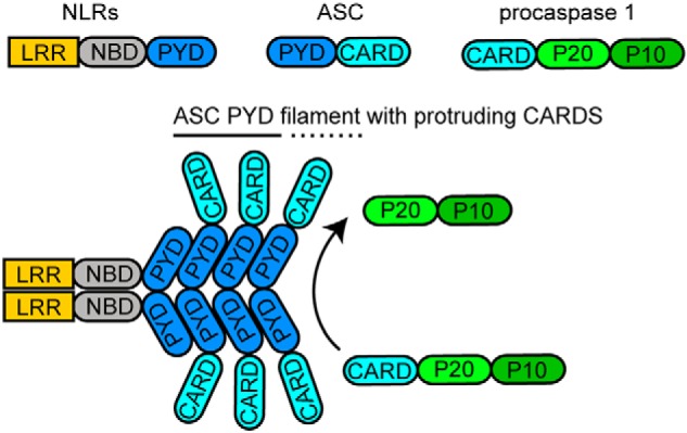
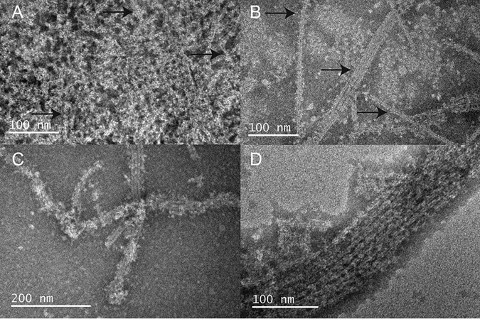
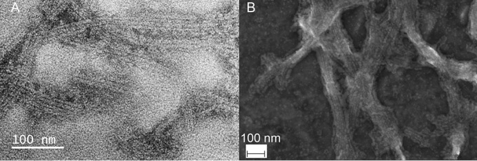
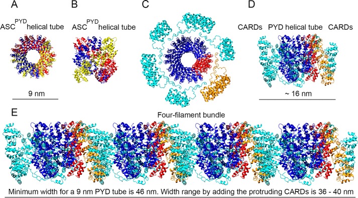
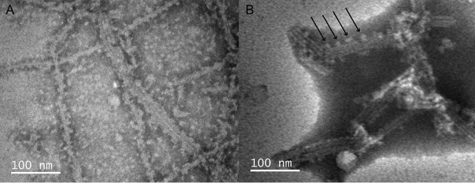
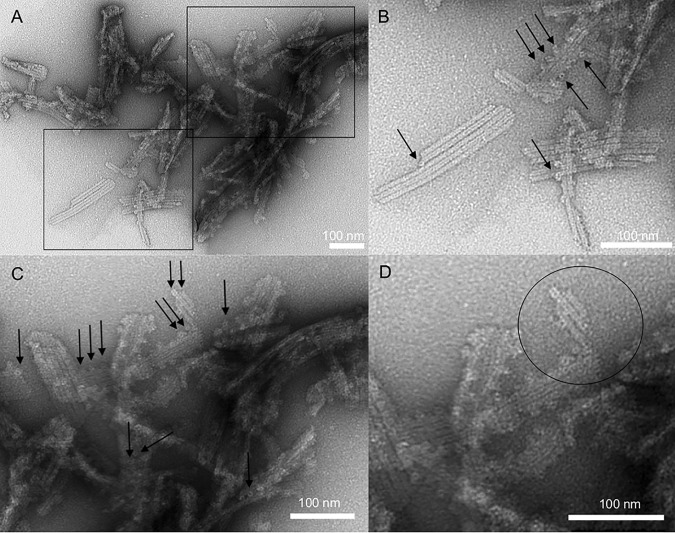
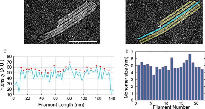
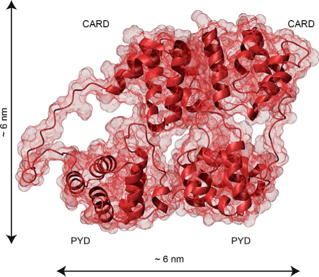
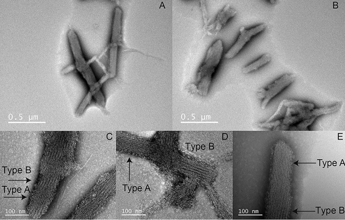
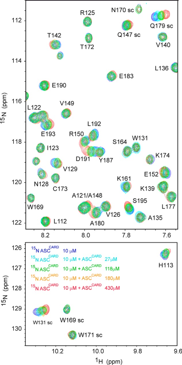
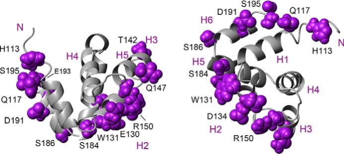
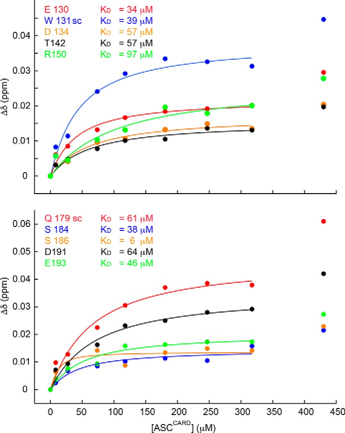
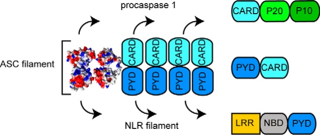
The inflammasome is a multiprotein complex necessary for the onset of inflammation. The adapter protein ASC assembles inflammasome components by acting as a molecular glue between danger-signal sensors and procaspase-1. The assembly is mediated by ASC self-association and protein interactions via its two Death Domains, PYD and CARD. Truncated versions of ASC have been shown to form filaments, but information on the filaments formed by full-length ASC is needed to construct a meaningful model of inflammasome assembly. To gain insights into this system, we used a combination of transmission EM, NMR, and computational analysis to investigate intact ASC structures. We show that ASC forms ∼6-7-nm-wide filaments that stack laterally to form bundles. The structural characteristics and dimensions of the bundles indicate that both PYD and CARD are integral parts of the filament. A truncated version of ASC with only the CARD domain (ASCCARD) forms different filaments (∼3-4-nm width), providing further evidence that both domains work in concert in filament assembly. Ring-shaped protein particles bound to pre-existing filaments match the size of ASC dimer structures generated by NMR-based protein docking, suggesting that the ASC dimer could be a basic building block for filament formation. Solution NMR binding studies identified the protein surfaces involved in the ASCCARD-ASCCARD interaction. These data provide new insights into the structural underpinnings of the inflammasome and should inform future efforts to interrogate this important biological system.

Keywords: ASC; CARD; Death Domain; NMR; PYD; apoptosis; inflammasome; inflammation; protein assembly; transmission electron microscopy.


Conflict of interest statement

The authors declare that they have no conflicts of interest with the contents of this article

Figures

Figure 1.
Figure 1.
Schematic representation of inflammasome assembly based on ASCPYD filament resulting in procaspase 1 activation. The main components of the ASC inflammasome are shown, including the sensor-signal NLR proteins, the adapter ASC, and the interleukin processing enzyme, procaspase 1.
Figure 2.
Figure 2.
Filament formation by full-length human ASC can be controlled by pH. Shown are TEM images of full-length human ASC filaments formed by slow pH increase using dialysis from pH value 3 to 4.1 (A), 6.5 (B), and 7.5 (C). Once assembled, the population of filaments is significantly reduced when the pH value is decreased back to 4.1, although some still persist (D). The number of filaments observed in 10 images at similar magnification (scale bars of 100–200 nm) under each condition of pH is as follows: pH 4.1, 0 filaments; pH 5.1 (image not shown), 22 filaments; pH 6.5, 76 filaments; pH decrease to 4.1 (after 6.5), 19 filaments. Arrows point to potentially forming or already formed filaments.
Figure 3.
Figure 3.
Human ASC filaments stack laterally, forming bundles with potential implications in micrometer-size inflammasomes. A, TEM image of full-length human ASC filament bundles. B, SEM image of ASC filament bundles of a width >100 nm, with potential implication in inflammasome assembly. Filaments were formed by slow pH increase from 3 to 8.2.
Figure 4.
Figure 4.
Stacked filaments of human ASC show dimensions that deviate significantly from the model based on the cryo-EM structure of the truncated protein. Shown are top (A) and lateral (B) views of the human ASCPYD helical tube (cryo-EM, PDB code 3J63) with the dimensions specified in the original published work (8) and containing the 15 ASCPYD domains of the PDB entry. Each group of five ASCPYD domains is colored yellow, red, and blue to show the helical arrangement (A and B). Shown are top (C) and side (D) views of the ASCPYD tube with protruding ASCCARD domains and linker to show CARD availability for potential interactions. The PYDs of 15 full-length NMR structures of ASC molecules (21) (PDB code 2KN6) are superimposed onto the 15 PYD domains of the cryo-EM structure. After superposition, the NMR PYDs were deleted, and only the cryo-EM PYDs and the NMR CARDs are shown. One side of the helical tube is colored red, and the corresponding CARDs are shown in orange to facilitate understanding of directionality. The dimension shown was calculated with MOLMOL (41) (C and D). Shown is a modeled bundle of an arrangement of four ASCPYD helical filaments with protruding CARDs and linker from D. The red side of the PYD tube and corresponding CARDs in orange (from C and D) are shown to follow directionality. CARD domains in filament interfaces are assumed to stack vertically (overlapping as a worst-case scenario), thus occupying approximately the dimension of the ASCCARD (∼3.5 nm). The dimensions shown are as follows: 1) the minimum width of the four-filament bundle, which is estimated by assuming a filament width of 9 nm from the cryo-EM structure and 2) the estimated width range according to the model shown in D for the four-filament bundle (E), which is calculated assuming a filament width of 6–7 nm from our TEM studies.
Figure 5.
Figure 5.
Protein particles ∼7 nm in diameter bind to pre-existing filaments, suggesting a mode for filament lateral growth. Shown are TEM images of full-length human ASC formed at pH 6.5 (A) and pH 8.2 (B). The arrows point to protruding protein material.
Figure 6.
Figure 6.
Filaments of human full-length ASC are formed by the stacking of ring-shaped particles of ∼7 nm in diameter. A, TEM images of ASC filaments formed using method 2 as described under “Experimental procedures.” Rectangles show enlarged areas of A that compose B (bottom rectangle) C (top rectangle). The arrows point to several ring-shaped structures laterally attached to pre-existing filaments. An additional enlargement of the top center region of C forms D. The circle shows stacked rings forming the filaments. Bar length is indicated for each panel.
Figure 7.
Figure 7.
Protomer spacing in bundled human ASC filaments. A, subsection of TEM micrograph of ASC filaments shown in Fig. 6A. Shown are line cuts of 3 pixels, corresponding to 3 nm, using ImageJ along the long axis of each filament in the region of interest. B, as an example, a line cut along filament number 3 is shown in cyan. C, resulting average of intensity profiles over this width. Red arrowheads indicate locations of pixel intensity values above the mean intensity along the filament (green dashed line). A.U., arbitrary units. D, mean values for the separation of maximum peaks from averaged intensity profiles for each of the filaments/line cuts across a total number of 22 filaments. The mean protomer separation is 5.0 nm ± 0.6 nm. Scale bar (A), 100 nm.
Figure 8.
Figure 8.
The dimensions of the ASC dimer agree with the size of the ring-shaped TEM particles. Shown is the contact surface of the ASC dimer structure derived from NMR-based protein docking with HADDOCK (26). The PYD and CARD domains are labeled. Dimensions were calculated with MOLMOL (41).
Figure 9.
Figure 9.
ASCCARD self-associates into filaments structurally different from full-length ASC filaments. ASCCARD forms ∼0.5–1-μm-long bundles with preferred directionality (A and B). Narrower bundles branch out from the thicker bundles at certain positions. ASCCARD forms thin filaments or fibrils of ∼4-nm width (labeled as Type A) and wider filaments probably composed of two narrower filaments (labeled as Type B) (C–E).
Figure 10.
Figure 10.
ASCCARD amino acids involved in the CARD–CARD interaction are mainly located in helices 2, 3, 5, and 6. Shown are superimposed SOFAST-HMQC spectra (27) resulting from the titration of 15N,13C-labeled ASCCARD (constant concentration) with unlabeled ASCCARD (increasing concentrations color-coded in the bottom spectrum). Some signals (e.g. NH side chain of Trp-169, amide NH of Arg-125, Thr-142, and Ser-164) remain unmodified, whereas others show clear changes in δ upon binding (e.g. NH side chain of Trp-131 and Gln-179, amide NH of Thr-142, Arg-150, and Asp-191).
Figure 11.
Figure 11.
Interacting surfaces in ASCCARD self-association. ASCCARD structure (21) is shown as a ribbon diagram in two different views. The amino acids that are exposed to the solvent and with the largest chemical shift perturbations are depicted in purple and labeled in black. Helices are numbered in purple.
Figure 12.
Figure 12.
ASCCARD self-associates with KD ∼50 μm. Shown are chemical shift perturbations of 13C,15N-uniformly labeled ASCCARD versus unlabeled ASCCARD concentration. Representative residues exposed to the solvent with the largest perturbations are shown in both the top and bottom panels, together with the corresponding KD values.
Figure 13.
Figure 13.
Model for ASC-dependent inflammasome assembly based on TEM data. ASC dimer is shown as the minimal building block. The ASC filament shows two interacting sides, one for recruiting procaspase-1 CARD and the other for interaction with the PYD of NLR sensor proteins.

References

    1. Park H. H., Logette E., Raunser S., Cuenin S., Walz T., Tschopp J., and Wu H. (2007) Death domain assembly mechanism revealed by crystal structure of the oligomeric PIDDosome core complex. Cell 128, 533–546 10.1016/j.cell.2007.01.019 - DOI - PMC - PubMed
    1. Park H. H., Lo Y. C., Lin S. C., Wang L., Yang J. K., and Wu H. (2007) The death domain superfamily in intracellular signaling of apoptosis and inflammation. Annu. Rev. Immunol. 25, 561–586 10.1146/annurev.immunol.25.022106.141656 - DOI - PMC - PubMed
    1. Schroder K., and Tschopp J. (2010) The inflammasomes. Cell 140, 821–832 10.1016/j.cell.2010.01.040 - DOI - PubMed
    1. Kersse K., Verspurten J., Vanden Berghe T., and Vandenabeele P. (2011) The death-fold superfamily of homotypic interaction motifs. Trends Biochem. Sci. 36, 541–552 10.1016/j.tibs.2011.06.006 - DOI - PubMed
    1. Chu L. H., Gangopadhyay A., Dorfleutner A., and Stehlik C. (2015) An updated view on the structure and function of PYRIN domains. Apoptosis 20, 157–173 10.1007/s10495-014-1065-1 - DOI - PMC - PubMed

Publication types

MeSH terms

LinkOut - more resources