. 2018 Dec 11;115(50):12757-12762.
doi: 10.1073/pnas.1815287115. Epub 2018 Nov 20.

Molecular structure of the ATP-bound, phosphorylated human CFTR

Affiliations

Molecular structure of the ATP-bound, phosphorylated human CFTR

Zhe Zhang et al. Proc Natl Acad Sci U S A. .

Abstract

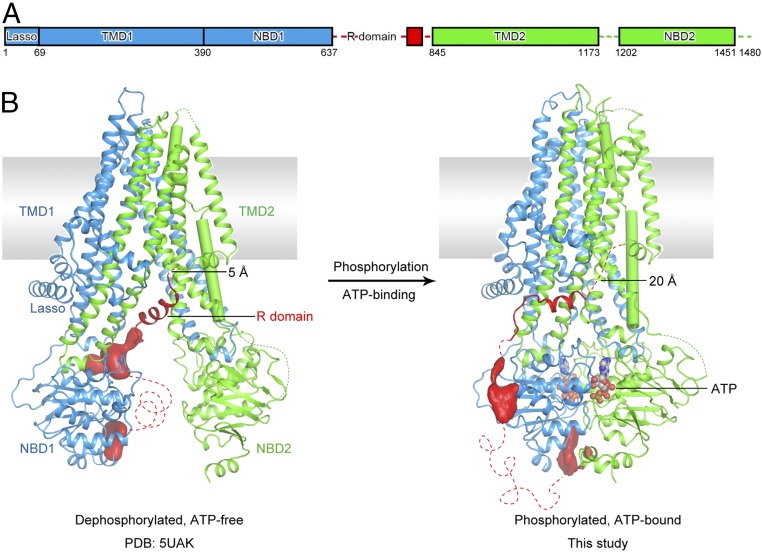
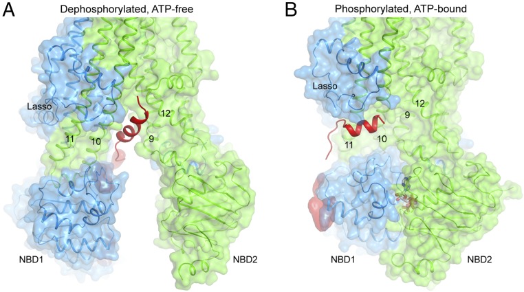
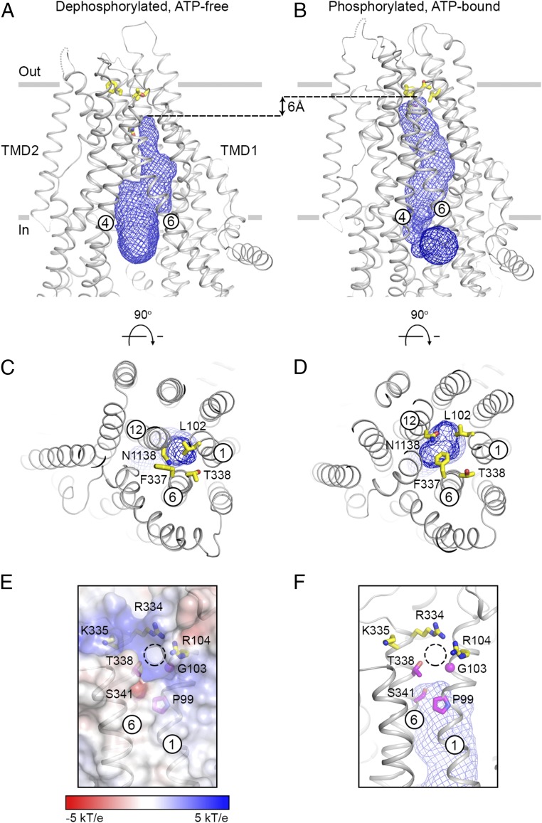
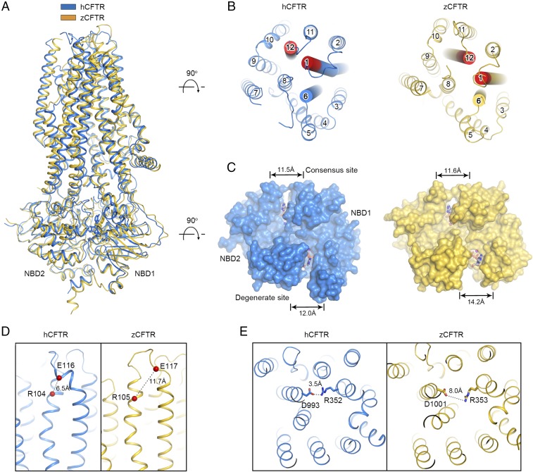
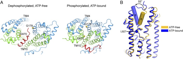
The cystic fibrosis transmembrane conductance regulator (CFTR) is an anion channel important in maintaining proper functions of the lung, pancreas, and intestine. The activity of CFTR is regulated by ATP and protein kinase A-dependent phosphorylation. To understand the conformational changes elicited by phosphorylation and ATP binding, we present here the structure of phosphorylated, ATP-bound human CFTR, determined by cryoelectron microscopy to 3.2-Å resolution. This structure reveals the position of the R domain after phosphorylation. By comparing the structures of human CFTR and zebrafish CFTR determined under the same condition, we identified common features essential to channel gating. The differences in their structures indicate plasticity permitted in evolution to achieve the same function. Finally, the structure of CFTR provides a better understanding of why the G178R, R352Q, L927P, and G970R/D mutations would impede conformational changes of CFTR and lead to cystic fibrosis.

Keywords: ABC transporter; anion channel; cryo-EM; human CFTR.


Conflict of interest statement

The authors declare no conflict of interest.

Figures

Fig. 1.
Fig. 1.
Two functional states of human CFTR. (A) Schematic diagram showing the domain structure of CFTR. The numbers represent the range of residues visible in the cryo-EM map, not the exact boundaries of different domains. (B) Ribbon diagrams of the dephosphorylated, ATP-free conformation (Left; PDB ID code 5UAK) and the phosphorylated, ATP-bound structure (Right). Regions not resolved in the structure are shown as dashed lines. The EM densities, shown in red, correspond to unstructured regions within the R domain. TM 8 is highlighted in cylinder. ATP is shown in ball-and-stick and colored by heteroatom. The distances between the last visible residue in the R domain and the first residue in TMD2 are indicated.
Fig. 2.
Fig. 2.
Conformational changes of the R domain. Close-up view of the intracellular opening in the dephosphorylated, ATP-free (A) and phosphorylated, ATP-bound (B) conformations. The R domain is shown in red. The lasso motif and TM helices 9–12 are indicated.
Fig. 3.
Fig. 3.
Conformational changes of the ion-permeation pathway. Two orthogonal views of the dephosphorylated, ATP-free (A and C) and the phosphorylated, ATP-bound (B and D) structures. The pore, shown as a blue mesh, is defined by a probe with the size of a dehydrated chloride ion (1.7-Å radius). It is connected to the cytosol through an opening between TMs 4 and 6. The side chains of residues L102, F337, T338, and N1138 are shown as yellow sticks. (E and F) The extracellular opening (shown as a dashed circle). CFTR is shown in ribbon along with the electrostatic surface (E) or the pore as blue mesh (F). Residues located around the external region of the opening are shown in stick models. In purple are potential gating residues; in yellow are positively charged residues influencing ion conduction.
Fig. 4.
Fig. 4.
Structural comparison of phosphorylated human and zebrafish CFTR. (A) Superposition of the overall structures. hCFTR is shown in blue, and zCFTR (PDB ID code 5W81) is in yellow. (B) An extracellular view showing the different locations of TMs 1, 6, and 12 (highlighted as cylinders) in the two structures. The extracellular region of TMs 1 and 6, where the residues have the largest displacement between these two structures, are colored in red (residues 107–109 and 1129–1134 of hCFTR, and 107–109 and 1137–1142 of zCFTR). (C) The symmetric NBD dimer in hCFTR versus the asymmetric dimer in zCFTR. The distances shown are between the Cα atoms of the conserved lysine in the Walker A motif and the glycine in the signature motif (LSGGQ) at each ATP-binding site. (D) The distance between R104 and E116 in the two structures. (E) The R352/D993 salt bridge is only observed in hCFTR.
Fig. 5.
Fig. 5.
Disease-causing mutations that obstruct conformational changes. (A) The positions of G178 and G970. R domain is shown in red. (B) Superposition of the hCFTR in the ATP-free and ATP-bound conformations, showing the rotation of the outer leaflet segment of TM 8 and the location of L927.

References

    1. Gadsby DC, Vergani P, Csanády L. The ABC protein turned chloride channel whose failure causes cystic fibrosis. Nature. 2006;440:477–483. - PMC - PubMed
    1. Boat TF, Welsh MJ, Beaudet AL. In: The Metabolic Basis of Inherited Disease. 6th Ed. Scriver CR, Beaudet Al, Sly WS, Valle D, editors. McGraw-Hill; New York: 1989. pp. 2649–2680.
    1. Rommens JM, et al. Identification of the cystic fibrosis gene: Chromosome walking and jumping. Science. 1989;245:1059–1065. - PubMed
    1. Elborn JS. Cystic fibrosis. Lancet. 2016;388:2519–2531. - PubMed
    1. Cheng SH, et al. Phosphorylation of the R domain by cAMP-dependent protein kinase regulates the CFTR chloride channel. Cell. 1991;66:1027–1036. - PubMed

Publication types

MeSH terms

Substances

Associated data

LinkOut - more resources