Review
. 2018 Nov 20;8(1):17081.
doi: 10.1038/s41598-018-35114-y.

p66Shc activation promotes increased oxidative phosphorylation and renders CNS cells more vulnerable to amyloid beta toxicity

Affiliations
Review

p66Shc activation promotes increased oxidative phosphorylation and renders CNS cells more vulnerable to amyloid beta toxicity

Asad Lone et al. Sci Rep. .

Abstract

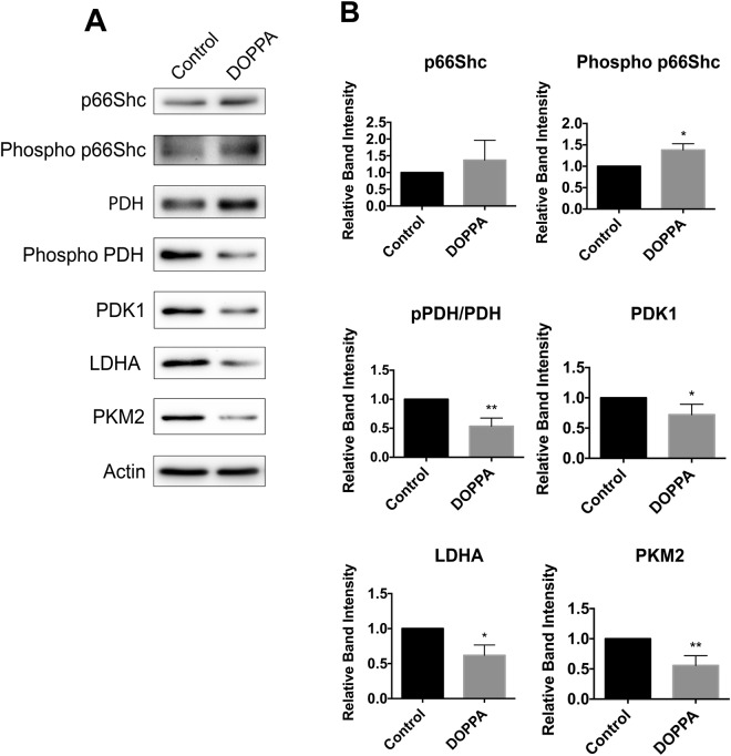
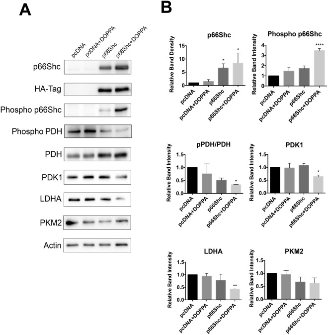
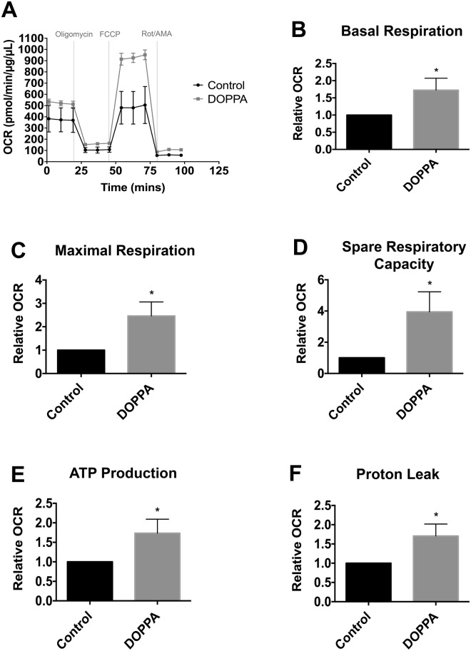
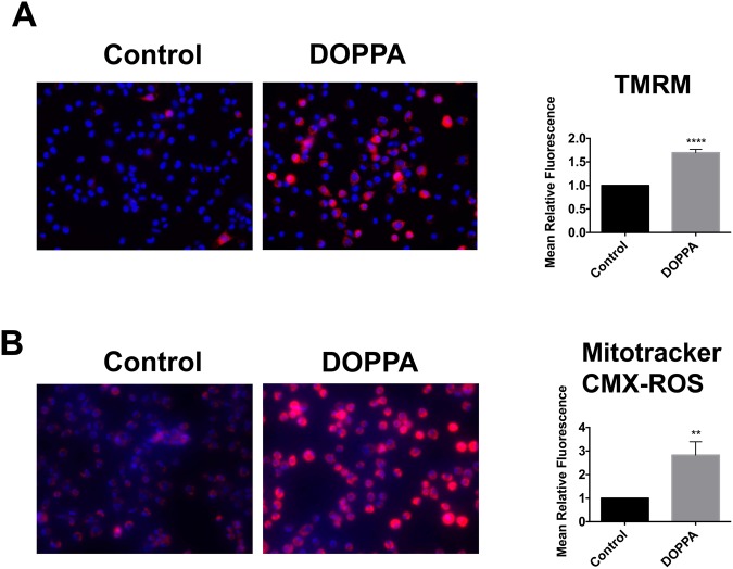
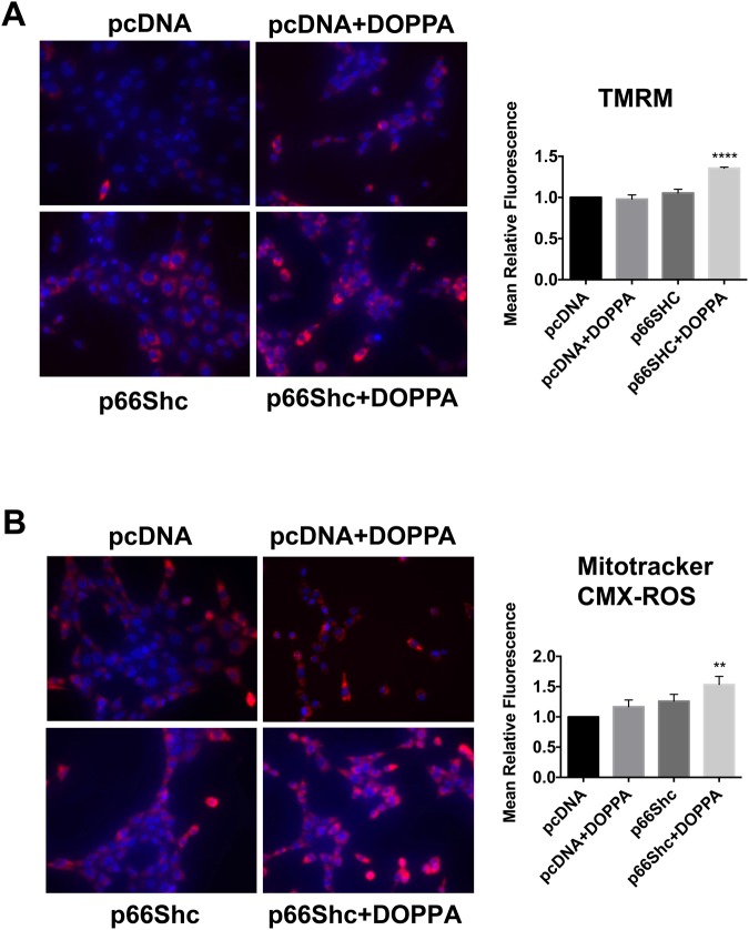
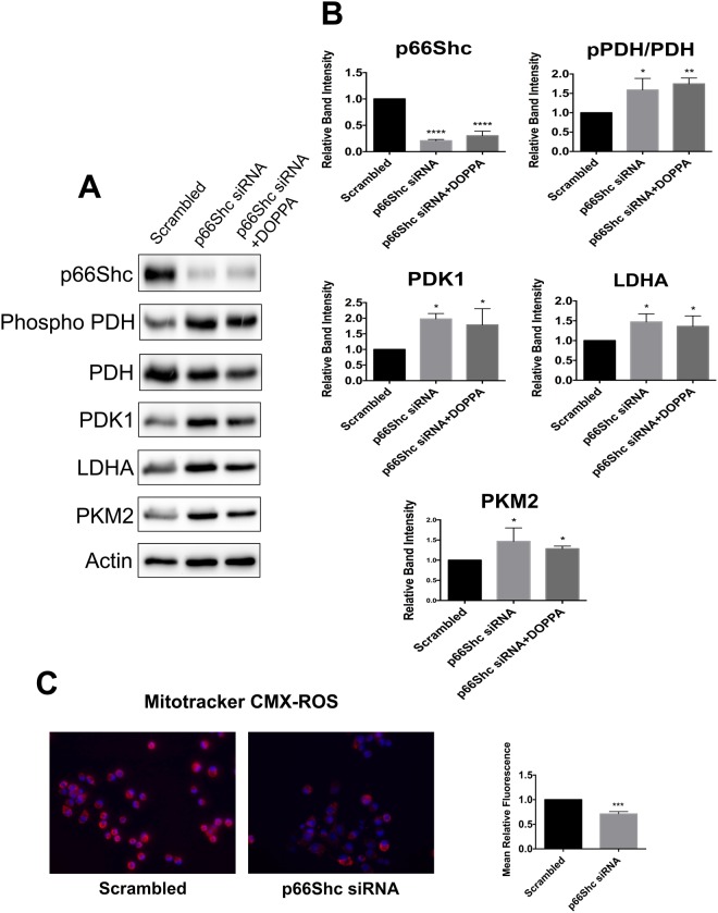
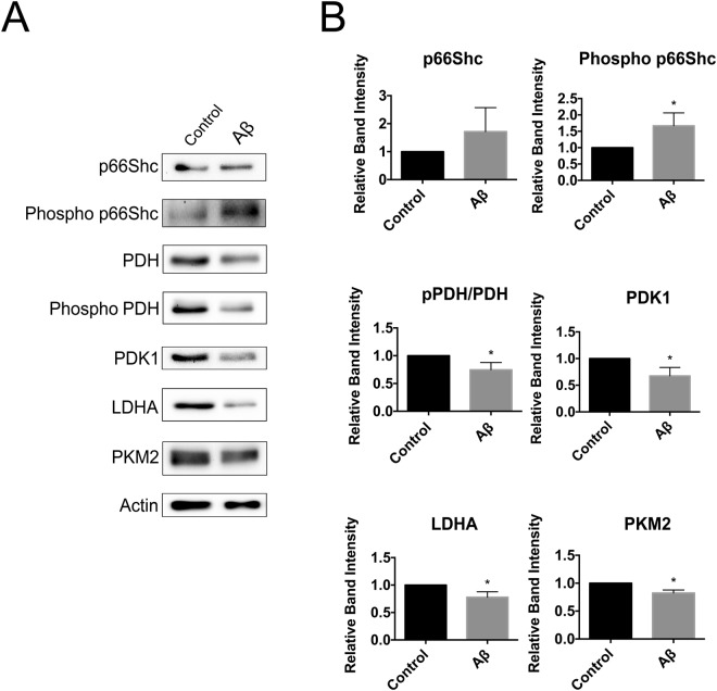
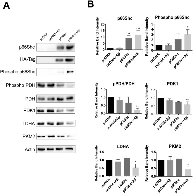
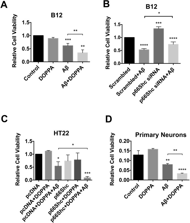
A key pathological feature of Alzheimer's disease (AD) is the accumulation of the neurotoxic amyloid beta (Aβ) peptide within the brains of affected individuals. Previous studies have shown that neuronal cells selected for resistance to Aβ toxicity display a metabolic shift from mitochondrial-dependent oxidative phosphorylation (OXPHOS) to aerobic glycolysis to meet their energy needs. The Src homology/collagen (Shc) adaptor protein p66Shc is a key regulator of mitochondrial function, ROS production and aging. Moreover, increased expression and activation of p66Shc promotes a shift in the cellular metabolic state from aerobic glycolysis to OXPHOS in cancer cells. Here we evaluated the hypothesis that activation of p66Shc in CNS cells promotes both increased OXPHOS and enhanced sensitivity to Aβ toxicity. The effect of altered p66Shc expression on metabolic activity was assessed in rodent HT22 and B12 cell lines of neuronal and glial origin respectively. Overexpression of p66Shc repressed glycolytic enzyme expression and increased both mitochondrial electron transport chain activity and ROS levels in HT22 cells. The opposite effect was observed when endogenous p66Shc expression was knocked down in B12 cells. Moreover, p66Shc activation in both cell lines increased their sensitivity to Aβ toxicity. Our findings indicate that expression and activation of p66Shc renders CNS cells more sensitive to Aβ toxicity by promoting mitochondrial OXPHOS and ROS production while repressing aerobic glycolysis. Thus, p66Shc may represent a potential therapeutically relevant target for the treatment of AD.


Conflict of interest statement

The authors declare no competing interests.

Figures

Figure 1
Figure 1
Activation of endogenous p66Shc in B12 cells promotes a reduction in the levels of aerobic glycolysis enzymes. (A) Immunoblot analysis of extracts from B12 cells revealed increased phosphorylation of p66Shc following 24-hour DOPPA (100 nM) exposure compared to untreated control cells. DOPPA exposure also promoted decreased phosphorylation of pyruvate dehydrogenase (PDH) and led to a reduction in levels of the aerobic glycolysis enzymes pyruvate dehydrogenase kinase 1 (PDK1), lactate dehydrogenase A (LDHA) and pyruvate kinase 2 (PKM2) compared to control cells. (B) Densitometric analysis of blots revealed a significant increase in S36 phosphorylation of p66Shc and a concomitant decrease in PDH phosphorylation and protein levels of PDK1, LDHA and PKM2 following DOPPA exposure. Data presented are the mean ± SEM of 3 independent experiments (*P < 0.05, **P < 0.01).
Figure 2
Figure 2
Ectopic expression and activation of p66Shc in HT22 cells promotes a reduction in aerobic glycolysis enzyme levels. (A) Immunoblot analysis of extracts from HT22 cells transiently transfected with either pcDNA control plasmid or a p66Shc-HA expression vector. DOPPA treatment (100 nM) promoted both increased p66Shc phosphorylation and repressed PDH phosphorylation in p66Shc-HA transfected cells. DOPPA exposure also led to a reduction in levels of the aerobic glycolysis enzymes PDK1, LDHA and PKM2 in p66Shc-HA expressing cells compared to control cells. (B) Densitometric analysis of blots revealed a significant increase in S36 phosphorylation of p66Shc and a concomitant decrease in PDH phosphorylation and protein levels of PDK1, LDHA and PKM2 in p66Shc-HA expressing cells following DOPPA exposure. Data presented are the mean ± SEM of 3 independent experiments (*P < 0.05, **P < 0.01).
Figure 3
Figure 3
Phosphorylation and activation of endogenous p66Shc in B12 cells leads to an increase in mitochondrial oxidative metabolism. (A) Oxygen consumption rate of B12 cells, with and without DOPPA (100 nM) treatment for 24 hours, was measured in real-time using a Seahorse XFe24 Flux Analyzer. After normalization to protein content, B12 cells treated with DOPPA displayed significant increases in (B) basal respiration, (C) maximal respiration, (D) spare respiratory capacity, (E) ATP production, and (F) proton leak when compared to untreated cells. Data presented are the mean ± SEM of 3 independent experiments (*P < 0.05).
Figure 4
Figure 4
p66Shc activation promotes an increase in mitochondrial membrane potential (∆𝜓m) and ROS production in B12 cells. (A) B12 cells were stained with the ∆𝜓m sensitive fluorochrome TMRM (red), while nuclei were stained with Hoechst stain (blue) and visualized by fluorescence microscopy. Quantification of TMRM fluorescence (right panel) revealed a significant elevation of ∆𝜓m in DOPPA (100 nM) treated B12 cells when compared to untreated control cells. (B) B12 cells were stained with Mitotracker CMX-ROS (Red) and visualized by fluorescence microscopy. Quantification of Mitotracker CMX-ROS (right panel) revealed a significant increase in mitochondrial ROS production following DOPPA treatment (100 nM) compared to control cells. Data presented are the mean ± SEM of 3 independent experiments (**P < 0.01; ****P < 0.001).
Figure 5
Figure 5
Ectopic expression of p66Shc in HT22 cells promotes increased mitochondrial membrane potential and ROS production following DOPPA exposure. (A) HT22 cells were transfected with either pcDNA or a p66Shc-HA expression plasmid, treated with DOPPA (100 nM) and stained with TMRM. Stained cells were visualized by fluorescence microscopy and fluorescence intensity was quantified (right panel). (B) HT22 cells transfected as indicated and treated with DOPPA (100 nM) were stained with Mitotracker CMX-ROS and visualized by fluorescence microscopy. Fluorescence intensity of stained cells was quantified (right panel). HT22 cells transfected with p66Shc and treated with DOPPA exhibited significantly higher TMRM and Mitotracker CMX-ROS staining compared to pcDNA control transfected cells. Nuclei were stained with Hoechst stain (blue). Data presented are the mean ± SEM of 3 independent experiments (**P < 0.01; ****P < 0.001).
Figure 6
Figure 6
Silencing p66Shc expression promotes aerobic glycolysis while reducing mitochondrial ROS production. (A) Immunoblot analysis of extracts from B12 cells transfected with p66Shc specific siRNA. Knockdown of p66Shc expression resulted in elevated levels of PDK1, LDHA and PKM2 in addition to increased phosphorylation of PDH. This effect was also observed in B12 cells with silenced p66Shc expression treated with DOPPA. (B) Densitometric analysis of immunoblots. (C) Mitotracker CMX-ROS (red) staining was significantly decreased in B12 cells with silenced p66Shc expression when compared to control cells. Nuclei were stained with Hoechst stain (blue). Data presented are the mean ± SEM of 3 independent experiments (*P < 0.05, **P < 0.01; ***P < 0.001).
Figure 7
Figure 7
Aβ exposure promotes p66Shc activation and a reduction in aerobic glycolysis enzyme levels in B12 cells. (A) Immunoblot analysis of B12 cells treated with Aβ1–42 (20 µM) for 24 hours. (B) Densitometric analysis of immunoblots revealed a significant increase in p66Shc phosphorylation and a concomitant decrease in PDH phosphorylation and levels of PDK1, LDHA, and PKM2 following Aβ exposure. Data presented are the mean ± SEM of 3 independent experiments (*P < 0.05).
Figure 8
Figure 8
Aβ exposure promotes activation of ectopically expressed p66Shc in HT22 cells and a reduction in aerobic glycolysis. (A) Immunoblot analysis of extracts from HT22 cells transfected with the indicated plasmids and treated with Aβ1–42 (20 µM) for 24 hours. (B) Densitometric analysis of immunoblots revealed that Aβ exposure promoted a significant increase in p66Shc phosphorylation while repressing PDH phosphorylation. Aβ treatment also promoted a significant decrease in the levels of PDK1, LDHA, and PKM2 in HT22 cells ectopically expressing p66Shc compared to pcDNA transfected control cells. Data presented are the mean ± SEM of 3 independent experiments (*P < 0.05; **P < 0.01; ***P < 0.001).
Figure 9
Figure 9
p66Shc activation enhances Aβ toxicity. (A) Treatment of B12 cells with both Aβ1–42 (20 µM) and DOPPA (100 nM) was significantly more toxic than Aβ treatment alone. (B) Silencing of p66Shc expression in B12 cells led to reduced Aβ-induced toxicity compared to B12 cells transfected with control siRNA and treated with Aβ. (C) HT22 cells ectopically expressing p66Shc and treated with DOPPA (100 nM) exhibited significantly decreased viability following Aβ treatment compared to pcDNA control cells treated with both agents. (D) DOPPA induced activation of p66Shc exacerbated Aβ toxicity in mouse primary cortical neurons. Data presented are the mean ± SEM of 3 independent experiments (*P < 0.05; **P < 0.01; ***P < 0.001; ****P < 0.0001).

Similar articles

Cited by

References

    1. Mosconi L, Pupi A, De Leon MJ. Brain glucose hypometabolism and oxidative stress in preclinical Alzheimer’s disease. Ann N. Y. Acad Sci. 2008;1147:180–95. doi: 10.1196/annals.1427.007. - DOI - PMC - PubMed
    1. Nistor M, et al. Alpha-and beta-secretase activity as a function of age and beta-amyloid in Down syndrome and normal brain. Neurobiol Aging. 2007;28:1493–1506. doi: 10.1016/j.neurobiolaging.2006.06.023. - DOI - PMC - PubMed
    1. Pedersen JT, Sigurdsson EM. Tau immunotherapy for Alzheimer’s disease. Trends Mol Med. 2015;21:394–402. doi: 10.1016/j.molmed.2015.03.003. - DOI - PubMed
    1. Masters CL, et al. Amyloid plaque core protein in Alzheimer disease and Down syndrome. Proc Natl Acad Sci. 1985;82:4245–4249. doi: 10.1073/pnas.82.12.4245. - DOI - PMC - PubMed
    1. Morris JC, et al. Cerebral amyloid deposition and diffuse plaques in “normal” aging: Evidence for presymptomatic and very mild Alzheimer’s disease. Neurol. 1996;46:707–719. doi: 10.1212/WNL.46.3.707. - DOI - PubMed

Publication types

MeSH terms

Substances