Review
. 2019 Mar;15(3):139-154.
doi: 10.1038/s41574-018-0126-x.

The obese adipose tissue microenvironment in cancer development and progression

Affiliations
Review

The obese adipose tissue microenvironment in cancer development and progression

Daniela F Quail et al. Nat Rev Endocrinol. 2019 Mar.

Abstract

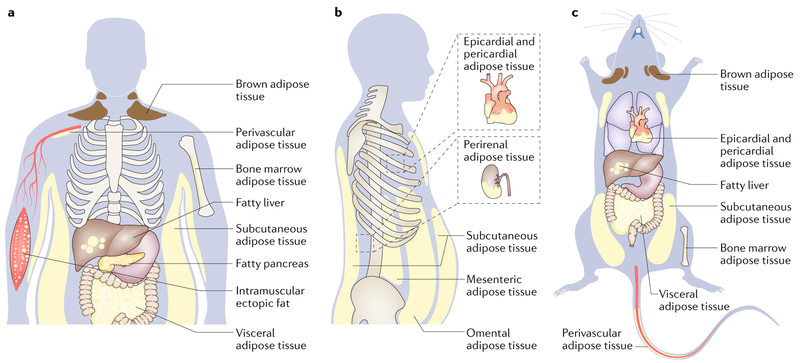
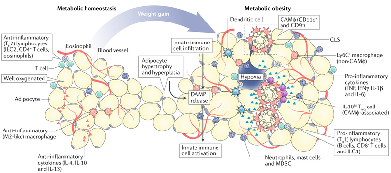
Obesity is associated with both increased cancer incidence and progression in multiple tumour types, and is estimated to contribute to up to 20% of cancer-related deaths. These associations are driven, in part, by metabolic and inflammatory changes in adipose tissue that disrupt physiological homeostasis both within local tissues and systemically. However, the mechanisms underlying the obesity-cancer relationship are poorly understood. In this Review, we describe how the adipose tissue microenvironment (ATME) evolves during body-weight gain, and how these changes might influence tumour initiation and progression. We focus on multiple facets of ATME physiology, including inflammation, vascularity and fibrosis, and discuss therapeutic interventions that have the potential to normalize the ATME, which might be translationally relevant for cancer prevention and therapy. Given that the prevalence of obesity is increasing on an international scale, translational research initiatives are urgently needed to provide mechanistic explanations for the obesity-cancer relationship, and how to best identify high-risk individuals without relying on crude measures, such as BMI.


Conflict of interest statement

Competing interests

The authors declare no competing interests.

Figures

Fig. 1 |
Fig. 1 |. Major adipose depots and anatomical locations in adult humans and mice.
There are two major types of adipose tissue, Lipid-rich white adipose tissue (WAT; energy storing) and mitochondria-rich brown adipose tissue (BAT; energy burning). Adipocytes from BAT and WAT emerge from distinct cell fate lineages; however, WAT can convert to metabolically active fat through the process of browning. WAT is found in many anatomical locations. The largest WAT depots are subcutaneous (under the skin; for example, inguinal, gluteal and femoral) and visceral (within the abdominal cavity, between the organs; for example, omental, mesenteric and epicardial). Smaller WAT depots are found around blood vessels (perivascular, which regulate vascular tone), within the bone marrow (which regulate bone remodelling), or as ectopic depots within specific organs (for example, non-alcoholic fatty liver disease, fatty pancreas or intramuscular fat). These adipose depots have unique molecular features; therefore, weight distribution has important biological consequences. For example, most visceral adipocytes have slightly higher mitochondrial density, are more lipolytic, are less sensitive to insulin, produce less leptin, and contribute more circulating free fatty acids compared to subcutaneous adipocytes, and therefore have a stronger association with the metabolic syndrome. Part a depicts the general distribution of fat in humans from a forward view and part b depicts types of visceral fat depots from a side view. Part c depicts the general distribution of fat in mice from a supine view.
Fig. 2 |
Fig. 2 |. Evolution of the adipose tissue microenvironment during obesity.
During healthy body-weight conditions (metabolic homeostasis), the adipose tissue microenvironment (ATME) is well-vascularized and rich in anti-inflammatory cytokines (such as IL-4, IL-10 and IL-13), and as a consequence, hosts a variety of type 2 immune cells, including alternatively activated (M2-like) macrophages, group 2 innate lymphoid cells, type 2 T helper (TH2) cells and IL-4-producing eosinophils. In response to body-weight gain or metabolic obesity, adipocytes undergo hyperplasia and hypertrophy; as the vascular supply becomes limited, these cells become stressed or die. This releases damage-associated molecular patterns (DAMP) into the microenvironment, which trigger the infiltration and activation of innate immunecells (for example, dendritic cells, macrophages and granulocytes). These effects promote the development of crown-like structures (CLS) and type 1 (pro-inflammatory) immune responses. This response includes accumulation of type 1 cytokines (for example TNF, IFNγ, IL-1β and IL-6), and pro-inflammatory immune cells, including various granulocytes, group 1 innate lymphoid cells, B cells and CD8+T cells, which perpetuate chronic inflammatory responses. Macrophages are highly diverse within the obese ATME; those associated with CLS (CLS-associated macrophage, CAMφ) proliferate and express the cell surface markers CD11c and CD9, while those that are further away (non-CAMφ) express lymphocyte antigen 6C (Ly6C). These inflammatory changes coincide with chronic fibrosis and vascular inflammation, which feed-forward to sustain inflammation. IL-10hi Treg cells, T regulatory cells producing high levels of IL-10; ILC1, group 1 innate lymphoid cells; MDSC, myeloid-derived suppressor cells.
Fig. 3 |
Fig. 3 |. Interactions between tumour cells and cells within the obese adipose tissue microenvironment.
Tumour cell biology is affected by multiple cellular players in the adipose tissue microenvironment (ATME). The enzyme neutrophil elastase cleaves insulin receptor substrate 1 (IRS1), leading to insulin resistance and elevated levels of free insulin, which can enhance phosphoinositide 3-kinase (PI3K) signalling within tumour cells. Mast cells produce various proteases such as cathepsin S, which can promote cancer progression and therapeutic resistance to cytotoxic therapies. Adipose stromal cells and myofibroblasts promote extracellular matrix (ECM) remodelling and facilitate tumour angiogenesis through vascular endothelial growth factor (VEGF) production. However, angiogenic vessels are dysfunctional, and when combined with adipocyte hypertrophy this triggers innate immune responses by macrophages and dendritic cells. Macrophages form crown-like structures (CLS) that not only serve as biomarkers for the metabolic syndrome, but also produce high levels of cytokines (promote vascular permeability) and cause desmoplasia, which together support tumour progression. At the same time, dendritic cells become chronically activated through the suppression of tolerogenic signalling pathways such as the canonical WNT and peroxisome proliferator-activated receptor-γ (PPARγ) pathways, eventually leading to T cell exhaustion. T cell activation is further suppressed by myeloid-derived suppressor cells (MDSCs), which engage co-inhibitory checkpoint molecules on T cells (such as programmed cell death 1; PD-1). Together, each of these cell types facilitates evolution of the tumour microenvironment and disease progression. MAPK, mitogen-activated protein kinase; TGFβ, transforming growth factor-β.
Fig. 4 |
Fig. 4 |. Vascular inflammation and fibrosis in the obese adipose tissue microenvironment.
During obesity, adipose tissue expands rapidly, leading to high demand for a vascular supply. Similar to a growing tumour, this effect results in regions where vascular supply is insufficient, creating areas of hypoxia. Hypoxia triggers angiogenesis through induction of pro-angiogenic factors (such as vascular endothelial growth factor (VEGF) and hypoxia-inducible factor 1α (HIF1α)); however, resulting blood vessels are poorly functional, and thus low oxygen levels persist. Chronic hypoxia eventually contributes to adipocyte death, and supports the formation of crown-like structures (CLS). Vascular integrity is in part reduced by downregulation of endothelial adhesions (such as zonula occludens 1 (ZO1) and ZO2) in responseto obesity-derived factors (for example, lipopolysaccharide and palmitate), while leukocyte adhesion molecules are increased to facilitate infiltration of immune cells (such as T cells). Adiponectin can reverse these effects and improve vascular integrity. In addition, prospero homeobox protein 1 (PROX1)+ lymphatic endothelial cells use lipids to stimulate lymphangiogenesis through fatty acid oxidation (FAO) and production of acetyl-CoA. The extracellular matrix is also aberrant; CLS-associated macrophages (CAMφ) and myofibroblasts contribute to increased tissue stiffness in the obese adipose tissue microenvironment, and adipocytes produce high levels of collagen VI, which further support adipocyte hyperplasia.

References

    1. James WPT & McPherson K The costs of overweight. Lancet Public Health 2, e203–e204 (2017). - PubMed
    1. Malik VS, Willett WC & Hu FB Global obesity: trends, risk factors and policy implications. Nat. Rev. Endocrinol 9, 13–27 (2013). - PubMed
    1. NCD Risk Factor Collaboration. Trends in adult body-mass index in 200 countries from 1975 to 2014: a pooled analysis of 1698 population-based measurement studies with 19.2 million participants. Lancet 387, 1377–1396 (2016). - PMC - PubMed
    1. Ng M et al. Global, regional, and national prevalence of overweight and obesity in children and adults during 1980–2013: a systematic analysis for the Global Burden of Disease Study 2013. Lancet 384, 766–781 (2014). - PMC - PubMed
    1. Hales CM, Fryar CD, Carroll MD, Freedman DS& Ogden CL Trends in obesity and severe obesity prevalence in US youth and adults by sex and age, 2007–2008 to 2015–2016. JAMA 319, 1723–1725 (2018). - PMC - PubMed

Publication types