. 2018 Oct 18:9:1187.
doi: 10.3389/fphar.2018.01187. eCollection 2018.

Astilbin Inhibits High Glucose-Induced Inflammation and Extracellular Matrix Accumulation by Suppressing the TLR4/MyD88/NF-κB Pathway in Rat Glomerular Mesangial Cells

Affiliations

Astilbin Inhibits High Glucose-Induced Inflammation and Extracellular Matrix Accumulation by Suppressing the TLR4/MyD88/NF-κB Pathway in Rat Glomerular Mesangial Cells

Fang Chen et al. Front Pharmacol. .

Abstract

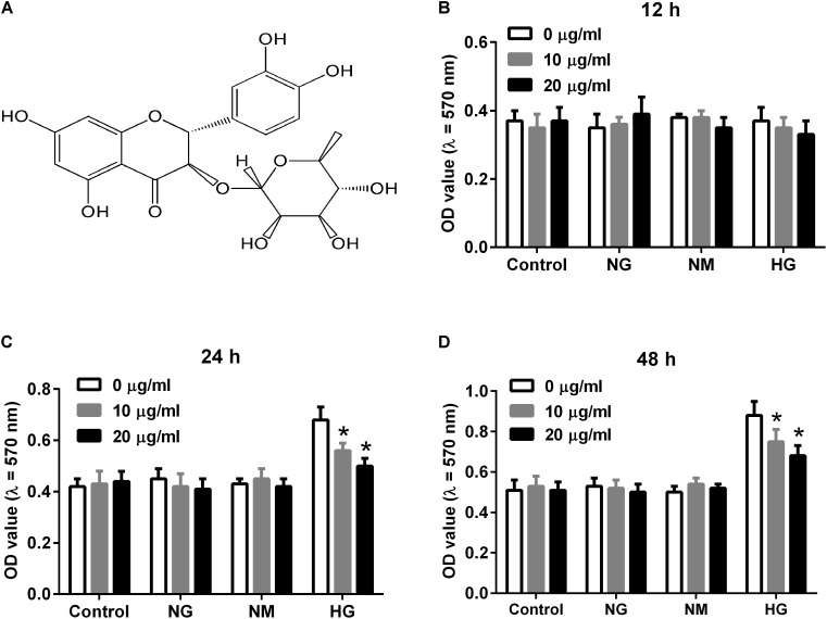
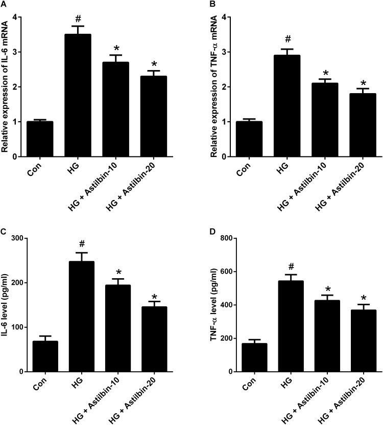
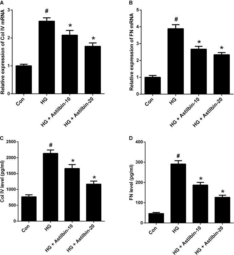
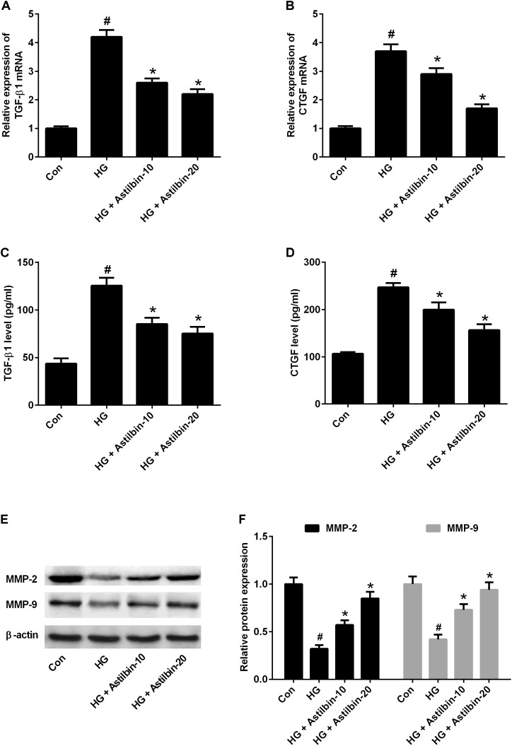
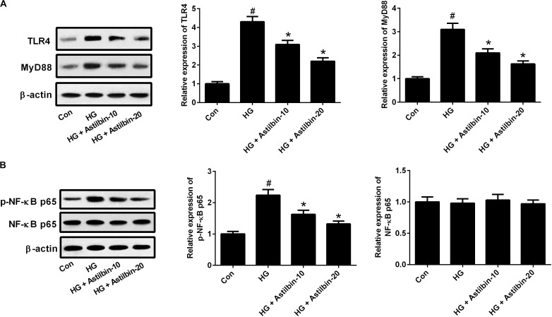
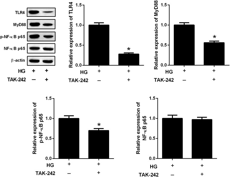
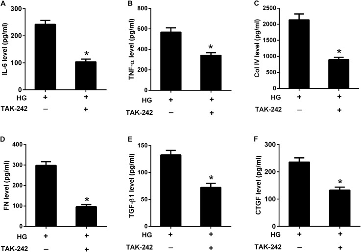
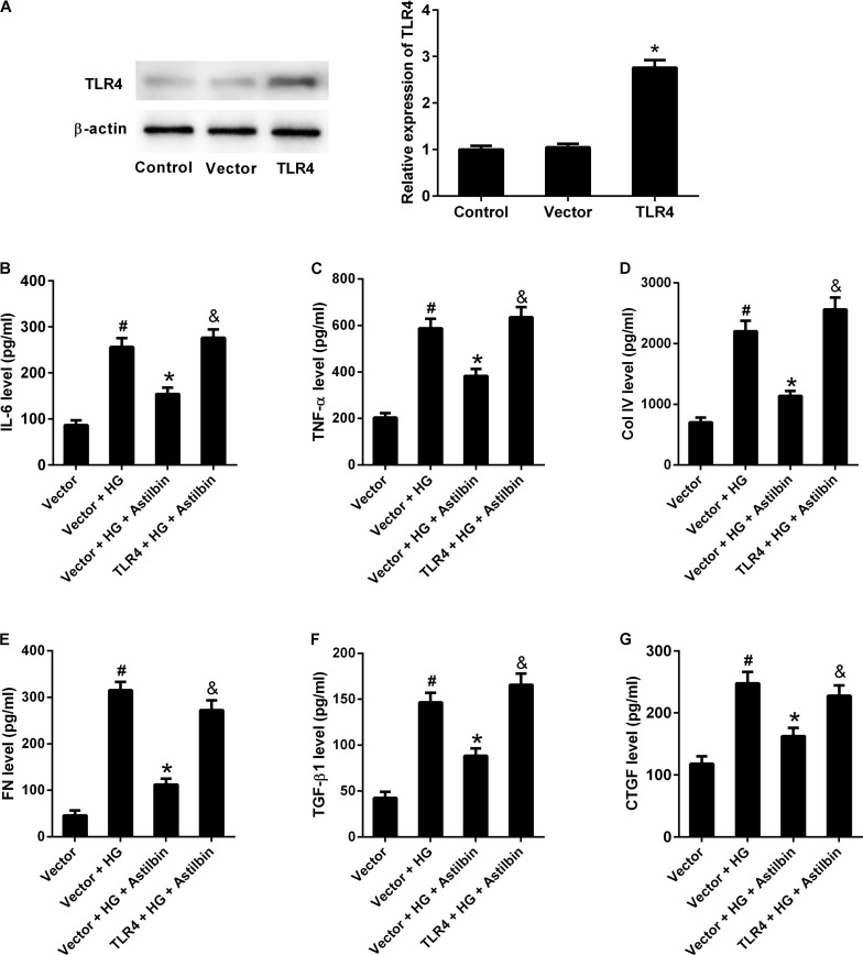
Diabetic nephropathy (DN) is characterized by inflammatory responses and extracellular matrix (ECM) accumulation. Astilbin is an active natural compound and possesses anti-inflammatory activity. The aim of this study was to evaluate the anti-inflammatory effect of astilbin on high glucose (HG)-induced glomerular mesangial cells and the potential mechanisms. The results showed that HG induced cell proliferation of HBZY-1 cells in a time-dependent manner, and astilbin inhibited HG-induced cell proliferation. The expression and secretion of inflammatory cytokines, including interleukin-6 (IL-6) and tumor necrosis factor alpha (TNF-α), and ECM components, including collagen IV (Col IV) and fibronectin (FN), were induced by HG. Moreover, TGF-β1 and CTGF were also induced by HG. The induction by HG on inflammatory response and ECM accumulation was inhibited after astilbin treatment. Astilbin treatment also attenuated HG-induced decrease in expression of matrix metalloproteinase (MMP)-2 and MMP-9. The TLR4/MyD88/NF-κB pathway was activated by HG, and the inhibitor of TLR4 exhibited the same effect to astilbin on reversing the induction of HG. TLR4 overexpression attenuated the effect of astilbin on HG-induced inflammatory cytokine production and ECM accumulation. The results suggested that astilbin attenuated inflammation and ECM accumulation in HG-induced rat glomerular mesangial cells via inhibiting the TLR4/MyD88/NF-κB pathway. This work provided evidence that astilbin can be considered as a potential candidate for DN therapy.

Keywords: TLR4/MyD88/NF-κB pathway; astilbin; diabetic nephropathy; extracellular matrix accumulation; inflammation.


Figures

FIGURE 1
FIGURE 1
Effect of astilbin on HG-induced proliferation of HBZY-1 cells. Cells were cultured under normal glucose condition (NG, 5.5 mM D-glucose), mannitol condition (NM, 5.5 mM D-glucose + 24.5 mM mannitol), or high glucose condition (HG, 30 mM D-glucose) in the presence of astilbin (0, 10, and 20 μg/ml) for 12, 24, or 48 h. (A) Chemical structure of astilbin. Cell proliferation was detected using MTT assay after incubation for 12 (B), 24 (C), and 48 h (D). P < 0.05 vs. HG-stimulated group (without astilbin). Statistical significance was determined by one-way ANOVA.
FIGURE 2
FIGURE 2
Effect of astilbin on HG-induced inflammatory cytokine production. HBZY-1 cells were treated with 10 and 20 μg/ml of astilbin under HG condition for 24 h. The mRNA levels of IL-6 (A) and TNF-α (B) in HBZY-1 cells were detected by qRT-PCR. The secretion levels of IL-6 (C) and TNF-α (D) in cell supernatant were detected by ELISA. #P < 0.05 vs. Con (control group). P < 0.05 vs. HG-stimulated group. Statistical significance was determined by one-way ANOVA.
FIGURE 3
FIGURE 3
Effect of astilbin on HG-induced ECM accumulation. HBZY-1 cells were treated with 10 and 20 μg/ml of astilbin under HG condition for 24 h. The mRNA levels of Col IV (A) and FN (B) in HBZY-1 cells were detected by qRT-PCR. The secretion levels of Col IV (C) and FN (D) in cell supernatant were detected by ELISA. #P < 0.05 vs. Con (control group). P < 0.05 vs. HG-stimulated group. Statistical significance was determined by one-way ANOVA.
FIGURE 4
FIGURE 4
Effect of astilbin on HG-induced the expression of TGF-β1, CTGF, MMP-2, and MMP-9. HBZY-1 cells were treated with 10 and 20 μg/ml of astilbin under HG condition for 24 h. The mRNA levels of TGF-β1 (A) and CTGF (B) in HBZY-1 cells were detected by qRT-PCR. The secretion levels of TGF-β1 (C) and CTGF (D) in cell supernatant were detected by ELISA. (E) The expression levels of MMP-2 and MMP-9 were determined by western blot. #P < 0.05 vs. Con (control group). P < 0.05 vs. HG-stimulated group. Statistical significance was determined by one-way ANOVA.
FIGURE 5
FIGURE 5
Effect of astilbin on HG-induced activation of the TLR4/MyD88 (A) and (B) NF-κB pathways. HBZY-1 cells were treated with 10 and 20 μg/ml of astilbin under HG condition for 24 h. The expression levels of TLR4, MyD88, p-NF-κB p65, and NF-κB p65 were measured by western blot. #P < 0.05 vs. Con (control group). P < 0.05 vs. HG-stimulated group. Statistical significance was determined by one-way ANOVA.
FIGURE 6
FIGURE 6
The TLR4/MyD88 and NF-κB pathways formed a signaling axis. HBZY-1 cells were treated with TAK-242 under HG condition for 24 h. The expression levels of TLR4, MyD88, p-NF-κB p65, and NF-κB p65 were measured by western blot. P < 0.05. Statistical significance was determined by Student’s t-test.
FIGURE 7
FIGURE 7
Inhibition of the TLR4/MyD88/NF-κB pathway suppressed HG-induced inflammation and ECM accumulation. HBZY-1 cells were treated with TAK-242 under HG condition for 24 h. The contents of IL-6 (A), TNF-α (B), Col IV (C), FN (D), TGF-β1 (E), and CTGF (F) in cell supernatant were measured by ELISA. P < 0.05. Statistical significance was determined by Student’s t-test.
FIGURE 8
FIGURE 8
TLR4 overexpression attenuated the effect of astilbin on HG-induced inflammation and ECM accumulation. HBZY-1 cells were transfected with empty pcDNA3.1 vector (Vector) or pcDNA3.1-TLR4 (TLR4) for 24 h, and treated with 20 μg/ml of astilbin under HG condition for another 24 h. (A) Transfection efficiency was examined by western blot 48 h after transfection. The contents of IL-6 (B), TNF-α (C), Col IV (D), FN (E), TGF-β1 (F), and CTGF (G) in cell supernatant were measured by ELISA. #P < 0.05 vs. Vector group (cells were transfected with pcDNA3.1 vector). *P < 0.05 vs. Vector + HG group. &P < 0.05 vs. Vector + HG + Astilbin group. Statistical significance was determined by one-way ANOVA.

References

    1. Barutta F., Bruno G., Grimaldi S., Gruden G. (2015). Inflammation in diabetic nephropathy: moving toward clinical biomarkers and targets for treatment. Endocrine 48 730–742. 10.1007/s12020-014-0437-1 - DOI - PubMed
    1. Chen F., Sun Z., Zhu X., Ma Y. (2018). Astilbin inhibits high glucose-induced autophagy and apoptosis through the PI3K/Akt pathway in human proximal tubular epithelial cells. Biomed. Pharmacother. 106 1175–1181. 10.1016/j.biopha.2018.07.072 - DOI - PubMed
    1. Chen X. M., Qi W., Pollock C. A. (2009). CTGF and chronic kidney fibrosis. Front. Biosci. 1 132–141. 10.2741/s13 - DOI - PubMed
    1. Del Prete D., Anglani F., Forino M., Ceol M., Fioretto P., Nosadini R., et al. (1997). Down-regulation of glomerular matrix metalloproteinase-2 gene in human NIDDM. Diabetologia 40 1449–1454. 10.1007/s001250050848 - DOI - PubMed
    1. Dugbartey G. J. (2017). Diabetic nephropathy: a potential savior with ’rotten-egg’ smell. Pharmacol. Rep. 69 331–339. 10.1016/j.pharep.2016.11.004 - DOI - PubMed