. 2018 Nov 6:9:2630.
doi: 10.3389/fmicb.2018.02630. eCollection 2018.

Lactobacillus crispatus BC5 Interferes With Chlamydia trachomatis Infectivity Through Integrin Modulation in Cervical Cells

Affiliations

Lactobacillus crispatus BC5 Interferes With Chlamydia trachomatis Infectivity Through Integrin Modulation in Cervical Cells

Carola Parolin et al. Front Microbiol. .

Abstract

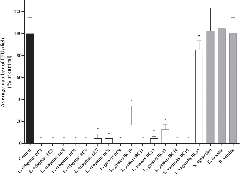
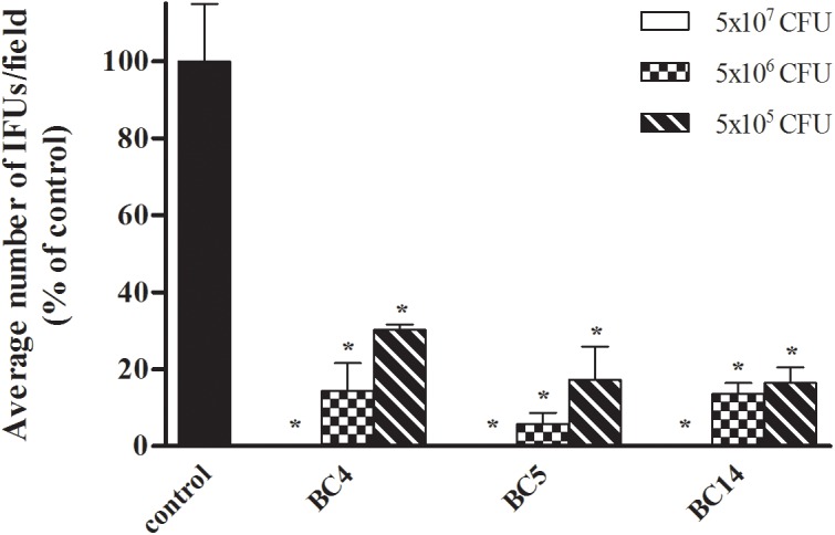
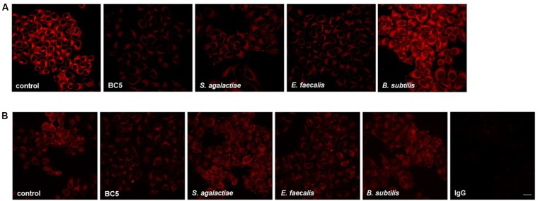
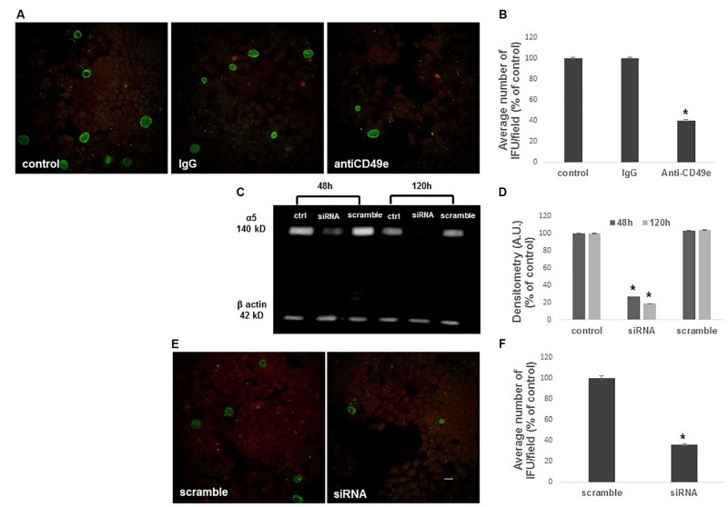
Lactobacilli play a crucial role in maintaining the ecological equilibrium of the vaginal niche, preventing the colonization of exogenous microorganisms. Although many studies have discussed the mechanisms displayed by lactobacilli in counteracting several urogenital pathogens, a few data are available on the interaction between lactobacilli and Chlamydia trachomatis. This study aimed to elucidate the molecular bases of the interaction among vaginal lactobacilli, the sexually transmitted pathogen C. trachomatis and the epithelial cervical cells. We evaluated the in vitro activity of 15 Lactobacillus strains, belonging to different species (i.e., L. crispatus, L. gasseri, L. vaginalis), against C. trachomatis. In particular, we evaluated the capability of lactobacilli cells to interfere with C. trachomatis infection in HeLa cells, by exclusion assays. Lactobacilli significantly reduced C. trachomatis infectivity, being L. crispatus the most active species. Although a dose-dependent effect was noticed, a significant antagonistic activity was maintained even at lower doses. As other Gram-positive bacteria (i.e., Streptococcus agalactiae, Enterococcus faecalis, and Bacillus subtilis) failed to interfere with C. trachomatis infectivity, Lactobacillus activity proved to be specific. The potential mechanism of protection was investigated in Lactobacillus crispatus BC5, chosen as the model strain. The incubation of HeLa cell line with BC5 cells induced important modifications in the epithelial plasma membrane, by altering lipid composition and α5 integrin subunit exposure. When α5 integrin subunits were masked by a specific blocking antibody or ITGA5 gene expression was silenced, Chlamydia infection was significantly reduced. It follows that α5 integrin subunit is crucial for the pathogen infection process, and the anti-Chlamydia activity can be directly linked to membrane properties modifications in cervical cells. The three Gram-positive bacteria used as controls failed to modify the expression of α5β1 integrin. In conclusion, we identified a potential molecular mechanism at the basis of the protection exerted by L. crispatus BC5 against C. trachomatis, getting insights into the role of the cervico-vaginal microbiota for the woman's health.

Keywords: Chlamydia trachomatis; HeLa cells; STIs; integrin; lactobacilli; probiotics; women health.


Figures

FIGURE 1
FIGURE 1
Antagonistic activity of vaginal lactobacilli cells against Chlamydia trachomatis infectivity in exclusion assays. Exclusion experiments were performed at the dose of 5 × 10⁷ lactobacilli cells in HeLa cells. Streptococcus agalactiae, Enterococcus faecalis, and Bacillus subtilis were used as reference Gram-positive bacteria. C. trachomatis infectivity was evaluated as number of IFUs/microscopic field. Results were expressed in percentage compared with control, taken as 100% (black bar). Bars represent median values, error bars represent median absolute deviations. Statistical significance was calculated vs. control. P ≤ 0.01.
FIGURE 2
FIGURE 2
Dose–response effect of Lactobacillus cells on C. trachomatis infectivity in HeLa cells. Exclusion experiments were performed at different doses (5 × 10⁷, 5 × 10⁶, and 5 × 10⁵ cells) for L. crispatus BC4, L. crispatus BC5, and L. gasseri BC14 strains. C. trachomatis infectivity was evaluated as number of IFUs/microscopic field. Results were expressed in percentage compared with control, taken as 100% (black bars). Bars represent median values, error bars represent median absolute deviations. Statistical significance was calculated vs. control. P ≤ 0.01.
FIGURE 3
FIGURE 3
Membrane lipid organization and α5 integrin exposure on HeLa cells incubated with microorganisms. (A) HeLa cells were incubated with L. crispatus BC5, S. agalactiae, E. faecalis, or B. subtilis for 1 h and then stained with NR. (B) HeLa cells were incubated with L. crispatus BC5, S. agalactiae, E. faecalis, B. subtilis for 1 h and then stained for α5 integrin subunit. IgG represents specificity staining control. Representative micrographs are shown. Experiments were repeated at least three times with similar results. Bar: 20 μm.
FIGURE 4
FIGURE 4
Inhibition of C. trachomatis infection by α5 integrin subunit blocking or ITGA5 gene silencing. HeLa cells were treated or not with an anti-CD49e antibody or control IgG for 1 h, then infected with CT EBs for 48 h. (A,B) Specimens were stained for chlamydial membrane lipopolysaccharide antigen. Representative micrographs are shown. C. trachomatis infectivity was evaluated as number of IFUs/microscopic field. Results were expressed in percentage compared with control, taken as 100%. Bars represent median values, error bars represent median absolute deviations. Statistical significance was calculated vs. control. P ≤ 0.01. (C,D) Western blotting of α5 integrin subunit expression in control, ITGA5 siRNA and scramble Hela cells, evaluated at 48 and 120 h post-siRNA. Quantification of α5 integrin subunit was normalized on β-actin. Bars represent mean values based on three independent experiments, error bars represent standard deviations. (E,F) HeLa cells treated with siRNA or scramble were infected with CT EBs for 48 h, and then stained for chlamydial membrane lipopolysaccharide antigen. Bar: 20 μm. Results were expressed in percentage compared with scramble, taken as 100%. Bars represent median values, error bars represent median absolute deviations. Statistical significance was calculated vs. control. P ≤ 0.01.

Similar articles

Cited by

References

    1. Abbai N. S., Reddy T., Ramjee G. (2015). Prevalent bacterial vaginosis infection - a risk factor for incident sexually transmitted infections in women in Durban, South Africa. Int. J. STD AIDS 27 1283–1288. 10.1177/0956462415616038 - DOI - PubMed
    1. Alcaide M. L., Chisembele M., Malupande E., Arheart K., Fischl M., Jones D. L. (2015). A cross-sectional study of bacterial vaginosis, intravaginal practices and HIV genital shedding; implications for HIV transmission and women’s health. BMJ Open 5:e009036. 10.1136/bmjopen-2015-009036 - DOI - PMC - PubMed
    1. Aroutcheva A., Gariti D., Simon M., Shott S., Faro J., Simoes J. A., et al. (2001). Defense factors of vaginal lactobacilli. Am. J. Obstet. Gynecol. 185 375–379. 10.1067/mob.2001.115867 - DOI - PubMed
    1. Barrons R., Tassone D. (2008). Use of Lactobacillus probiotics for bacterial genitourinary infections in women: a review. Clin. Ther. 30 453–468. 10.1016/j.clinthera.2008.03.013 - DOI - PubMed
    1. Bolton M., Van Der Straten A., Cohen C. R. (2008). Probiotics: potential to prevent HIV and sexually transmitted infections in women. Sex. Transm. Dis. 35 214–225. 10.1097/OLQ.0b013e31815b017a - DOI - PubMed