. 2018 Nov 6:9:2641.
doi: 10.3389/fmicb.2018.02641. eCollection 2018.

Suppression of Fluconazole Resistant Candida albicans Biofilm Formation and Filamentation by Methylindole Derivatives

Affiliations

Suppression of Fluconazole Resistant Candida albicans Biofilm Formation and Filamentation by Methylindole Derivatives

Jin-Hyung Lee et al. Front Microbiol. .

Abstract

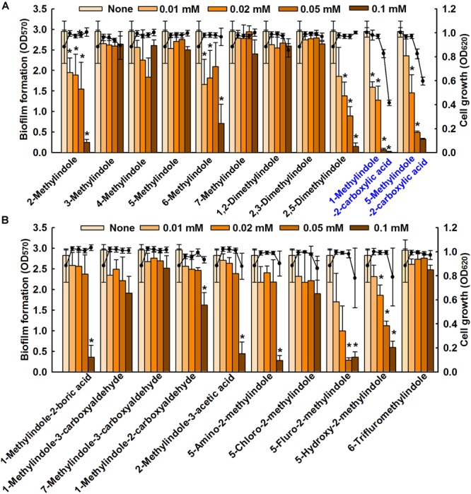
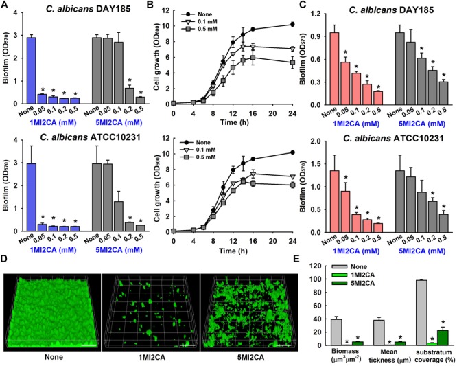
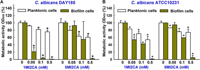
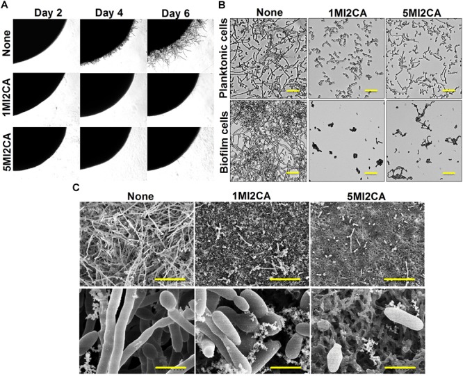
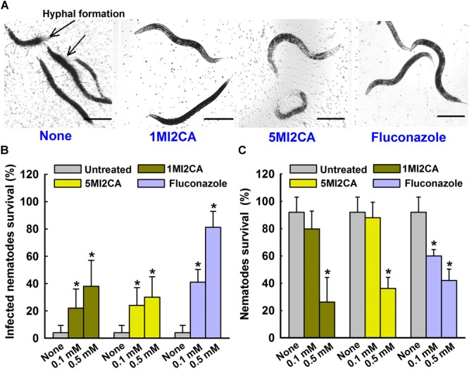
Candida albicans is an opportunistic fungal pathogen and most prevalent species among clinical outbreaks. It causes a range of infections, including from mild mucosal infections to serious life-threatening candidemia and disseminated candidiasis. Multiple virulence factors account for the pathogenic nature of C. albicans, and its morphological transition from budding yeast to hyphal form and subsequent biofilm formation is regarded as the most important reason for the severity of Candida infections. To address the demanding need for novel antifungals, we investigated the anti-biofilm activities of various methylindoles against C. albicans using a crystal violet assay, and the metabolic activity was assessed by using a 2,3-bis (2-methoxy-4-nitro-5-sulfo-phenyl)-2H-tetrazolium-5-carboxanilide reduction assay. Changes in biofilm morphologies and thicknesses were determined by confocal laser scanning microscopy and scanning electron microscopy, respectively. Of the 21 methylindoles tested, 1-methylindole-2-carboxylic acid (1MI2CA) at 0.1 mM (17.5 μg ml-1) and 5-methylindole-2-carboxylic acid (5MI2CA) at 0.1 mM effectively inhibited biofilm formation by C. albicans DAY185 and ATCC10231 strains. Moreover, 1MI2CA and 5MI2CA both effectively inhibited hyphal formation, and thus, improved C. albicans infected nematode survival without inducing acute toxic effects. Furthermore, our in silico molecular modeling findings were in-line with in vitro observations. This study provides information useful for the development of novel strategies targeting candidiasis and biofilm-related infections.

Keywords: C. albicans; C. elegans; biofilm; filamentation; methylindoles.


Figures

FIGURE 1
FIGURE 1
Inhibition of biofilm formation by methylindoles. The anti-biofilm activities of 21 methylindoles against C. albicans DAY185 were examined after incubation for 24 h (A,B). The chemical structures of methylindoles are shown in Supplementary Table S1. At least two independent experiments were conducted (six wells per sample); error bars indicate standard deviations. p < 0.05 vs. untreated controls. Bars indicate biofilm formation and lines indicate planktonic cell growth.
FIGURE 2
FIGURE 2
Effect of methylindoles on C. albicans biofilm formation and cell growth. The anti-biofilm activities of 1-methylindole-2-carboxylic acid (1MI2CA) and 5-methylindole-2-carboxylic acid (5MI2CA) were examined against C. albicans DAY185 and C. albicans ATCC10231 in PDB medium (A) and YPD medium (C) after incubation for 24 h. At least two independent experiments were conducted (six wells per sample). Planktonic cell growths of C. albicans DAY185 after incubation with 1MI2CA or 5MI2CA at their biofilm inhibitory concentrations of 0.1 and 0.5 mM for 24 h (B). Confocal laser scanning microscopic observations of the effects of methylindoles on C. albicans biofilms (D). Biofilm formation by C. albicans on polystyrene plates was observed in the presence of 1MI2CA or 5MI2CA at 0.1 mM by confocal laser microscopy. Scale bars represent 100 μm. Biofilm formation was quantified using COMSTAT (E). Two independent experiments were conducted (six wells per sample). Error bars indicate standard deviations. p < 0.05 vs. untreated controls.
FIGURE 3
FIGURE 3
Effects of 1MI2CA and 5MI2CA on the metabolic activity of C. albicans. The metabolic activities of C. albicans DAY185 (A) and C. albicans ATCC10231 (B) planktonic cells and biofilms were quantified in the presence of methylindoles using an XTT reduction assay after incubation for 24 h. Results are presented as mean percentages of metabolic activity vs. untreated controls. Two independent experiments were conducted (six wells per sample); error bars indicate standard deviations. None indicates untreated samples. Error bars indicate standard deviations. p < 0.05 vs. untreated controls.
FIGURE 4
FIGURE 4
Effects of 1MI2CA and 5MI2CA on C. albicans DAY185 morphology and hyphal growth. C. albicans morphology on solid media (A). C. albicans was streaked on PDA agar plates supplemented with 10% fetal bovine serum in the absence or presence of 1MI2CA or 5MI2CA (0.1 mM). Colony morphologies were photographed every 2 days at 37°C. Inhibition of the hyphal growths of planktonic and biofilm cells of C. albicans (B). C. albicans was grown in PDB medium in the absence or presence of 1MI2CA or 5MI2CA (0.1 mM) at 37°C. Cultures were sampled after 24 h and photographed under a bright field microscope. The scale bar represents 100 μm. Microscopic observations (SEM) of the effects of the two methylindoles (0.1 mM) on biofilms and hyphal formation (C). C. albicans cells were grown on nylon filter paper in the presence or absence of 1MI2CA or 5MI2CA and visualized by SEM. Scale bars represent 50 and 5 μm, respectively. At least two independent experiments were conducted. None indicates untreated control cells.
FIGURE 5
FIGURE 5
Molecular docking of the interaction between 1MI2CA or 5MI2CA and adenylate cyclase. The binding orientations of 1MI2CA and 5MI2CA within the active site of adenylate cyclase were similar to that of 2′,5′-dideoxyadenosine 3′-polyphosphate as a positive control.
FIGURE 6
FIGURE 6
Effects of methylindoles on C. albicans virulence in the model of C. elegans. Microscopic images of C. albicans infected C. elegans in absence or presence of methylindoles (0.5 mM) or fluconazole as a positive control (0.1 mM) (A). The arrows indicate hyphal formation in untreated infected nematodes and the scale bar represents 100 μm. The bar graph shows nematode percentage survival after exposure of C. albicans for 4 days to methylindoles (0.1 or 0. 5 mM) (B). The toxicities of 1MI2CA and 5MI2CA were studied on non-infected nematodes by calculating survival rates after exposure for 4 days (C). Fluconazole (0.1 or 0.5 mM) was used as the positive control. None indicates untreated controls. Worm survival was determined based on movement. At least two independent experiments were conducted. Error bars indicate standard deviations. p < 0.05 vs. untreated controls.

Similar articles

Cited by

References

    1. Alastruey-Izquierdo A., Melhem M. S., Bonfietti L. X., Rodriguez-Tudela J. L. (2015). Susceptibility test for fungi: clinical and laboratorial correlations in medical mycology. Rev. Inst. Med. Trop. Sao Paulo 57 57–64. 10.1590/S0036-46652015000700011 - DOI - PMC - PubMed
    1. Almeida R. S., Brunke S., Albrecht A., Thewes S., Laue M., Edwards J. E., et al. (2008). The hyphal-associated adhesin and invasin Als3 of Candida albicans mediates iron acquisition from host ferritin. PLoS Pathog. 4:e1000217. 10.1371/journal.ppat.1000217 - DOI - PMC - PubMed
    1. Brown G. D., Denning D. W., Gow N. A., Levitz S. M., Netea M. G., White T. C. (2012). Hidden killers: human fungal infections. Sci. Transl. Med. 4:165rv113. 10.1126/scitranslmed.3004404 - DOI - PubMed
    1. CDC (2013). Centers for Disease Control and Prevention. Antibiotic Resistance Threats in the United States. Available at: https://www.cdc.gov/drugresistance/biggest_threats.html
    1. Desai N. C., Patel B. Y., Dave B. P. (2017). Synthesis and antimicrobial activity of novel quinoline derivatives bearing pyrazoline and pyridine analogues. Med. Chem. Res. 26 109–119. 10.1007/s00044-016-1732-6 - DOI