. 2018 Oct 16:9:2387.
doi: 10.3389/fimmu.2018.02387. eCollection 2018.

Extended Cleavage Specificity of Human Neutrophil Elastase, Human Proteinase 3, and Their Distant Ortholog Clawed Frog PR3-Three Elastases With Similar Primary but Different Extended Specificities and Stability

Affiliations

Extended Cleavage Specificity of Human Neutrophil Elastase, Human Proteinase 3, and Their Distant Ortholog Clawed Frog PR3-Three Elastases With Similar Primary but Different Extended Specificities and Stability

Zhirong Fu et al. Front Immunol. .

Abstract

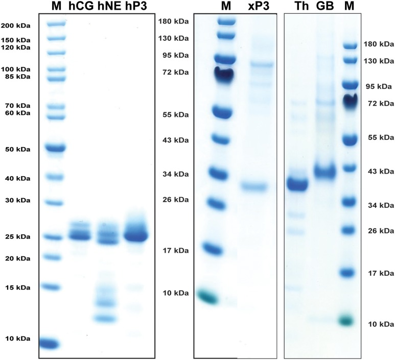
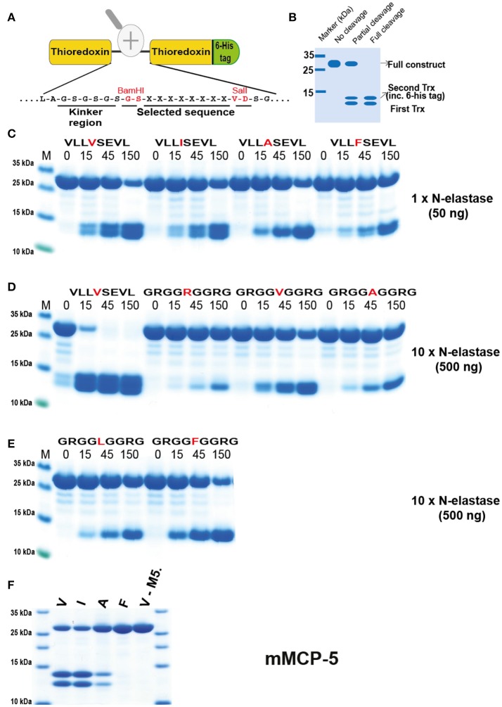
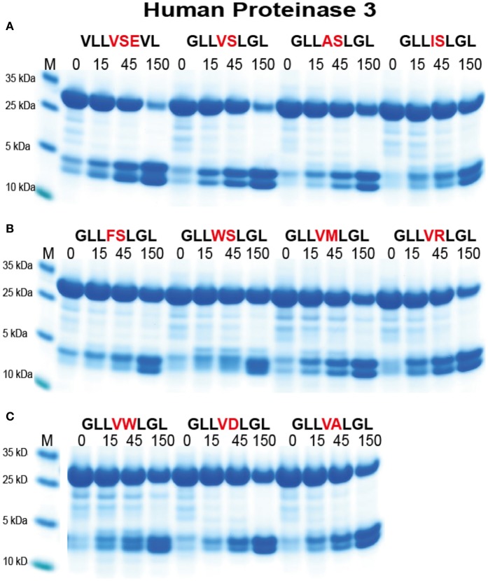
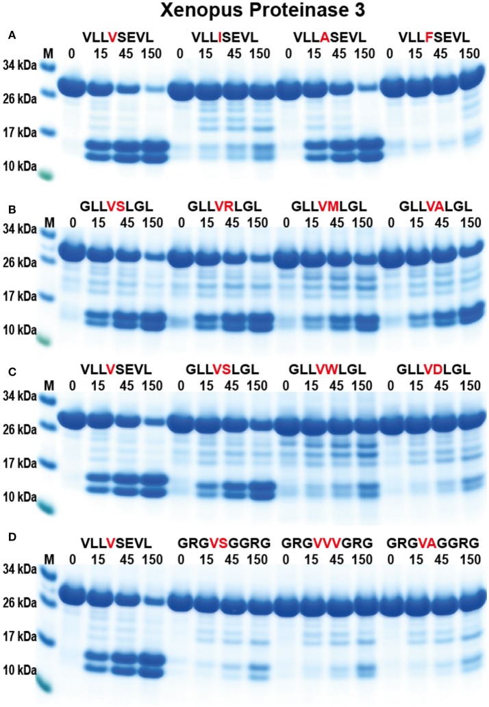
Serine proteases are major granule constituents of several of the human hematopoietic cell lineages. Four proteolytically active such proteases have been identified in human neutrophils: cathepsin G (hCG), N-elastase (hNE), proteinase 3 (hPR-3), and neutrophil serine protease 4 (hNSP-4). Here we present the extended cleavage specificity of two of the most potent and most abundant of these enzymes, hNE and hPR-3. Their extended specificities were determined by phage display and by the analysis of a panel of chromogenic and recombinant substrates. hNE is an elastase with a relatively broad specificity showing a preference for regions containing several aliphatic amino acids. The protease shows self-cleaving activity, which results in the loss of activity during storage even at +4°C. Here we also present the extended cleavage specificity of hPR-3. Compared with hNE, it shows considerably lower proteolytic activity. However, it is very stable, shows no self-cleaving activity and is actually more active in the presence of SDS, possibly by enhancing the accessibility of the target substrate. This enables specific analysis of hPR-3 activity even in the presence of all the other neutrophil enzymes with addition of 1% SDS. Neutrophils are the most abundant white blood cell in humans and one of the key players in our innate immune defense. The neutrophil serine proteases are very important for the function of the neutrophils and therefore also interesting from an evolutionary perspective. In order to study the origin and functional conservation of these neutrophil proteases we have identified and cloned an amphibian ortholog, Xenopus PR-3 (xPR-3). This enzyme was found to have a specificity very similar to hPR-3 but did not show the high stability in the presence of SDS. The presence of an elastase in Xenopus closely related to hPR-3 indicates a relatively early appearance of these enzymes during vertebrate evolution.

Keywords: N-elastase; amphibian; hematopoiesis; neutropenia; neutrophilic granulocyte; phage display; proteinase 3; serine protease.


Figures

Figure 1
Figure 1
Phylogenetic analysis of the hematopoietic serine proteases. A phylogenetic tree of a large number of different vertebrate hematopoietic serine proteases using the MrBayes program. The proteases encoded within the metase locus are enlarged and highlighted and the three proteases analyzed in this communication, hNE, hPR-3, and Xenopus PR-3 are marked by red arrows.
Figure 2
Figure 2
Analysis of the purified hNE, hPR-3, hCG, Xenopus PR-3, human thrombin (Th) and human granzyme B (GB) used in the chromogenic substrate assay and in the determination of the extended cleavage specificity. The three human neutrophil enzymes were commercial preparations purified from peripheral blood neutrophils. The Xenopus PR-3 was produced in the human cell line HEK293-EBNA. The xPR-3 proenzyme was first purified on Ni-NTA beads (–EK) and then activated by removal of the His6-tag by enterokinase digestion (+EK). The enzymes were analyzed by separation on SDS-PAGE and visualized with Coomassie Brilliant Blue staining.
Figure 3
Figure 3
Chromogenic substrate assay. (A–K) A panel of different chromogenic substrates was used to determine the primary specificity of hNE, HPR-3, xPR-3, hCG, human granzyme B, and human thrombin. The panel included different chymase, elastase, tryptase and aspase substrates. The amino acid sequences of the substrates are listed on the left side of the figure. Human thrombin, hCG, and human granzyme B were included as reference enzymes for tryptase, chymase, and asp-ase activities, respectively.
Figure 4
Figure 4
Phage displayed nonamers susceptible to cleavage by hNE, hPR-3 and hCG after five biopannings. After the last selection step, phages released by proteolytic cleavage of the three proteases were isolated and the sequences encoding the nonamers were determined. The general sequence of the T7 phage capsid proteins are PGG(X)9HHHHHH, where (X)9 indicates the randomized nonamers. The protein sequences were aligned into a P5-P4′ consensus, where cleavage occurs between positions P1 and P1′. Sequences occurring more than once are marked by the corresponding number to the right of the sequence. The aa are color coded according to the side chain properties as indicated in the legend.
Figure 5
Figure 5
Analysis of the cleavage specificity of hNE by the use of recombinant protein substrates. (A) Shows the overall structure of the recombinant protein substrates used for analysis of the efficiency in cleavage by the different enzymes in Figures 5–10. In these substrates two thioredoxin (trx) molecules are positioned in tandem and the adjacent trx has a His6-tag positioned in its C terminus. The different cleavable sequences are inserted in the linker region between the two trx molecules by the use of two unique restriction sites, one Bam HI and one Sal I site, which are indicated in A. (B) A hypothetical cleavage is shown to highlight possible cleavage patterns. (C–E) Cleavage of recombinant substrates by hNE at different enzyme concentrations (50 or 500 ng). The name and sequence of the different substrates are indicated above the pictures of the gels. The time of cleavage in minutes is also indicated above the corresponding lanes of the different gels. The uncleaved substrates have a molecular weight of ~25 kDa and the cleaved substrates appear as two closely located bands with a size of 12–13 kDa. The cleavage of a panel of substrates with a more strict elastase, as represented by mouse mast cell protease 5, was included in (F).
Figure 6
Figure 6
Analysis of the cleavage specificity of hNE by the use of recombinant protein substrates. (A–D) Shows the cleavages of a number of substrates by hNE. The sequences of the different substrates are indicated above the pictures of the gels. The time of cleavage in minutes is also indicated above the corresponding lanes of the different gels. The uncleaved substrates have a molecular weight of ~25 kDa and the cleaved substrates appear as two closely located bands with a size of 12–13 kDa.
Figure 7
Figure 7
Analysis of the cleavage specificity of hPR-3 by the use of recombinant protein substrates. (A–C) Shows the cleavages of a number of substrates by hPR-3. The sequences of the different substrates are indicated above the pictures of the gels. The time of cleavage in minutes is also indicated above the corresponding lanes of the different gels. The uncleaved substrates have a molecular weight of ~25 kDa and the cleaved substrates appear as two closely located bands with a size of 12–13 kDa.
Figure 8
Figure 8
Analysis of the cleavage specificity of Xenopus PR-3 by the use of recombinant protein substrates. (A–D) Shows the cleavages of a number of substrates by hPR-3. The sequences of the different substrates are indicated above the pictures of the gels. The time of cleavage in minutes is also indicated above the corresponding lanes of the different gels. The uncleaved substrates have a molecular weight of ~25 kDa and the cleaved substrates appear as two closely located bands with a size of 12–13 kDa.
Figure 9
Figure 9
Analysis of the effect of different concentrations of SDS on the cleavage efficiency by five different enzymes, hPR-3, hNE, hCG, human thrombin and xPR-3. Consensus substrate sequences for the different enzymes were inserted in the 2x Trx substrates as shown in Figure 5. These substrates were then were used to analyze the effect of different SDS concentration on the cleavage efficiency by the five different enzymes. We used the substrate (VLLVSEVL) for hNE, the substrate (VLLFSEVL) for hCG, the substrate (VLLVSEVL) for hPR-3, the substrate (LTPRGVRL) for human thrombin and the substrate (VLLVSEVL) for Xenopus PR-3. Lane C for the different enzymes is a control without SDS. The concentrations of the SDS are then listed above the corresponding lane from 0.1 to 8%. The uncleared substrates have a molecular weight of ≈25 kDa and the cleaved substrates appear as two closely located bands with a size of 12–13 kDa. Due to the effect of the SDS to open the protein structure of the Trx molecules cleavage also occur within the Trx molecules, which results in additional bands of different molecular weights.
Figure 10
Figure 10
Analysis of the difference in efficacy in cleavage of a number of consensus substrates identified through phage display and an MS-based method for four human neutrophil proteases. The cleavage of a number of consensus cleavage sites for four different human neutrophil proteases (N-elastase, hCG, hPR3, and NSP4) were studied with the 2x-Trx system. The “A” consensus sites come from phage display analyses performed in our lab and the substrates B and C comes from a proteomics study by O'Donoghue et al. (39). The substrates originating from the two different methods for N-elastase and proteinase 3 both show very good cleavage, whereas for hCG the consensus sites obtained by the proteomics method shows only minor cleavage, indicating a relatively poor site. No phage display has yet been performed on hNSP4, therefore only a proteomics site was studied where minor cleavage after using a relatively high concentration of the enzyme was seen.
Figure 11
Figure 11
Alignment of a panel of NE, PR-3, and NSP-4 sequences. The sequences of a panel of NSP-4, NE, and PR3 sequences from mammals and amphibians were aligned using the Clustal W algorithm in the DNASTAR program together with a small subfamily of fish proteases that cluster between NSP-4 and NE and PR-3 in the phylogenetic tree shown (from Figure 1). The conserved cysteines are marked with red dots and the three residues of the catalytic triad, His-Asp-Ser, with larger green dots. The three residues with positions 189, 216, and 226 in bovine chymotrypsin are marked with purple arrows. These three residues, are in many trypsin related serine proteases, located in the bottom and the sides of the pocket of the protease where the P1 residue of the substrate is positioned upon cleavage.

References

    1. Pham CT. Neutrophil serine proteases: specific regulators of inflammation. Nat Rev Immunol. (2006) 6:541–50. 10.1038/nri1841 - DOI - PubMed
    1. Cowland JB, Borregaard N. Granulopoiesis and granules of human neutrophils. Immunol Rev. (2016) 273:11–28. 10.1111/imr.12440 - DOI - PubMed
    1. Kobayashi SD, Voyich JM, Burlak C, DeLeo FR. Neutrophils in the innate immune response. Arch Immunol Ther Exp. (2005) 53:505–17. 10.1055/s-2005-870318 - DOI - PubMed
    1. Papayannopoulos V, Zychlinsky A. NETs: a new strategy for using old weapons. Trends Immunol. (2009) 30:513–21. 10.1016/j.it.2009.07.011 - DOI - PubMed
    1. Yang H, Biermann MH, Brauner JM, Liu Y, Zhao Y, Herrmann M. New insights into neutrophil extracellular traps: mechanisms of formation and role in inflammation. Front Immunol. (2016) 7:302. 10.3389/fimmu.2016.00302 - DOI - PMC - PubMed

Publication types