Review
. 2018 Oct 29:9:2492.
doi: 10.3389/fimmu.2018.02492. eCollection 2018.

Schistosoma "Eggs-Iting" the Host: Granuloma Formation and Egg Excretion

Affiliations
Review

Schistosoma "Eggs-Iting" the Host: Granuloma Formation and Egg Excretion

Christian Schwartz et al. Front Immunol. .

Abstract

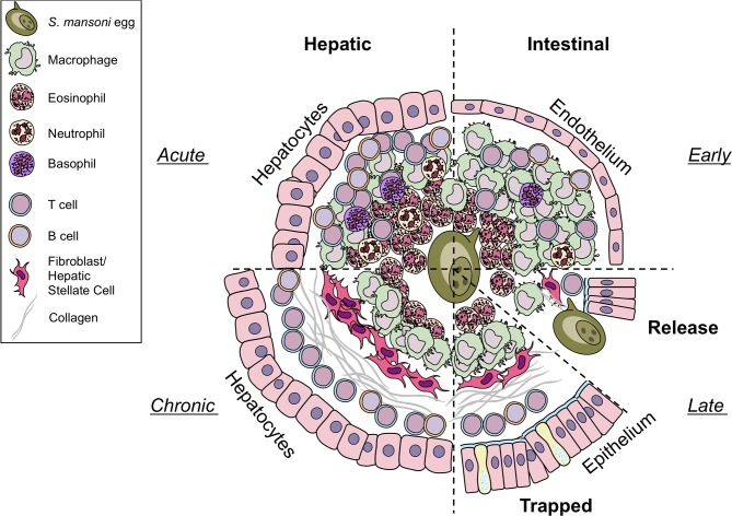
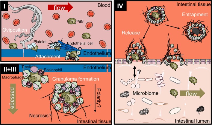
Schistosomiasis is a major cause of morbidity in humans invoked by chronic infection with parasitic trematodes of the genus Schistosoma. Schistosomes have a complex life-cycle involving infections of an aquatic snail intermediate host and a definitive mammalian host. In humans, adult male and female worms lie within the vasculature. Here, they propagate and eggs are laid. These eggs must then be released from the host to continue the life cycle. Schistosoma mansoni and Schistosoma japonicum reside in the mesenteric circulation of the intestines with egg excreted in the feces. In contrast, S. haematobium are present in the venus plexus of the bladder, expelling eggs in the urine. In an impressive case of exploitation of the host immune system, this process of Schistosome "eggs-iting" the host is immune dependent. In this article, we review the formation of the egg granuloma and explore how S. mansoni eggs laid in vasculature must usurp immunity to induce regulated inflammation, to facilitate extravasation through the intestinal wall and to be expelled in the feces. We highlight the roles of immune cell populations, stromal factors, and egg secretions in the process of egg excretion to provide a comprehensive overview of the current state of knowledge regarding a vastly unexplored mechanism.

Keywords: egg; excretion; granuloma; inflammation; intestine; liver; regulation; schistosoma.


Figures

Figure 1
Figure 1
Differences in hepatic and intestinal granuloma composition. Cellular granuloma composition in the liver (left) and the intestine (right). While early (upper half) granulomas may appear similar, intestinal granulomas harbor less eosinophils, T cells, and B cells than hepatic granulomas, more macrophages are present. Only few neutrophils and basophils can be observed in both sites. During later stages (lower half) eggs in the liver become trapped and fibrosis develops. In contrast, eggs deposited in the gut must be released to the intestinal lumen by yet to define mechanisms. However, they may also become trapped and may resemble chronic liver granulomas in shape and collagen content. Illustrations modified from Servier Medical Art, licensed under a Creative Common Attribution 3.0 Unported License.
Figure 2
Figure 2
Release of smCKBP from S.mansoni live eggs under in vitro conditions and detection in vivo adjacent to eggs within granulomas in the liver and intestine of infected mice. (A) Live eggs were cultured in vitro with anti-smCKBP rabbit sera or normal rabbit sera (NRS), with SmCKBP-antibody precipitate formation (arrows) when cultured with anti-smCKBP sera. (B) Immohistochemistry detection of smCKBP (brown stain) within the granulomas surrounding eggs in liver or intestines of infected mice. Bar, 50 μm. 2015 Smith et al. Originally published in The Journal of Experimental Medicine. https://doi.org/10.1084/jem.20050955. (C) Smaller granulomas forming around eggs within intestines of S. mansoni-infected mice treated with anti-CD4 mAb compared to control mice, as described (44). H&E-stained sections. Bar, 100 μm.
Figure 3
Figure 3
Proposed four-stage process of intestinal egg excretion. I: Adult schistosomes deposit eggs into the vasculature close to the lamina propria. Platelets and fibrinogen adhere to the eggs and activate the endothelium. Endothelial cells actively grow over the egg supporting its extravasation. Eggs that do not cross the endothelial border are disseminated by the blood flow and become trapped mostly in the liver portal system. II + III: Immune cells, such as macrophages, T cells and eosinophils start to encapsulate the egg. Granuloma formation occurs around the egg and together with other processes, such as fibrinolysis, egg secretions-induced necrosis, leads to the passage of the egg toward the intestinal lumen. IV: Entrapped eggs become fibrotic and calcified. Interaction with the microbiome, epithelial cell death and remodeling may lead to the active release of eggs, which are then released to the environment with host feces. Illustrations modified from Servier Medical Art, licensed under a Creative Common Attribution 3.0 Unported License.

Similar articles

Cited by

References

    1. Vos T, Flaxman AD, Naghavi M, Lozano R, Michaud C, Ezzati M, et al. . Years lived with disability (YLDs) for 1160 sequelae of 289 diseases and injuries 1990–2010: a systematic analysis for the Global Burden of Disease Study 2010. Lancet (2012) 380:2163–96. 10.1016/S0140-6736(12)61729-2 - DOI - PMC - PubMed
    1. Who_Fact_Sheet WHO Schistosomiasis Fact Sheet (2018). Available online at: http://www.who.int/news-room/fact-sheets/detail/schistosomiasis (Accessed June 27, 2018).
    1. Berry A, Mone H, Iriart X, Mouahid G, Aboo O, Boissier J, et al. . Schistosomiasis haematobium, Corsica, France. Emerg Infect Dis. (2014) 20:1595–7. 10.3201/eid2009.140928 - DOI - PMC - PubMed
    1. Berry A, Fillaux J, Martin-Blondel G, Boissier J, Iriart X, Marchou B, et al. . Evidence for a permanent presence of schistosomiasis in Corsica, France, 2015. Euro Surveill. (2016) 21. 10.2807/1560-7917.ES.2016.21.1.30100 - DOI - PubMed
    1. Colley DG, Andros TS, Campbell CH, Jr. Schistosomiasis is more prevalent than previously thought: what does it mean for public health goals, policies, strategies, guidelines and intervention programs? Infect Dis Poverty (2017) 6:63. 10.1186/s40249-017-0275-5 - DOI - PMC - PubMed

Publication types