Review
. 2018 Nov 6:9:1612.
doi: 10.3389/fpls.2018.01612. eCollection 2018.

Pectin Methylesterases: Cell Wall Remodeling Proteins Are Required for Plant Response to Heat Stress

Affiliations
Review

Pectin Methylesterases: Cell Wall Remodeling Proteins Are Required for Plant Response to Heat Stress

Hui-Chen Wu et al. Front Plant Sci. .

Abstract

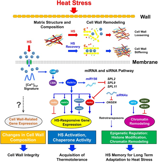
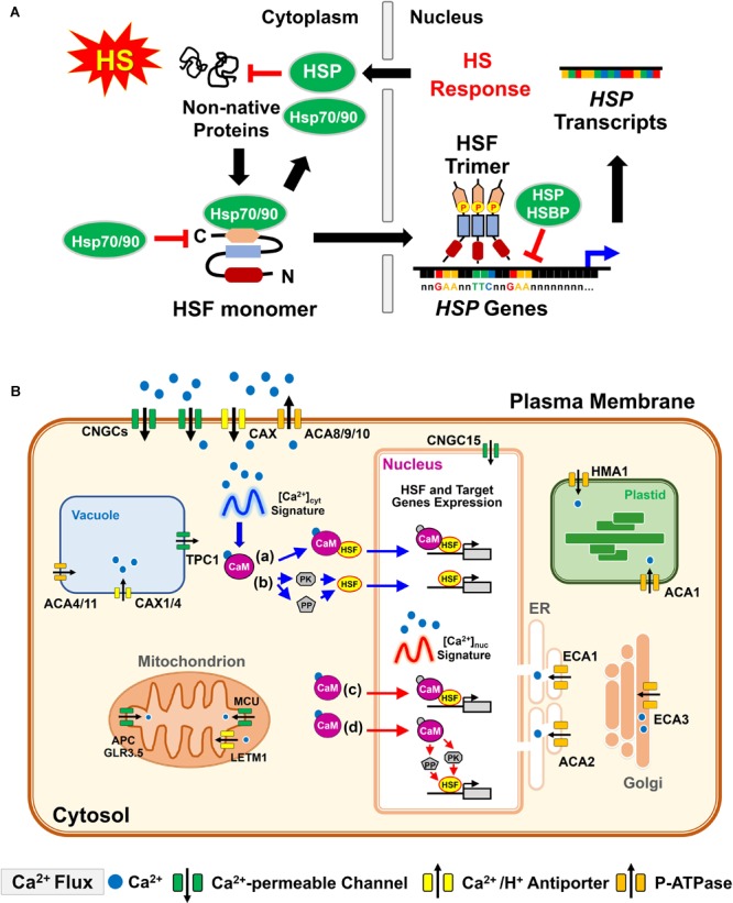
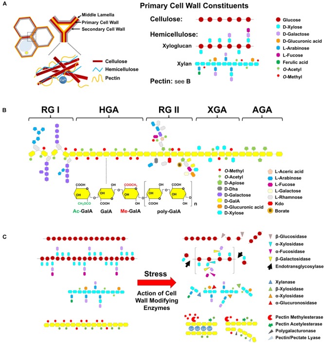
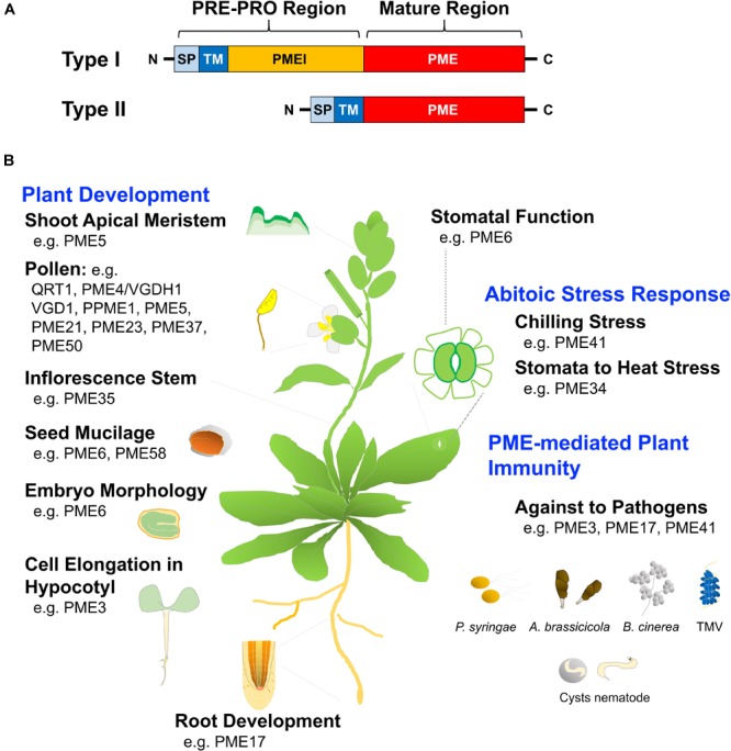
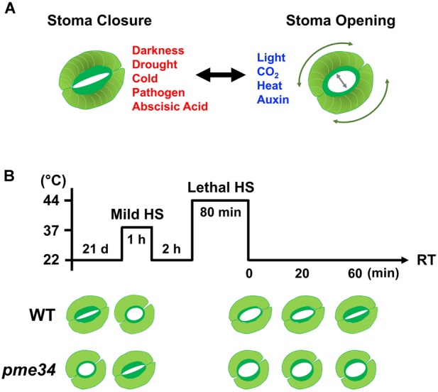
Heat stress (HS) is expected to be of increasing worldwide concern in the near future, especially with regard to crop yield and quality as a consequence of rising or varying temperatures as a result of global climate change. HS response (HSR) is a highly conserved mechanism among different organisms but shows remarkable complexity and unique features in plants. The transcriptional regulation of HSR is controlled by HS transcription factors (HSFs) which allow the activation of HS-responsive genes, among which HS proteins (HSPs) are best characterized. Cell wall remodeling constitutes an important component of plant responses to HS to maintain overall function and growth; however, little is known about the connection between cell wall remodeling and HSR. Pectin controls cell wall porosity and has been shown to exhibit structural variation during plant growth and in response to HS. Pectin methylesterases (PMEs) are present in multigene families and encode isoforms with different action patterns by removal of methyl esters to influencing the properties of cell wall. We aimed to elucidate how plant cell walls respond to certain environmental cues through cell wall-modifying proteins in connection with modifications in cell wall machinery. An overview of recent findings shed light on PMEs contribute to a change in cell-wall composition/structure. The fine-scale modulation of apoplastic calcium ions (Ca2+) content could be mediated by PMEs in response to abiotic stress for both the assembly and disassembly of the pectic network. In particular, this modulation is prevalent in guard cell walls for regulating cell wall plasticity as well as stromal aperture size, which comprise critical determinants of plant adaptation to HS. These insights provide a foundation for further research to reveal details of the cell wall machinery and stress-responsive factors to provide targets and strategies to facilitate plant adaptation.

Keywords: cell wall remodeling; guard cell wall; heat stress response; pectin; pectin methylesterase.


Figures

FIGURE 1
FIGURE 1
Integration of cell wall remodeling and the heat response network. Plant perception of heat involves several pathways in different compartments. The cell wall is the first protective barrier in plants that is exposed to heat. Heat stress (HS)-triggered pectin methylesterases (PME) activity, accompanied by Ca2+ mobilization from apoplastic sources, is involved in cell wall remodeling and is crucial for plant thermotolerance. During recovery time after HS, PME performs linear demethylesterification on highly esterified pectin residues and interacts with Ca2+ to form a pectate gel lawn, which causes cell-wall stiffening. During non-lethal HS, acidic PME acts randomly on pectin and promotes the action of endo-polygalacturonases (PG) to contribute to cell-wall loosening and the release of Ca2+ through Ca2+-permeable channels (green oval) in the plasma membrane, thus causing a transient increase in [Ca2+]cyt oscillation. This is followed by induction of a Ca2+/calmodulin (CaM)-dependent pathway to activate the master HS regulator HsfA1s, which directly triggers HS-responsive transcription factors, including HsfA2, HsfA7s, HsfBs, and dehydration-responsive element-binding protein 2A for downstream HsfA3 and HSP gene expression involved in the acquisition of thermotolerance. Histone modification and several epigenetic regulators, including small RNAs and transposons, are involved in the HSR and HS memory. MicroRNA156 targets the SQUAMOSA promoter-binding protein-like gene family, which downregulates HS-inducible genes and therefore maintains the expression of HsfA2 and HSP genes during recovery from HS for long-term adaptation to HS. The retrotransposon ONSEN, as a target of HsfA1s and HsfA2, can be modulated by siRNAs for the regulation of HS memory. Through heat-intolerant 4 (HIT4), HS can relax the silencing of transposons, whereas they can be silenced by deficient in DNA methylation 1 (DDM1) and Morpheus’ molecule 1 (MOM1). However, the HS-induced cell wall-related transcript profile needs to be further explored with regard to the maintenance and modification of cell wall integrity.
FIGURE 2
FIGURE 2
Basic function of plant HSF and HS-induced [Ca2+]cyt/nuc oscillation interpretation by CaM in response to heat. (A) Under unstressed conditions, Hsp70/HspP90 can directly regulate the function of HSF by blocking its transcriptional activity. Upon HS, non-native proteins induce the conversion of monomeric HSF into an active trimeric form, which is phosphorylated and translocated into the nucleus. HSF trimer, with high-affinity DNA binding capacity to the HSE (5′-nGAAnnTTCnnGAAn-3′) of the HSP gene promoter region, activates HSP gene expression, whereas it is downregulated by the interaction of HSP and HSBP with the HSF trimer to attenuate HSR in plants. HSP production and relocation to the cytoplasm inhibits non-native protein misfolding and aggregation. (B) Cellular Ca2+ transport is tightly controlled within all membrane-bound organisms during heat stress. An increase in [Ca2+]cyt is manifested by Ca2+ influx to the cytosol, mediated by Ca2+-permeable ion channels, either from the apoplast across the plasma membrane, or from intracellular stores such as the endoplasmic reticulum or vacuole. In contrast, Ca2+-ATPases and the Ca2+/H+ antiporter systems are responsible for Ca2+ extrusion out of the cytosol. HS-elevated Ca2+ occurs from apoplast entry to the cytosol or nucleus (either diffused from the cytosol or released from nuclear Ca2+ reservoirs). The CaM responds to the elevation of [Ca2+]cyt signature to modulate the activity of numerous target proteins. (a) and (c) The Ca2+/CaM complex interacts with the HS transcription factors (HSFs) and modulates either HSF DNA-binding or transcriptional activities. (b,d) The Ca2+/CaM complex regulates the activation of HSF by modulating the phosphorylation status. The regulation is achieved by CaM-binding protein kinase (PK) or CaM binding protein phosphatase (PP). (c,d) CaM recognizes a high frequency and magnitude of the cytosolic Ca2+ signature and is translocated into the nucleus for responding to the nuclear [Ca2+] ([Ca2+]nuc) to bind or regulate the status of HSF phosphorylation in the nucleus. ACAs, autoinhibited Ca2+-ATPases; APC, adenine nucleotide/phosphate carrier; CAXs, Ca2+/H+ cation antiporters; CNGC, cyclic nucleotide-gated ion channels; ECAs, ER-type calcium ATPases; GLR3.5, glutamate receptor 3.5; HMA1, heavy metal translocating P-type ATPase; LETM1; leucine zipper-EF-hand-containing transmembrane protein 1; MCU, mitochondrial calcium uniporter; TPC1, two-pore voltage-gated channel 1.
FIGURE 3
FIGURE 3
Cell wall composition and enzymatic modification in response to heat. (A) The cell wall is a complex structure that is composed of cellulose and non-cellulosic neutral polysaccharides embedded in a pectin matrix. Pectins are located in the middle lamella and primary and secondary cell wall. Major primary cell wall are constituted of cellulose microfibrils (multiple chains of β-glucose with β-1,4 glycosidic bonds) which are cross-linked to hemicelluloses and to pectin. Xyloglucan is a major hemicellulose molecule that is composed of β-1,4-linked glucose residues with α-1,6-linked xylosyl side chains. In turn, these side chains can be decorated with either galactose, or fucose residues to create a complex pattern of branches. Xylan consists a backbone of β-1,4-linked xylose (Xyl) residues that can be substituted with glucuronic acid and/or arabinose. Additional substitutions such as acetyl and methyl groups can be also presented. And (B) pectins are highly complex class of polysaccharides that comprise galacturonic acid-rich, consisting of five major classes, namely: homogalacturonan (HGA), rhamnogalacturonan I (RG-I), rhamnogalacturonan II (RG-II), xylogalacturonan (XGA), and apiogalacturonan (AGA) form a structurally diverse glue which provides stiffness or flexibility relying on the chemical modification. (C) Based on the action of hydrolysis and substrate specificity, the degradation of cellulose is cleaved by β-glucosidase into two molecules of glucose; for breaking down hemicellulose, xyloglucan endotransglycosylase/hydrolase (XTH), and expansin proteins (not shown) associated with disassembly of cellulose and xyloglucan matrix may play a role in the cell wall remodeling in different aspects of plant development and stress responses. Xylanase is responsible for degrading xylan by cleaving β-1,4 xylose linkages in the backbone. β-xylosidases cleave xylose from the non-reducing end of the xylan chain, and glucuronidases cleave the α-1,2 linked glucuronic acid, and α-arabinosidases cleave the α-1,2 and α-1,3 linked arabinose from the backbone. Pectinolytic enzymes such as PME, PAE, PG, PL, and Arabinanase by hydrolysis of pectic substances, are important for cell wall remodeling. The HGA, a polysaccharide of α-1,4-linked galacturonic acid (GalA) residues, is the predominant form of pectin. A critical feature of HGA that influences its properties is the methyl-esterification and acetylation of specific carbons on GalA during synthesis of the backbone. HGA is de-methylesterified by the activity of PME, which results in random and contiguous patterns of free carboxylic residues. De-methyl-esterification randomly releases protons, which become a target for pectin-degrading enzymes such as PG, which act by hydrolyzing the α-1,4 link between GalA. The contiguous de-methylesterified HGA binds with Ca2+ to induce gel formation, which can rigidify the cell wall.
FIGURE 4
FIGURE 4
Structural motif and function of pectin methylesterase (PME). (A) Types I and II PMEs contain a conserved PME domain, as the active part of the proteins. Type I includes the N-terminal extension of the PRE-PRO region, with the PRE domain containing a signal peptide (SP) or a transmembrane domain (TM) that is required for PME targeting to the cell wall. The N-terminal PRO region shows homology with pectin methylesterase inhibitors (PMEI), whereas type II is characterized by the absence of the PRO region. (B) Deficiency in specific PME genes reveals multiple roles of PME that have been linked to alteration of plant growth development and the response of plant defenses and abiotic stresses, respectively; related references are indicated in Table 1.
FIGURE 5
FIGURE 5
Arabidopsis PME34 regulates the stomatal aperture under heat stress. (A) The inner wall of a guard cell is thicker and more elastic than the outer cell wall to facilitate the opening of the stomatal pore. The elastic property of the guard cell wall acts reversibly during stomatal opening and closing owing to differential thickening and the orientation of cellulose microfibrils (expressed in threads). The openings and closures of the stomata pore are strictly regulated by the integration of environmental stimuli and endogenous hormonal signals. (B) Comparison of elevated temperature stimulated-stomatal opening in wild-type (WT; Col) and pme34 mutant plants. Leaves (21-day-old) of WT and pme34 plants were treated with 37°C-mild and 44°C-lethal heat stress (LHS), respectively, as indicated. The pictogram shows the HS regime and the schematic diagram in the lower panel indicates the response of the stomatal aperture. Under normal condition, pme34 plants had a larger stomata aperture compared with that of WT plants. Under mild-HS treatment, the stomatal apertures in WT plants increased for aiding the dissipation of heat, whereas those of pme34 plants did not. Following the further 44°C LHS at recovery time (RT), stomatal apertures of pme34 were opened wider than those of WT plants, indicating greater water loss than that in WT plants.

Similar articles

Cited by

References

    1. Ali O., Traas J. (2016). Force-driven polymerization and turgor-induced wall expansion. Trends Plant Sci. 21 398–409. 10.1016/j.tplants.2016.01.019 - DOI - PubMed
    1. Amsbury S., Hunt L., Elhaddad N., Baillie A., Lundgren M., Verhertbruggen Y., et al. (2016). Stomatal function requires pectin de-methyl-esterification of the guard cell wall. Curr. Biol. 26 2899–2906. 10.1016/j.cub.2016.08.021 - DOI - PMC - PubMed
    1. An S. H., Sohn K. H., Choi H. W., Hwang I. S., Lee S. C., Hwang B. K. (2008). Pepper pectin methylesterase inhibitor protein CaPMEI1 is required for antifungal activity, basal disease resistance and abiotic stress tolerance. Planta 228 61–78. 10.1007/s00425-008-0719-z - DOI - PMC - PubMed
    1. Anthon G. E., Barrett D. M. (2006). Characterization of the temperature activation of pectin methylesterase in green beans and tomatoes. J. Agric. Food Chem. 54 204–211. 10.1021/jf051877q - DOI - PubMed
    1. Aragunde H., Biarnés X., Planas A. (2018). Substrate recognition and specificity of chitin deacetylases and related family 4 carbohydrate esterases. Int. J. Mol. Sci. 19:412. 10.3390/ijms19020412 - DOI - PMC - PubMed